Chapter 7: Microscopic Examination
 Call Kai
 Learn
Learn Practice Test
 Spaced Repetition
 Match
Match Flashcards
 Knowt Play
1/53

There's no tags or description
Looks like no tags are added yet.
Name | Mastery | Learn | Test | Matching | Spaced | Call with Kai | Chat |
|---|
No analytics yet
Send a link to your students to track their progress
54 Terms
purulence
pus, or the condition of containing or discharging pus
gram postive bacteria
purple
-thick peptidoglycan wall

Gram-negative bacteria
pink/red
-thin peptidoglycan wall

smears from swabs
-Don't use swab from inoculated media
-Collect two swabs
-Don't rub swab on slide
-Roll back and forth

smears from thick liquids or semisolids
such as feces
-swab immersed in the specimen for several seconds
-thin spread of material on the slide
Smears from thick, granular, or mucoid materials
-Two-slide technique
-Place slide on top of other slide and pull slides away from each other
smears from thin fluids
-dropped, not spread
-done on well-marked area

cytocentrifugation
mechanical process in which centrifugal force is used in a machine to deposit cells on a glass slide for staining and viewing

Protein stains
gram-negative
cytocentrifuge technique
1. small aliquots of fluid are placed in the cytocentrifuge holders
2. material is spun for 10 minutes
3. slide is removed; if deposit of cells is too heavy, a portion of the cellular deposition can be smeared
4. the sediment is fixed and decontaminated in 70% alcohol for 5 minutes

simple stains
one dye is used; reveals shape, size, and arrangement
-directed toward coloring the forms and shapes present

differential stains
directed toward coloring specific components of the elements present

probe-mediated stains
are directed specifically at identification of an organism
Wright-Giemsa stain
visualizes bacteria, yeasts, parasites, and viral inclusions

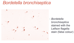
Leifson stain
flagella
Methylene blue stain
metachromatic granules
-Corynebacterium diphtheriae

Acid-fast stains
differential stain used to identify acid-fast organisms such as members of the genus Mycobacterium

Ziehl-Neelsen stain
Use to stain Acid-fast bacteria
-sediments for mycobacteria
-partial acid-fastness of Nocardia spp.

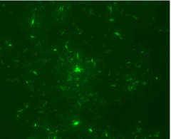
Fluorochrome stain
preferred acid-fast stain
-sediments for mycobacteria
Kinyoun stain
This stain is a modified Ziehl-Neelson stain, with no heating step
-for Crytosporidium and Cyclospora parasites in stool specimens

Calcofluor white stain
A fluorochrome that stains chitin found in the cell wall of fungi. Viewed under UV light. Fungi appear white to blue.
-for bronchoalveolar fungi and some parasitic cysts
-differentiates them from background materials of similar morphology

gross examination
Viewing specimens with the naked eye
-eyes, magnifying glass, and dissecting microscope
bright-field microscope
most widely used; specimen is darker than surrounding field; used for live and preserved stained specimens

dark-field microscope
brightly illuminated specimens surrounded by dark field; used for live and unstained specimens
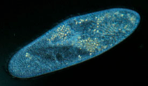
phase-contrast microscope
light microscope that enhances contrast; useful in examining living, unstained cells

fluorescence microscope
Instrument used to visualize a specimen that has been labeled with a fluorescent dye

electron microscope
microscope that forms an image by focusing beams of electrons onto a specimen
transmission electron microscope
An electron microscope used to study the internal structure of thin sections of cells

scanning electron microscope
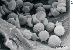
An electron microscope used to study the fine details of cell surfaces
Background material: amorphous debris
Debris without form or shape that is made up of necrotic tissue or cells, protein fluids, or mucus

Background material: black particulate debris
smoke inhalation, crack cocaine
Background material: Charcot-Leyden crystals
Bronchial asthma (eosinophil membranes)

Background material: Epithelials with contaminating bacteria
passage of specimen through contaminated area during collection
Background material: Curschmann's spirals present (sputum)
Bronchospasm, obstruction, asthma

Background material: Epithelial cells
epithelial surface involved or adjacent to collection site
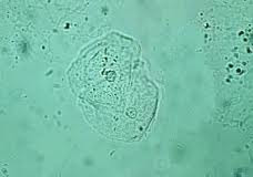
Background material: Intracellular organisms
inferred association in infection

Background material: Local material
reflection of collection site
Background material: mononuclear cells present
chronic inflammation

Background material: mononuclear cells
blood cells that have a single, round nucleus, such as lymphocytes and monocytes
Background material: mucus
irritation of glandular surface

Background material: purulence
acute inflammation, exudation
Background material: red blood cells
trauma, hemorrhage

colony-forming units
A number of separable cells on the surface of a semisolid agar medium that create a visible colony.

monomicrobial
infection caused by a single species
polymicrobial
infection caused by multiple species
Direct smear examination of clinical samples is a rapid means to identify presumptively the etiologic agents of infectious disease. (True or False)
True
The presence of an infectious disease process can be assessed on a direct smear based on which of the following?
a. the presence of numerous inflammatory cells
b. the morphology of the bacteria present
c. gram stain reaction of bacteria present
d. the presence of numerous epithelial cells
A. the presence of numerous inflammatory cells
Which of the following stains may be used for direct smear examination?
a. gram stain
b. acid-fast stain
c. wright or giemsa stain
d. calcofluor white stain
e. all of the above
E. all of the above
Which of the following stains is best used to detect mycobacterial organisms in clinical samples?
a. gram
b. giemsa
c. acid-fast
d. india ink
C. acid-fast
Calcofluor white is a colorless dye that binds with which of the following structures?
a. cell wall containing mycolic acid
b. chitin
c. peptidoglycan layer
d. metachromatic granules
B. chitin
chitin
Polysaccharide found in arthropod exoskeletons and fungal cell walls.

Cytocentrifugation is an excellent method for which of the following types of samples?
a. heavily contaminated
b. nonviscous fluids
c. thick purulent material
d. filled with mucus
B. nonviscous fluids
A monobacterial type of infection can be immediately suspected based on the direct microscopic examination of the clinical sample. (True or False)
True
In a Wright-Giemsa-stained smear, bacteria would appear as which of the following colors?
a. red
b. blue
c. purple
d. orange
B. blue