Bones & Muscles of Trunk and Pelvic Floor
 Call Kai
 Learn
Learn Practice Test
 Spaced Repetition
 Match
Match Flashcards
 Knowt Play
1/91

There's no tags or description
Looks like no tags are added yet.
Name | Mastery | Learn | Test | Matching | Spaced | Call with Kai | Chat |
|---|
No analytics yet
Send a link to your students to track their progress
92 Terms
Cervical vertebrae
7 vertebrae located in the neck region.
Thoracic vertebrae
12 vertebrae located in the thorax; articulate with the ribs.
Lumbar vertebrae
5 vertebrae located in the small of the back.
Sacrum
5 fused vertebrae that articulate with the hip bones.
Coccyx
Usually 4 fused coccygeal vertebrae; commonly called the tailbone.
Vertebral column regions
Cervical → thoracic → lumbar → sacrum → coccyx.
Ribs
12 pairs of bones that articulate with the thoracic vertebrae and sternum.
Sternum
Bone of the anterior thorax consisting of the manubrium, body, and xiphoid process.
Manubrium
Superior portion of the sternum.
Body of sternum
Large middle portion of the sternum.
Xiphoid process
Inferior portion of the sternum.
Three regions of the sternum
Manubrium, body, and xiphoid process.
Hip bones
Part of the appendicular skeleton; important attachment sites for muscles.
Three regions of the hip bone
Ilium, ischium, and pubis.
Ilium
Superior region of the hip bone.
Iliac crest
Superior border of the ilium.
Ischium
Posteroinferior portion of the hip bone.
Ischial tuberosity
Part of the ischium commonly called the 'sits bone.'
Pubis
Anterior portion of the hip bone.
Superficial back muscles
Back muscles primarily involved in moving the upper limbs.
Deep back muscles
Back muscles that move and stabilize the vertebral column.
Quadratus lumborum
Deep back muscle that extends and laterally flexes the lumbar vertebral column.
Bilateral action of quadratus lumborum
Both quadratus lumborum muscles together extend the lumbar vertebral column.
Unilateral action of quadratus lumborum
One quadratus lumborum laterally flexes the vertebral column.
Quadratus lumborum location
Extends from the iliac crest of the hip bone toward the lumbar region/rib.
Erector spinae
Group of deep back muscles that extend the vertebral column and help maintain erect posture.
Function of erector spinae
Extends the vertebral column and maintains erect posture.
Three muscles of erector spinae
Iliocostalis, longissimus, and spinalis.
Iliocostalis
Lateral muscle of the erector spinae group.
Longissimus
Intermediate muscle of the erector spinae group.
Spinalis
Medial muscle of the erector spinae group.
Erector spinae arrangement
Lateral to medial: iliocostalis → longissimus → spinalis.
Superficial thorax muscles
Primarily move the upper limbs and are not primarily involved in respiration.
Deep thorax muscles
Include the diaphragm and intercostal muscles involved in respiration.
Diaphragm
Dome-shaped muscle whose contraction pulls its central portion downward, increasing the vertical dimension of the thorax.
Diaphragm attachments
Attaches to the ribs, sternum, and vertebrae.
Diaphragm during inhalation
Contracts and flattens, increasing the vertical dimension of the thoracic cavity.
Diaphragm during exhalation
Relaxes and returns to its dome shape.
External intercostals
Muscles located between adjacent ribs that elevate the ribs during inhalation.
External intercostal fiber direction
Fibers run inferomedially.
Internal intercostals
Muscles between the ribs that depress the ribs during forced exhalation.
Internal intercostal fiber direction
Fibers run inferolaterally.
Main muscle actions during inhalation
Diaphragm contracts and external intercostals elevate the ribs, increasing the dimensions of the thoracic cavity.
Abdominal muscles
Muscles that bilaterally flex the vertebral column and compress the abdominal wall.
Bilateral action of abdominal muscles
Flex the vertebral column and compress the abdominal wall.
Unilateral action of abdominal obliques
External and internal abdominal obliques laterally flex the vertebral column when acting unilaterally.
External abdominal oblique
Abdominal muscle whose fibers run inferomedially; acts in flexion, compression, and lateral flexion of the vertebral column.
External abdominal oblique fiber direction
Fibers run inferomedially, similar to the external intercostals.
Internal abdominal oblique
Abdominal muscle whose fibers run inferolaterally; acts in flexion, compression, and lateral flexion of the vertebral column.
Internal abdominal oblique fiber direction
Fibers run inferolaterally, similar to the internal intercostals.
Transversus abdominis
Abdominal muscle whose fibers run horizontally or straight across the abdomen.
Transversus abdominis fiber direction
Fibers run straight across the abdomen.
Rectus abdominis
Abdominal muscle with fibers running vertically up and down; contributes to flexion of the vertebral column.
Rectus abdominis fiber direction
Fibers run vertically up and down.
"Wash-board abs"
Rectus abdominis.
Muscles that flex the vertebral column
Rectus abdominis and other abdominal muscles; the notes also identify sternocleidomastoid as flexing the head/neck.
Muscles that laterally flex the vertebral column
External abdominal oblique, internal abdominal oblique, and quadratus lumborum.
Pelvis
Bony ring formed by the left and right hip bones and the sacrum.
Pelvic floor muscles
Muscles that attach to the bones of the pelvis and help form the floor of the pelvic cavity.
Pelvic diaphragm
Part of the pelvic floor; formed from 3 separate muscles and described as funnel-shaped.
Shape of pelvic diaphragm
Funnel-shaped.
Functions of the pelvic floor
Closes the inferior opening of the pelvis, supports pelvic organs, and assists with defecation, voluntary urination, and childbirth.
Pelvic floor and pelvic organs
Pelvic floor muscles support the organs located within the pelvis.
Pelvic floor and urination
Pelvic floor muscles assist with the voluntary part of urination.
Pelvic floor and defecation
Pelvic floor muscles assist with defecation.
Pelvic floor and childbirth
Pelvic floor muscles assist with childbirth.
Perineum
Diamond-shaped region between the thighs, commonly described as the crotch.
Two regions of the perineum
Urogenital triangle and anal triangle.
Urogenital triangle
Region of the perineum containing the external genitalia and urethra.
Anal triangle
Region of the perineum containing the anus.
Pelvic organ prolapse
Occurs when damage to the pelvic floor causes pelvic organs to herniate or 'fall.'
Cause of pelvic organ prolapse
Damage or weakening of the pelvic floor.
Possible causes of pelvic floor weakening
Pregnancy, childbirth, older age, obesity, and heavy lifting.
Pelvic organ prolapse treatments
Exercises to strengthen the muscles, devices that help hold the organs in place, or surgery.

Name most superior part of sternum
Manubrium

Name of middle part of sternum
Body

Name of most inferior part of sternum
Xiphoid process

Name yellow part
Ilium
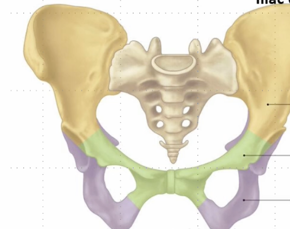
Name green part
Pubis

Name purple part
Ischium

Name this muscle and bone
Quadratus Lumborum (Deep back muscle) and Iliac crest of hip bone

Name blue muscle
Iliocostalis (part of erector spinae)
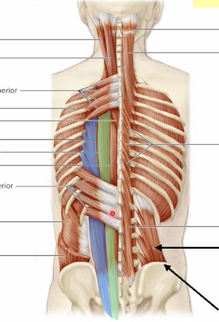
Name green muscle
Longissimus (part of erector spinae)
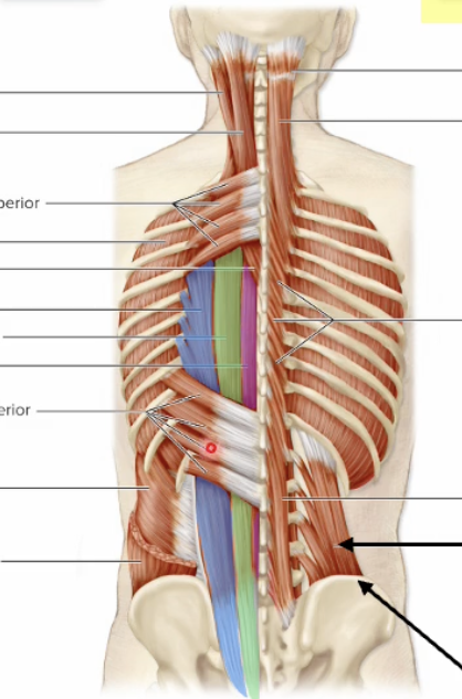
Name purple muscle
Spinalis (part of erector spinae)
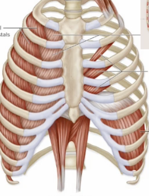
Name lateral muscle on ribs
External Intercostals


Name medial muscle on ribs
Internal Intercostals
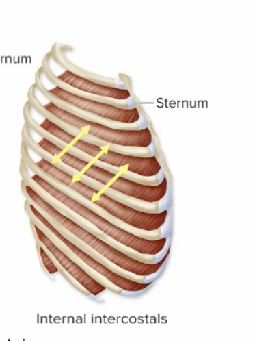

Name muscle
External Abdominal Obliques

Name muscle
Internal Abdominal Obliques
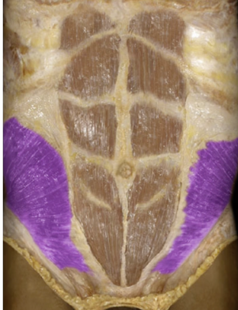
Name muscle that gives six pack abs
Rectus Abdominis

Name muscle
Transversus abdominis

Name muscle in green
Urogenital triangle
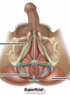
Name muscle in purple
Anal triangle