Muscular System
1/69
There's no tags or description
Looks like no tags are added yet.
Name | Mastery | Learn | Test | Matching | Spaced |
|---|
No study sessions yet.
70 Terms
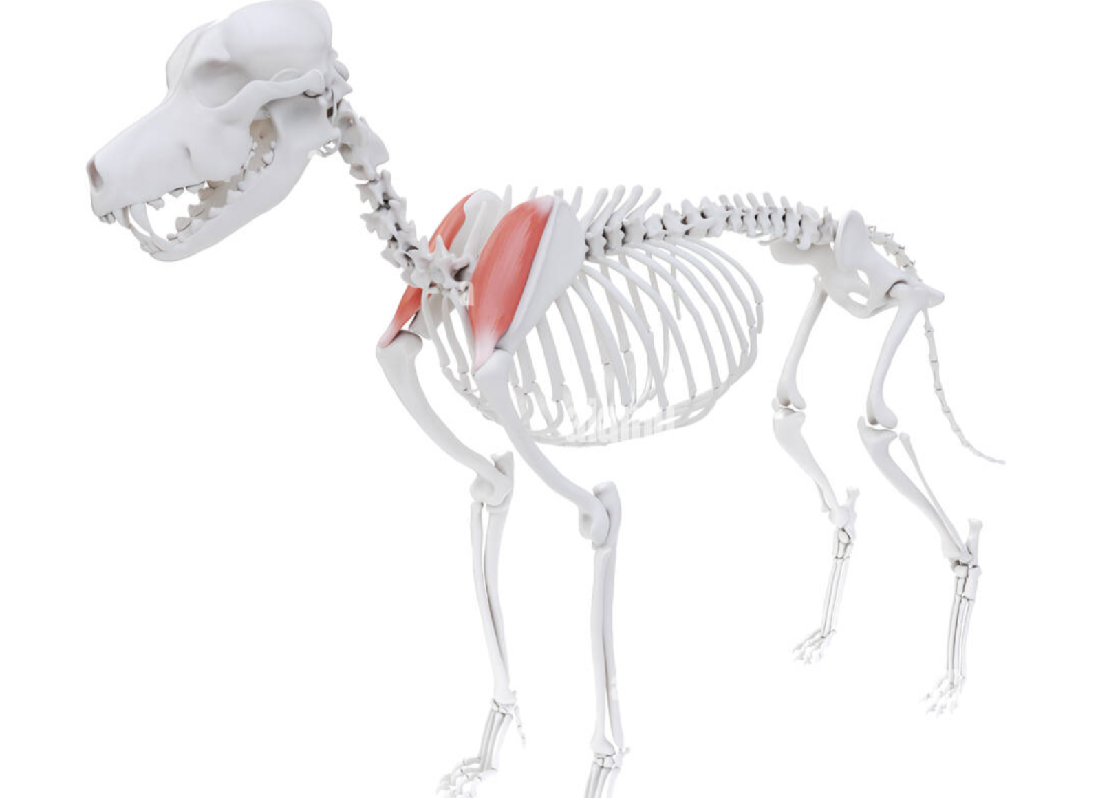
Supraspinatus Muscle
Extensor muscle. Cranial to the spine of the scapula. Extends and stabilizes the shoulder joint.
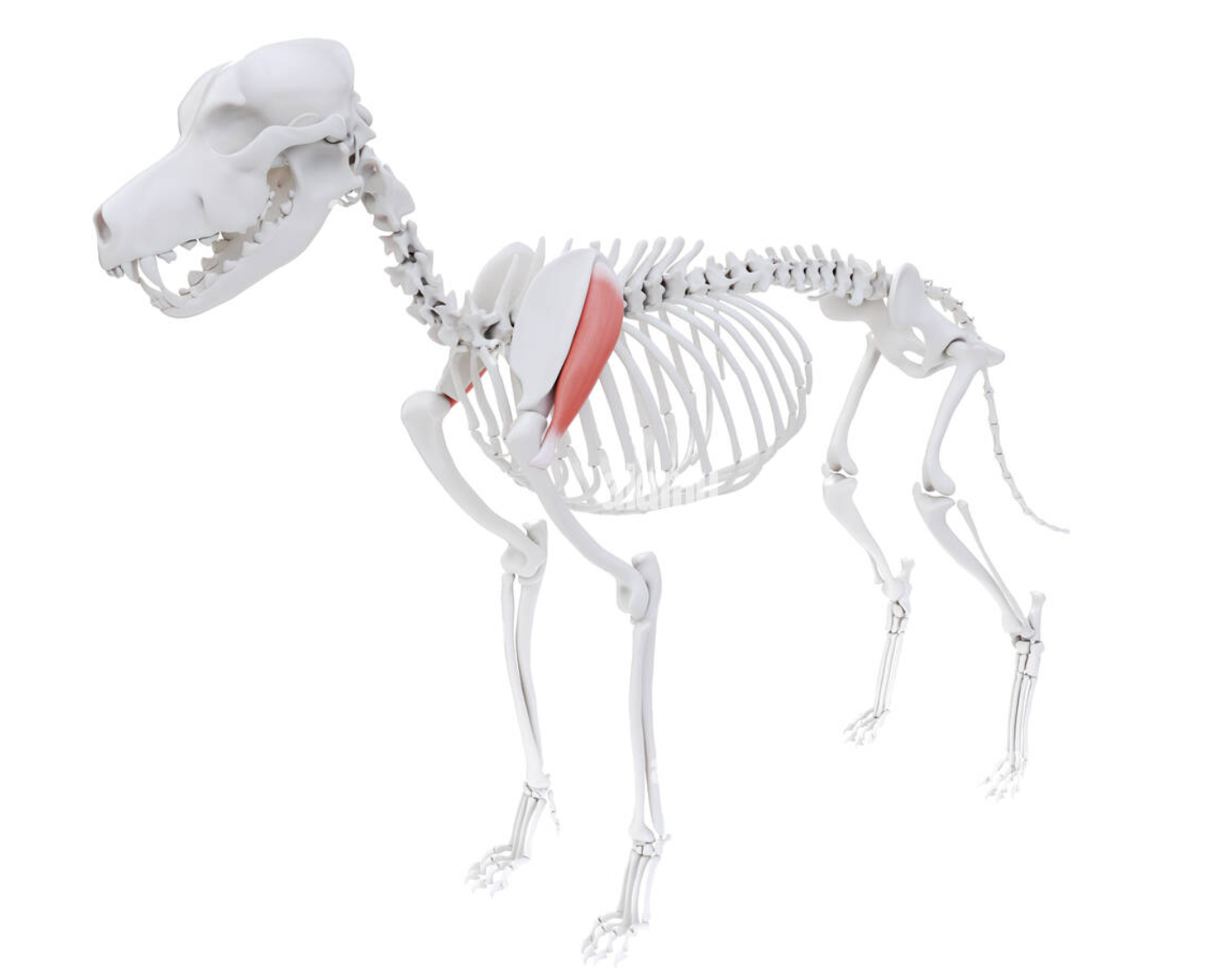
Infraspinatus Muscle
Caudal to the scapular spine. Flexes the shoulder joint in conjunction with muscles that attach the humerus to the spine or sternum
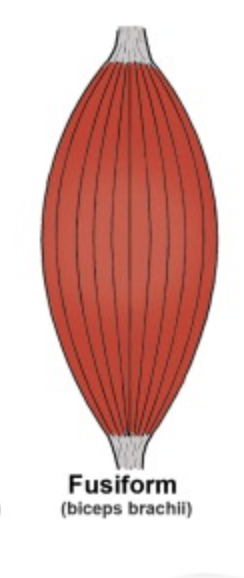
Fusiform Muscle
(Strap Muscle) Muscle with parallel fiber arrangement generally associated with greater range of motion and joint velocity than muscles with a different fiber arrangement.
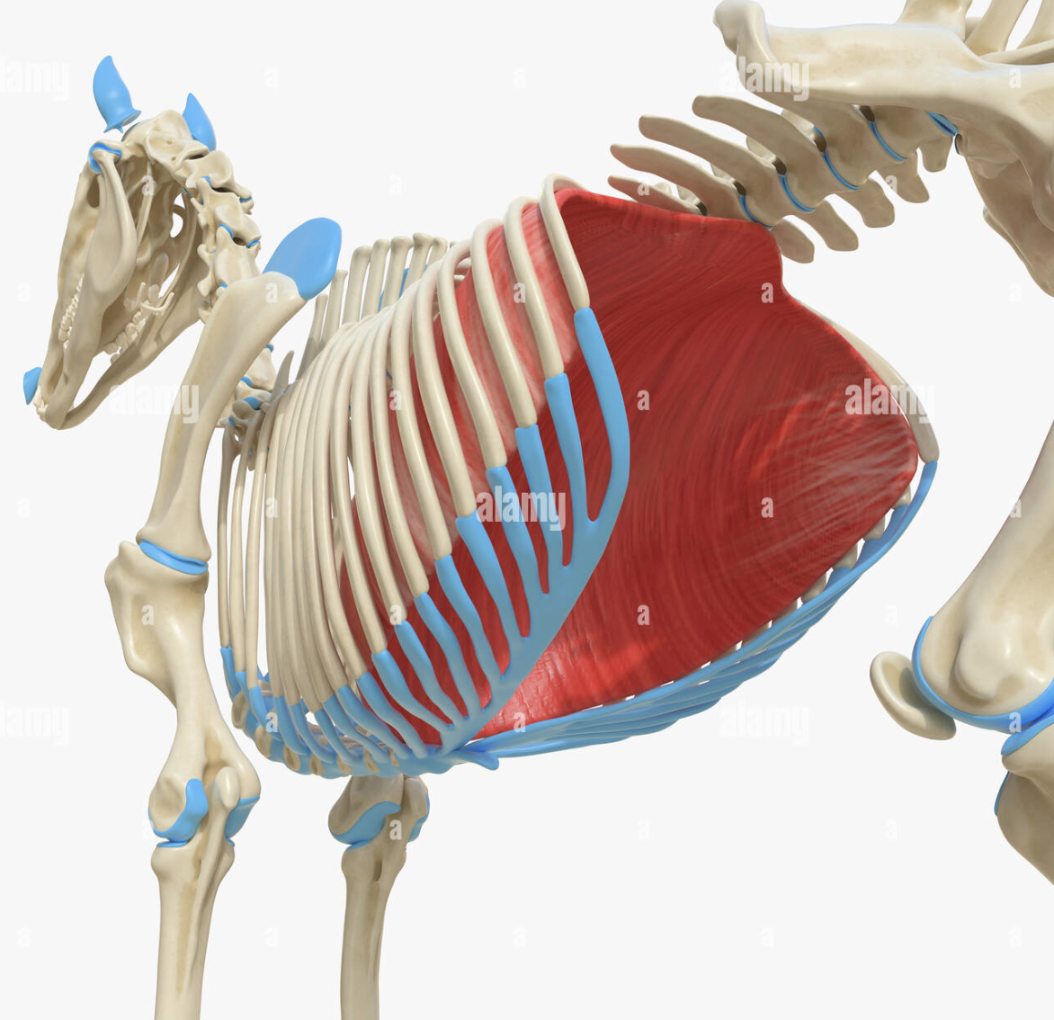
Pectoral Muscles
These muscles form a thick mass, which occupy the space between the lower part of the chest wall and the medial face of the shoulder and arm. They are divisible into two layers – superficial and deep. The superficial layer is again divisible into anterior and posterior muscles.
Aponeurosis
A thin sheath of connective tissue that helps connect muscles to bones. Similar to tendons. They support muscles and give the body strength and stability and absorb energy when muscles move.
Diaphragm
A very large, flat, dome-shaped, aponeurosis muscle that complete separates the thoracic cavity from the abdominal cavity. From its thoracic side, the diaphragm is highly convex, while its abdominal side is concave.
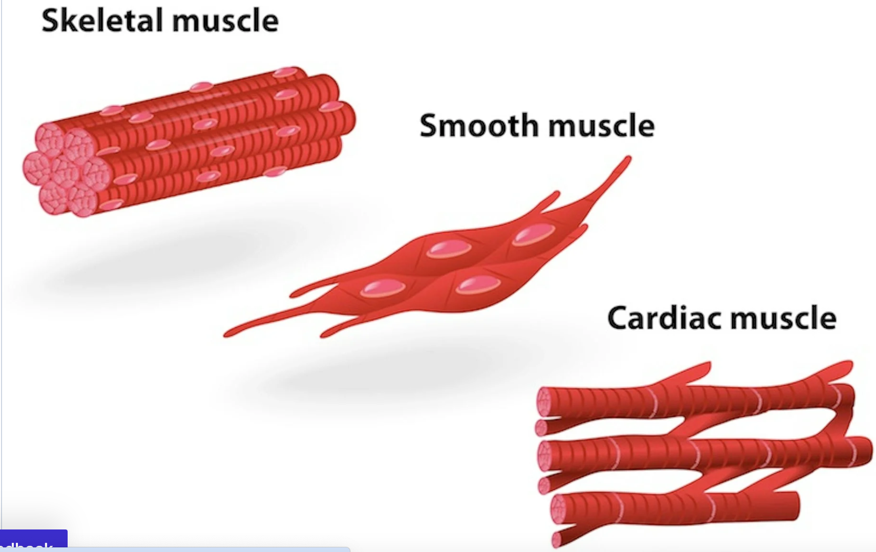
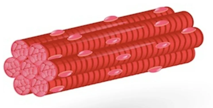
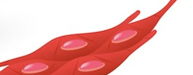
Three types of muscle cells
Skeletal, cardiac, and smooth muscles. Skeletal muscles are voluntary and striated, cardiac muscles are involuntary and striated, and smooth muscles are involuntary and non-striated.
Skeletal Muscle
Skeletal (striated) cells are responsible for practically all movements that are under voluntary control. These cells can be very large and are often referred to as muscle fibers because of their highly elongated shape. Skeletal muscle fibers are multinucleated and have a striated appearance due to the arrangement of actin and myosin filaments, allowing for coordinated contraction.
Actin
A protein that forms the thin filaments in muscle cells and plays a key role in muscle contraction by interacting with myosin. Actin facilitates the step of cytokinesis in the cell cycle. At the end of mitosis, the duplicated DNA has been divided into two daughter cells, and the cells must be separated. Actin forms a contractile ring with the protein myosin, which constricts and separates the membranes of the two daughter cells.It serves as a principal component of the sarcomere and is involved in cellular motility and structure.
Myosin
Myosin is the prototype of a molecular motor—a protein that converts chemical energy in the form of ATP to mechanical energy, thus generating force and movement. Myosin interacts with actin filaments to enable muscle contraction. It plays a critical role in muscle tissue and is an essential component of the sacromere
Sacromere
The basic contractile unit of muscle tissue. It consists of overlapping actin and myosin filaments. It is responsible for muscle contraction and the striated appearance of skeletal muscle.
Sliding Filament Theory
The mechanism explaining muscle contraction, where actin filaments slide over myosin filaments shortening the muscle fiber. The process is fundamental to the contraction of all types of muscles.
5 Stages of Sliding Filament Theory
Resting, Excitement-Contraction, Contraction, Recharge, Relaxation
Multinucleated
containing more than one nucleus per cell
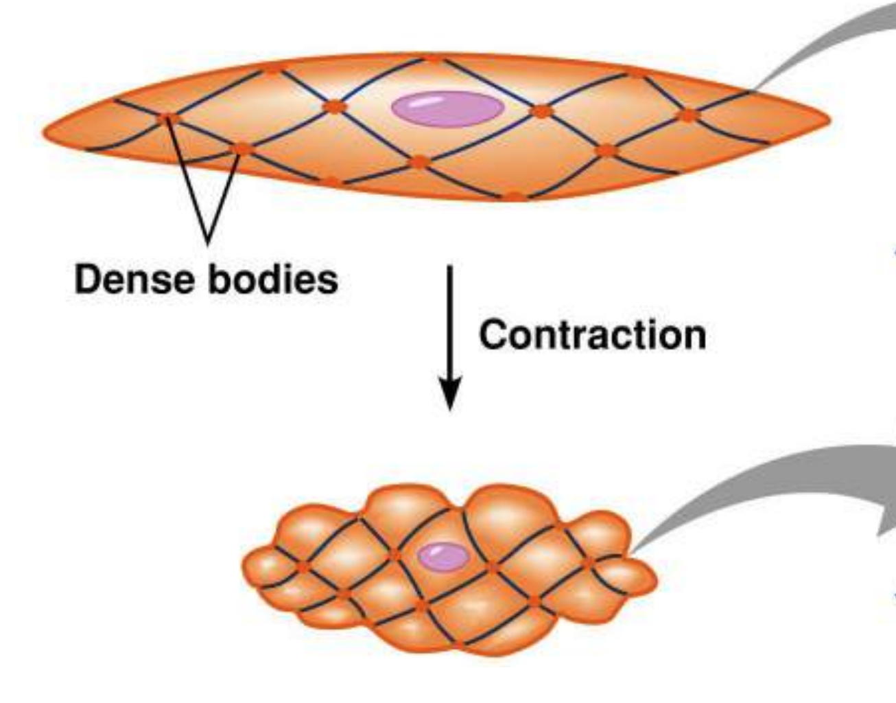
Visceral Muscle
“Smooth Muscle” Involuntary muscle located in organs such as the stomach and intestines, as well as in blood vessels. It is called smooth muscle because it is not striated like skeletal or cardiac muscle. Visceral muscle cells have a single nucleus.
Cardiac Muscle
“Myocardium”. Makes up the thick middle layer of the heart. The myocardium is surrounded my a thin outer layer called the EPICARDIUM (Visceral Pericardium) and inner ENDOCARDIUM. Cells contain 1 to 2 nuclei.
Epicardium
Innermost layer of the pericardium and outermost layer of the heart itself. Composed of mesothelial cells, fat and connective tissue.
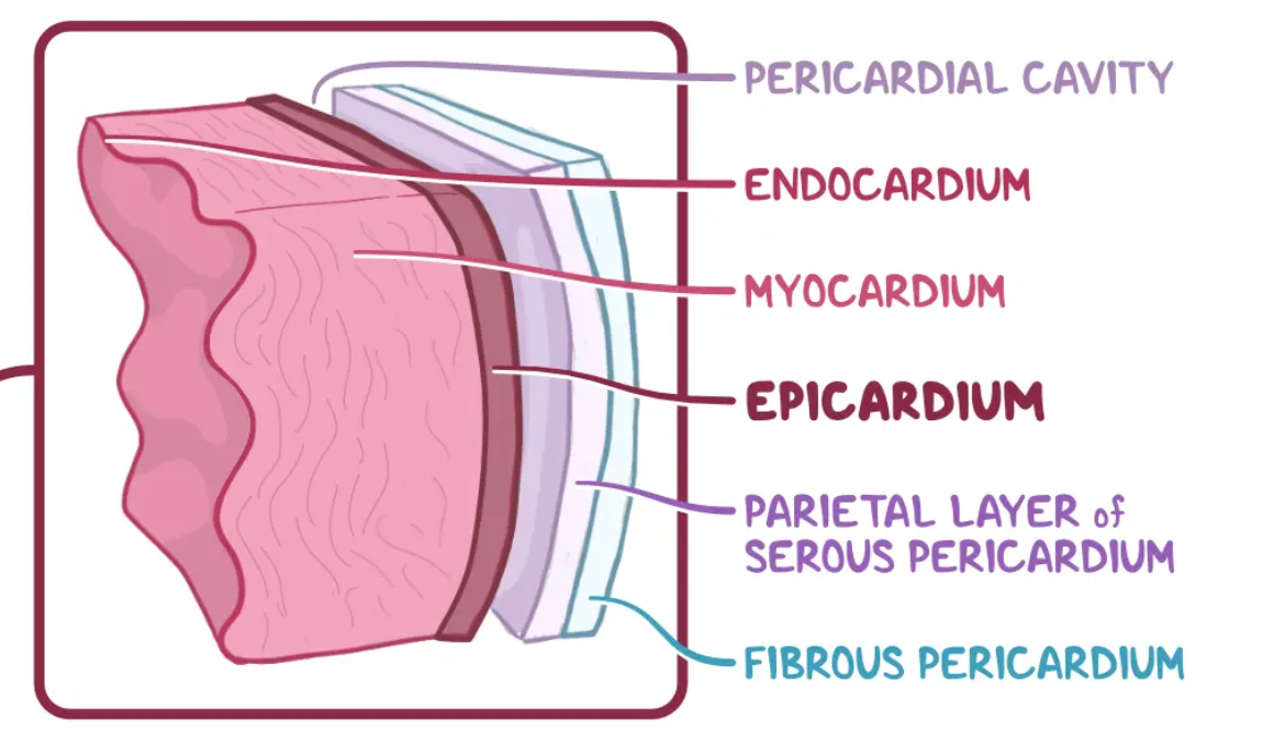
Mesothelial Cells
Form a layer of specialized “pavement” like cells that line the body’s serous cavities and internal organs. The primary function of this layer (the MESOTHELIUM) is to provide a slipper, non-adhesive and protective surface.
Endocardium
The innermost layer of tissue that lines the chambers of the heart. Its cells are similar to the endothelial cells that line blood vessels. The endocardium also provides protection to the valves and heart chambers.

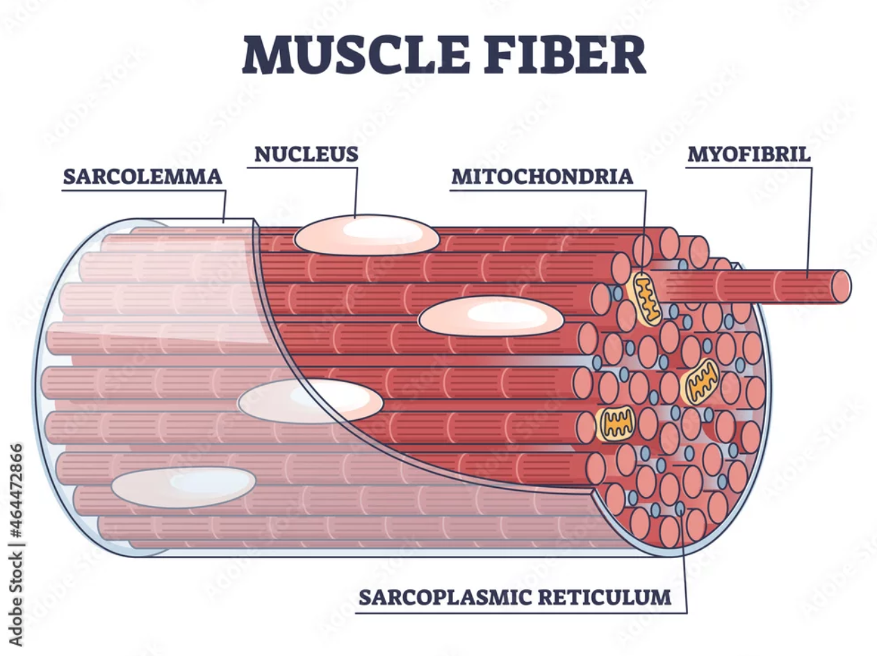
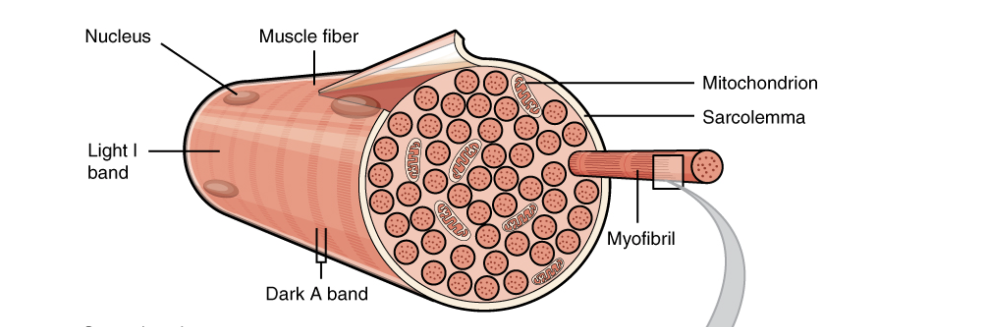
Sarcolemma
The membrane surrounding muscle fibers, exhibiting excitability to transmit electrical impulses and containing proteins for various functions like transport and enzyme activity.
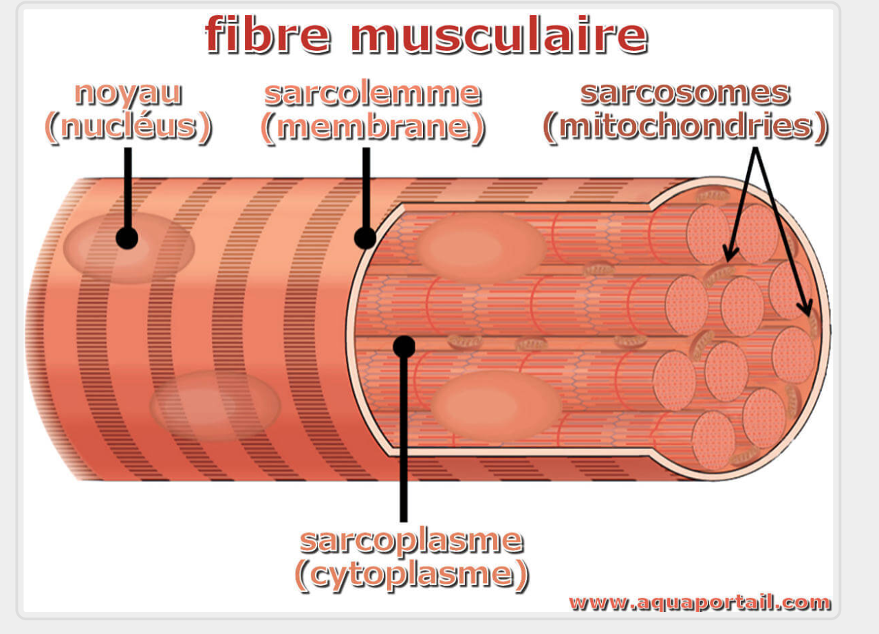
Sarcoplasm
They cytoplasm of the muscle cell. Contains elements such as mitochondria, glycogen granules and fat droplets. Located beneath the sarcolemma throughout the length of the fiber.

Sarcoplasmic Reticulum
Specialized form of the endoplasmic reticulum found in muscle cells. Cardia SR membranes are specialized in the regulation of calcium iron (Ca2) transport and control of “excitation - contraction” coupling. Skeletal SR is a highly ordered structure consisting of an intricate network of tubules and cisternae specialized for regulating Ca2 homeostasis in the context of muscle contraction
Cisterna / Cisternae
A flattened membrane vesicle found in the endoplasmic (and sarcoplasmic) reticulum and Golgi apparatus. Cistenae are an integral part of the packaging and modification process of proteins occurring in the Golgi.
Sarcosomes
Large mitochondrion within the cell of a striated muscle fiber

Trapezius Muscle
Broad, thin, triangular muscle that lies superficially over the shoulder blade. Consists of cervical and thoracic parts. Elevates the shoulder and draws the scapula up and towards the head and up and towards the tail. Starts in the middle of the neck at the 3rd cervical vertebrae (C3) and along the spine up to the 9th thoracic vertebrae (T9).
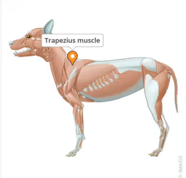
Cervical Trapezius Muscle
Starts at the cervical portion of the spine. Helps to move the shoulder up and forward (cranio-dorsal movement) in protraction (forward extension) of the limb.
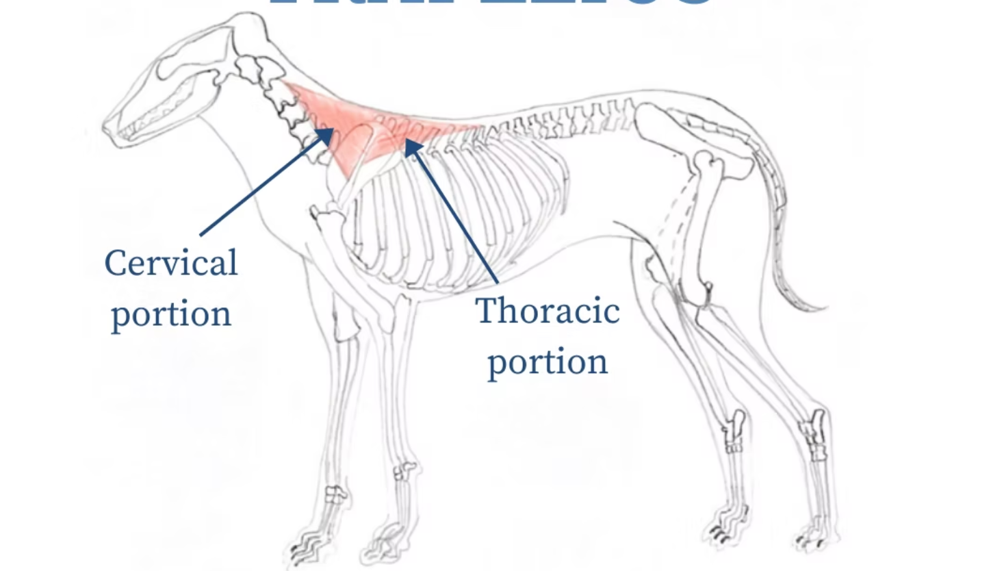
Thoracic Trapezius Muscle
Helps to move the shoulder up and backwards (caudo-dorsal movement) in retraction (backward movement) of the limb.

Adduction
To move a limb or body part toward the center of the body.
Abduction
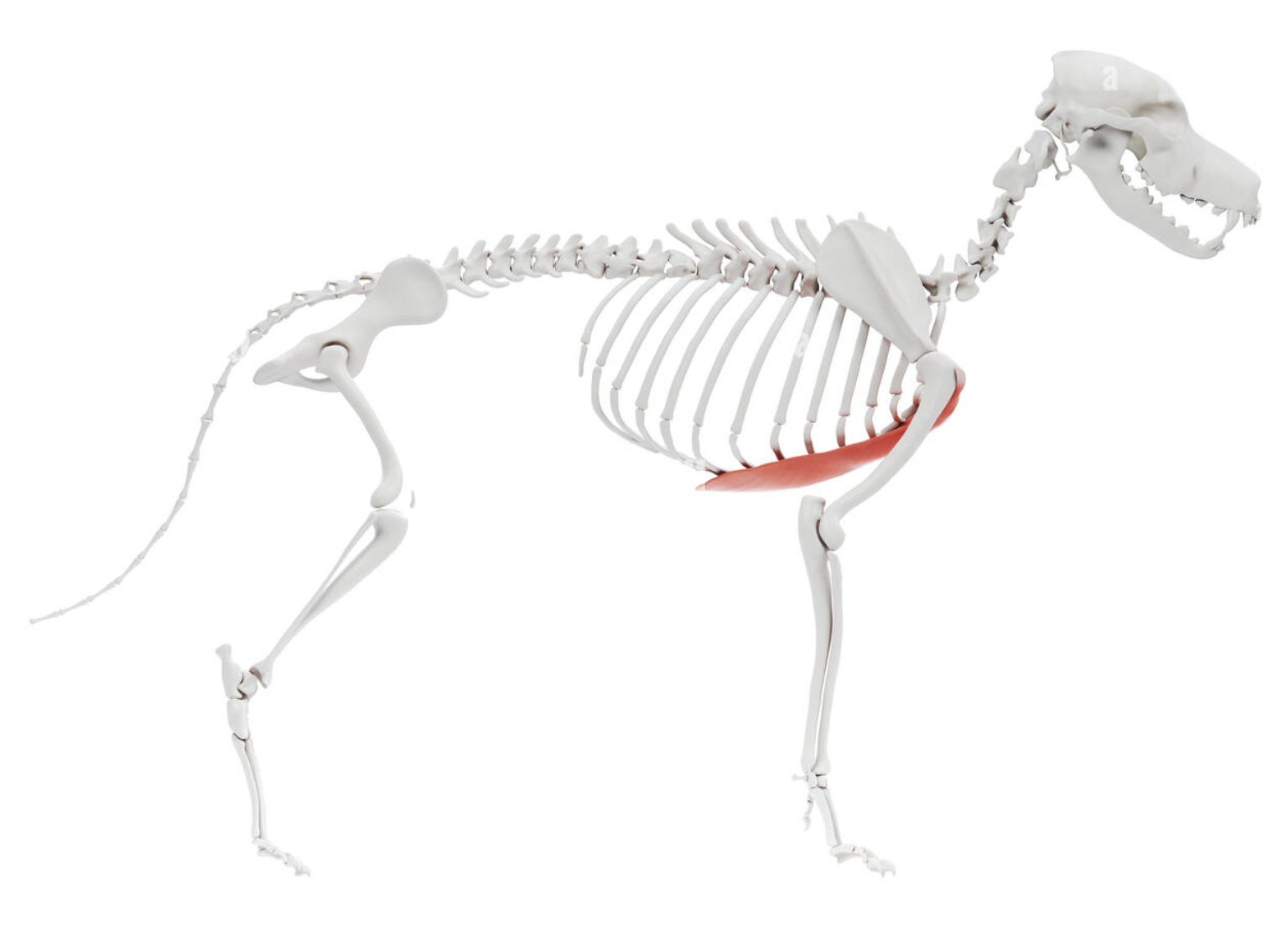
Biceps Brachii
Located in the proximal forelimb. Muscle is biarticular (crosses two joints) shoulder and elbow. It has a double pennate fiber structure. Originates at supraglenoid tubercle and inserts (ends) at two points - the radial tubercle and the ulna tuberosity. Stabilizes the shoulder joint at the cranial border. Causes shoulder to extend and elbow to flex.
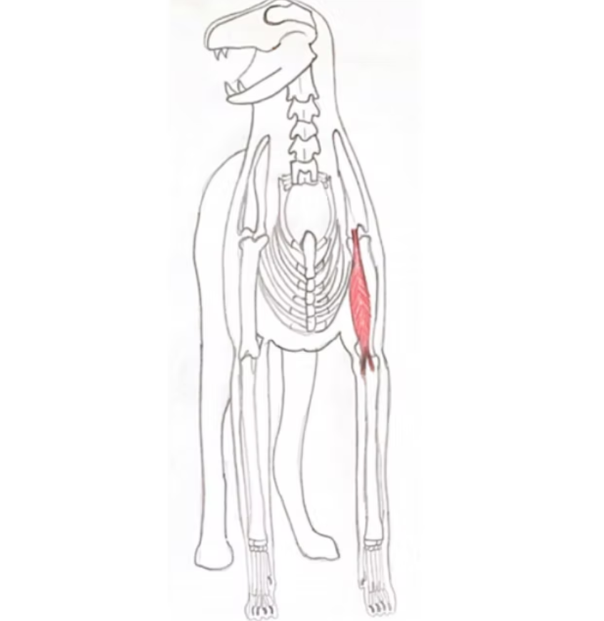
Biarticular
Muscles that cross two joints rather than one
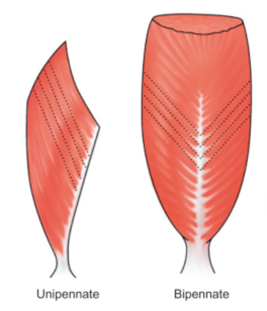
Pennate Muscle
Muscle that resembles a feather with muscle fibers approaching a central tendon at an oblique angle. This structure maximizes the muscle’s force potential making it well suited for generating large forces to support or propel the weight of the body.
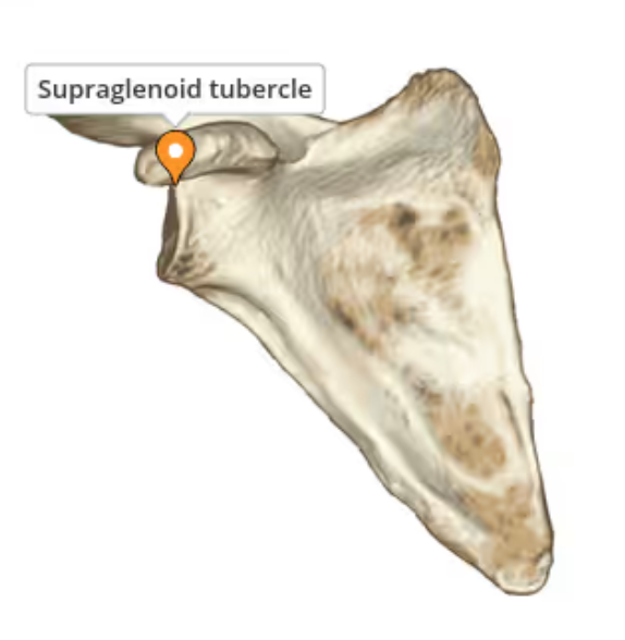
Supraglenoid Tubercle
Small, bony prominence of the scapula located at the superior aspect of the glenoid cavity. It serves as an attachment point for the tendon of the long head of the biceps brachii muscle.
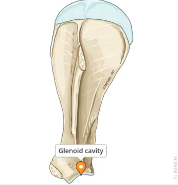
Glenoid Cavity
For the articulation of the head of the humerus. Located at the ventral angle of the scapula
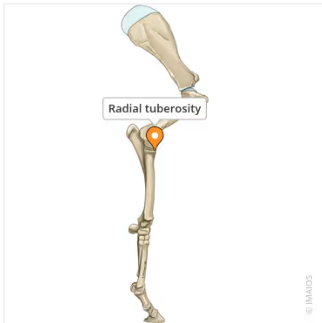
Radial Tuberosity
Marks the insertion of the biceps brachii muscle
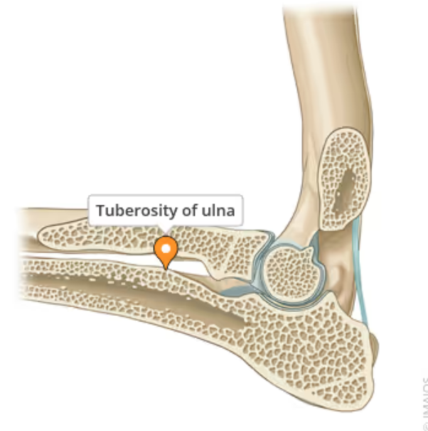
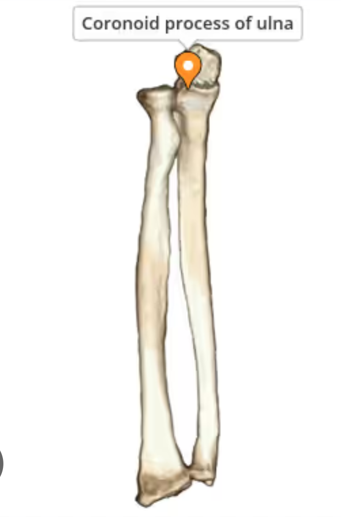
Ulnar Tuberocity
A roughened area locates near the proximal end of the medial border of the ulna, just distal to the menial coronoid process
Coronoid Process
A prominence at the front of the ulna which articulates with the humerus and radius. It has an outside, lateral part and an inside medial part. The medial part (Medial Coronoid Process) can become diseased.
Triceps Brachii
Located in the forelimb on the upper arm. Made of 4 heads working together to extend or straighten the elbow joint. The long head also works in isolation to flex the shoulder joint bringing the limb upwards and toward the body
4 Heads of the Triceps Brachii
Long Head, Medial Head, Lateral Head, Accessory Head

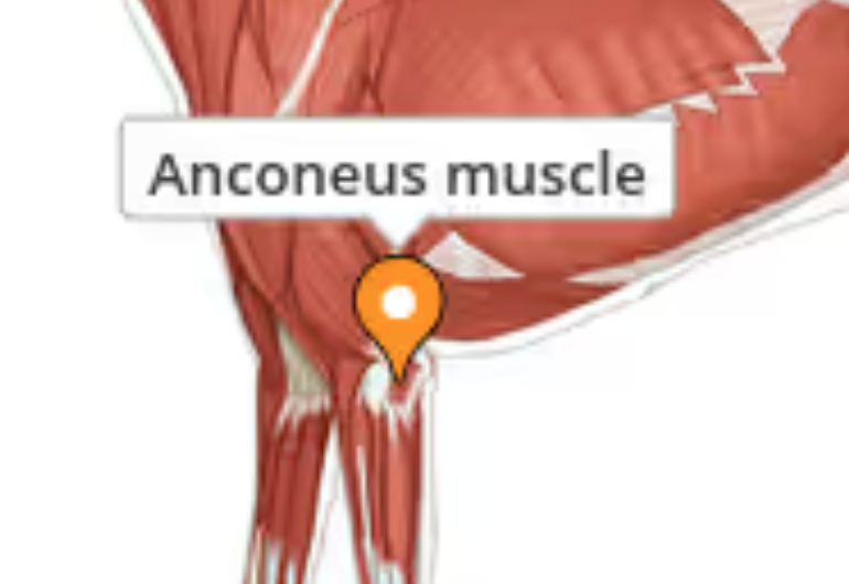
Anconeus Muscle
Extends the elbow and tenses the antebrachial fascia. Origin : lateral epicondylar crest, the lateral epicondyle and the olecranon fossa.
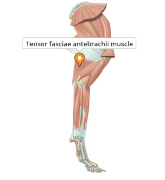
Tensor Fascie Antebrachii
Supports the action fo the triceps brachii and is the chief tensor of the antebrachial fascia
Fascia
A thin casing of connective tissue that surrounds and holds every organ, blood vessel, bone, nerve fiber and muscle in place. It has nerves that make it almost as sensitive as skin
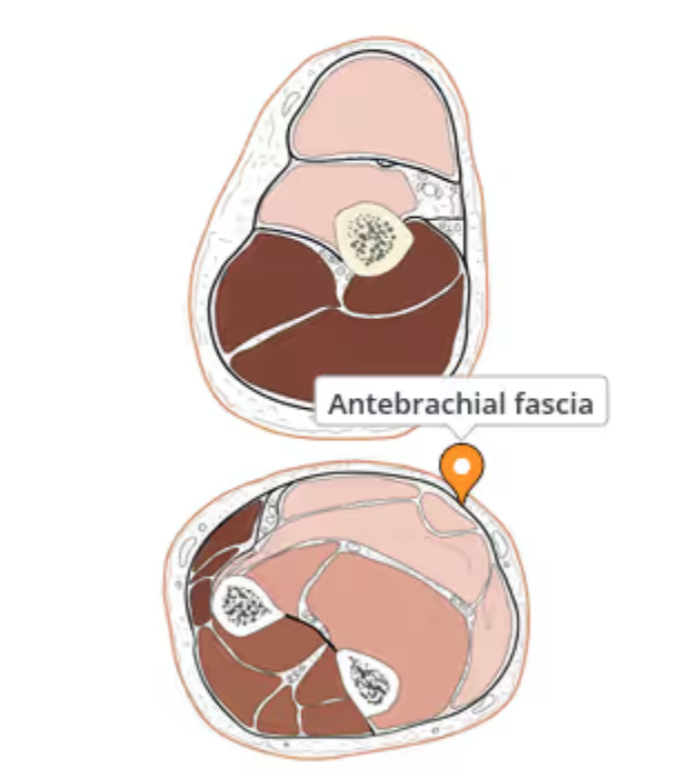
Antebrachial Fascia
Fascia of the thoracic limb which covers the muscles of the forearm. It follows the brachial fascia at the elbow joint and is continues by the dorsal fascia and palmar fascia of the hand at the carpal joint.
Condyle
The smooth surface area at the end of a bone where one bone connects to another and forms part of a joint
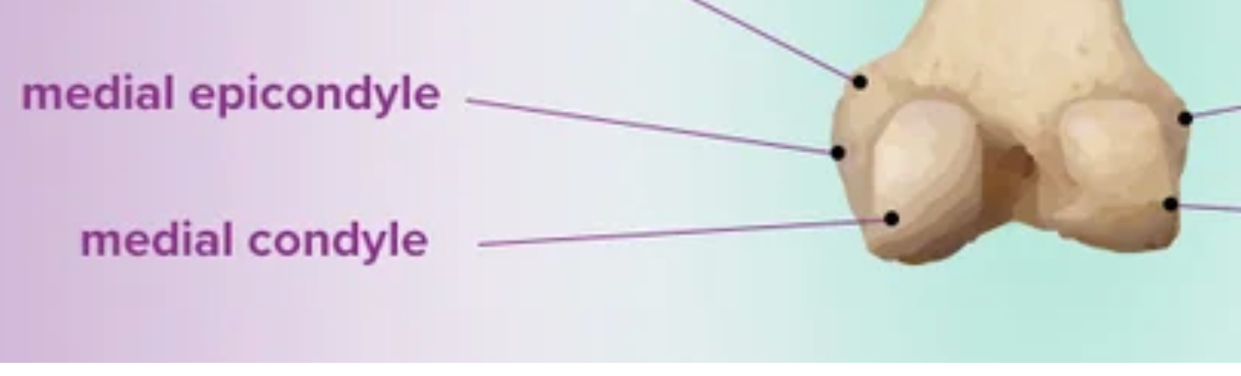
Epicondyle
A rounded protuberance at the end of a bone serving as a place of attachment for ligaments, tendons and muscles.

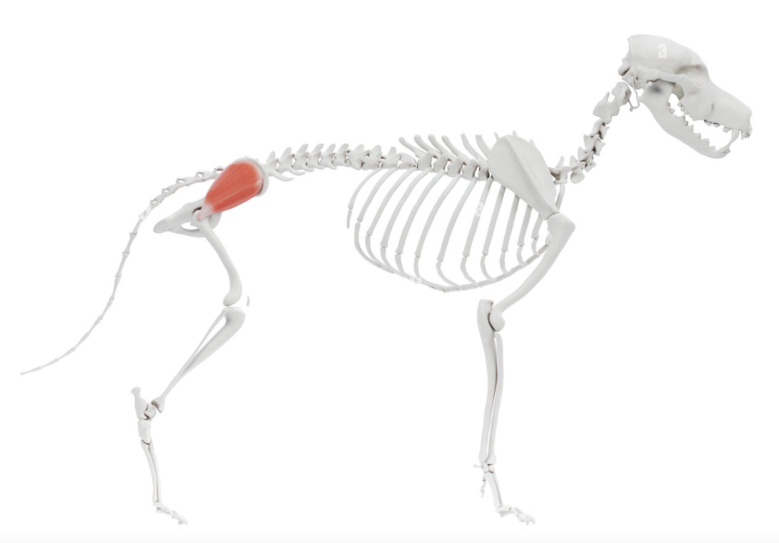
Gluteal Muscles
Help extend and move the hip joint outward from the body. Originate on the pelvic bones and end at proximal femur
Gluteus Medius
A major muscle in the hip responsible for hip extension and abduction. Also helps stabilize the pelvis during locomotion especially during single leg stance.
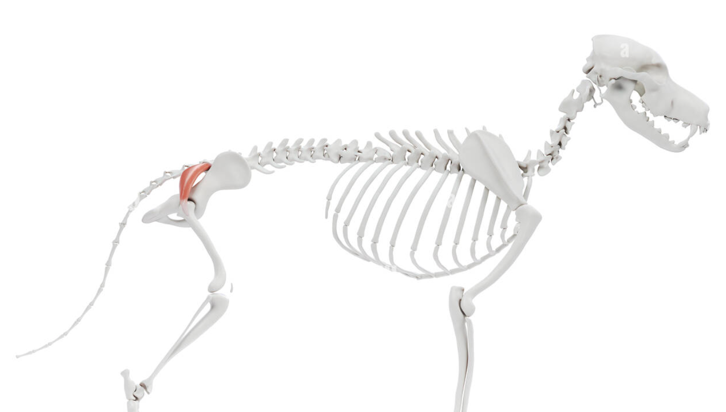
Gluteus Superficialis
Thin, flat muscle just under the skin and fascia covering the caudal part of the gluteus medius. Its primary function is to extend the hip joint and assist in elevating the hind limb.
Gluteus Profundus
The deepest muscle of the gluteal region, located beneath the gluteus medius. It originates from the ilium and ischiatic spine and insterts on the greater trochanter of teh femur Primarily functions in extending and abduction the hip joint
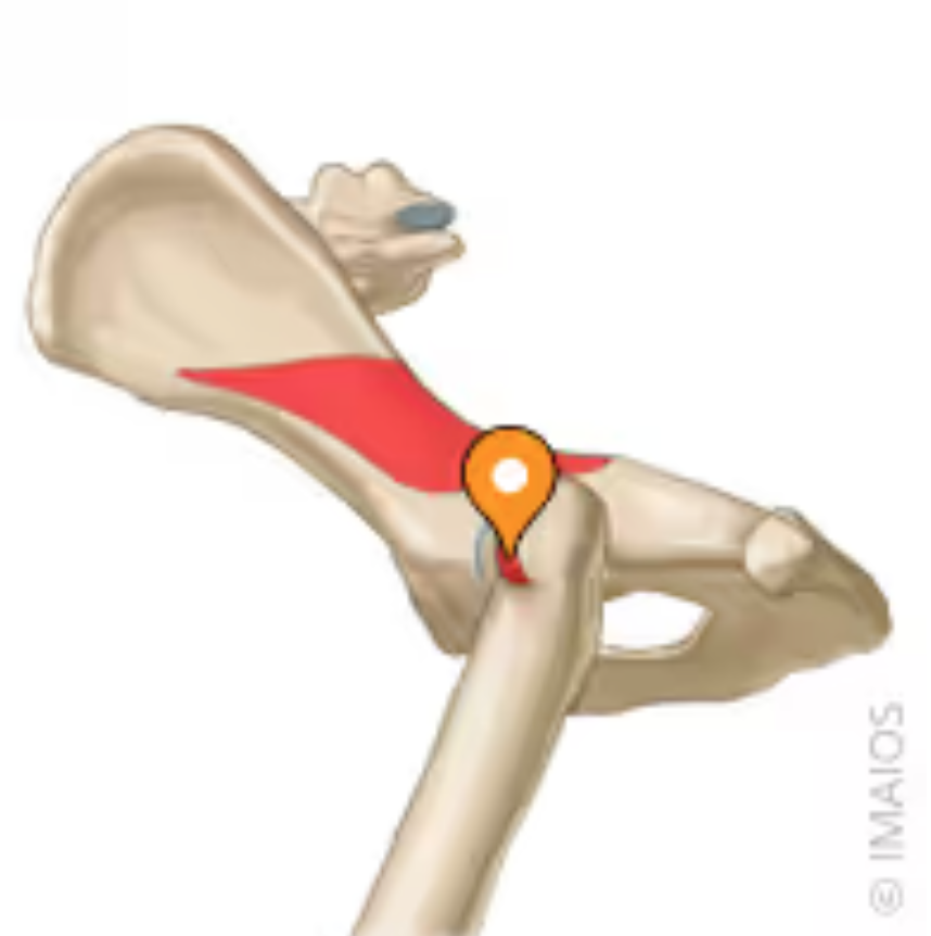
Biceps Femoris
Largest muscle in the muscle group that makes up the hamstring. Covered only by fascia and skin and can usually be easily palpated.
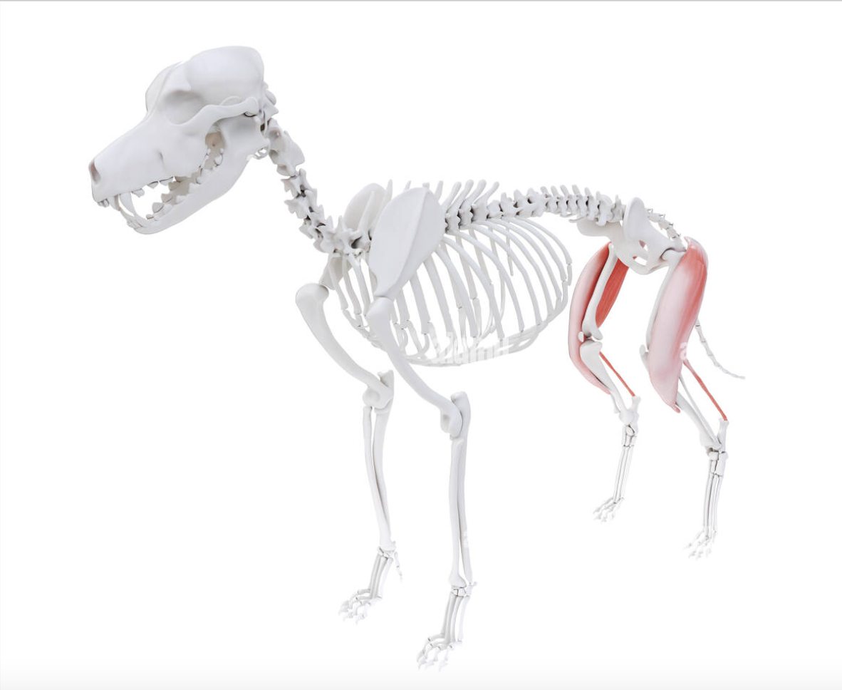
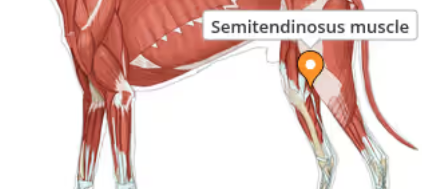
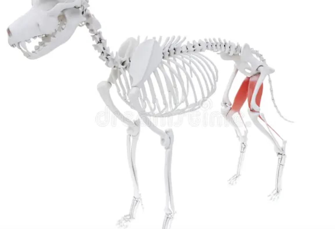
Semitendinosis Muscle
Located on the back and inner aspects of the thigh. Functions to extend the hip, stifle and tarsus and flex the stifle when in a non weight bearing position.
Gracilis Muscle
Located on inner aspects of the thigh. Allows adduction of the thigh, extension of the hip and extension of the hock
Semimembranous Muscle
Extends teh hip when the foot is placed on a solid substrate. The cranial head extends the hip and is active during the stance phase o locomotion. The caudal head is a two joint muscle and can flex the stifle and contribute to hip extension
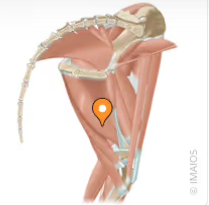
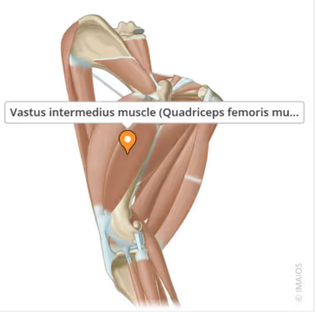
Quadriceps Femoris
Well developed extensor muscle of the thigh. Extends the stifle while also flexing the hip. Opposes the hamstring . Originates on both the pelvis and femur and ends at the patella (and extends directly to the tibia via the patellar ligament
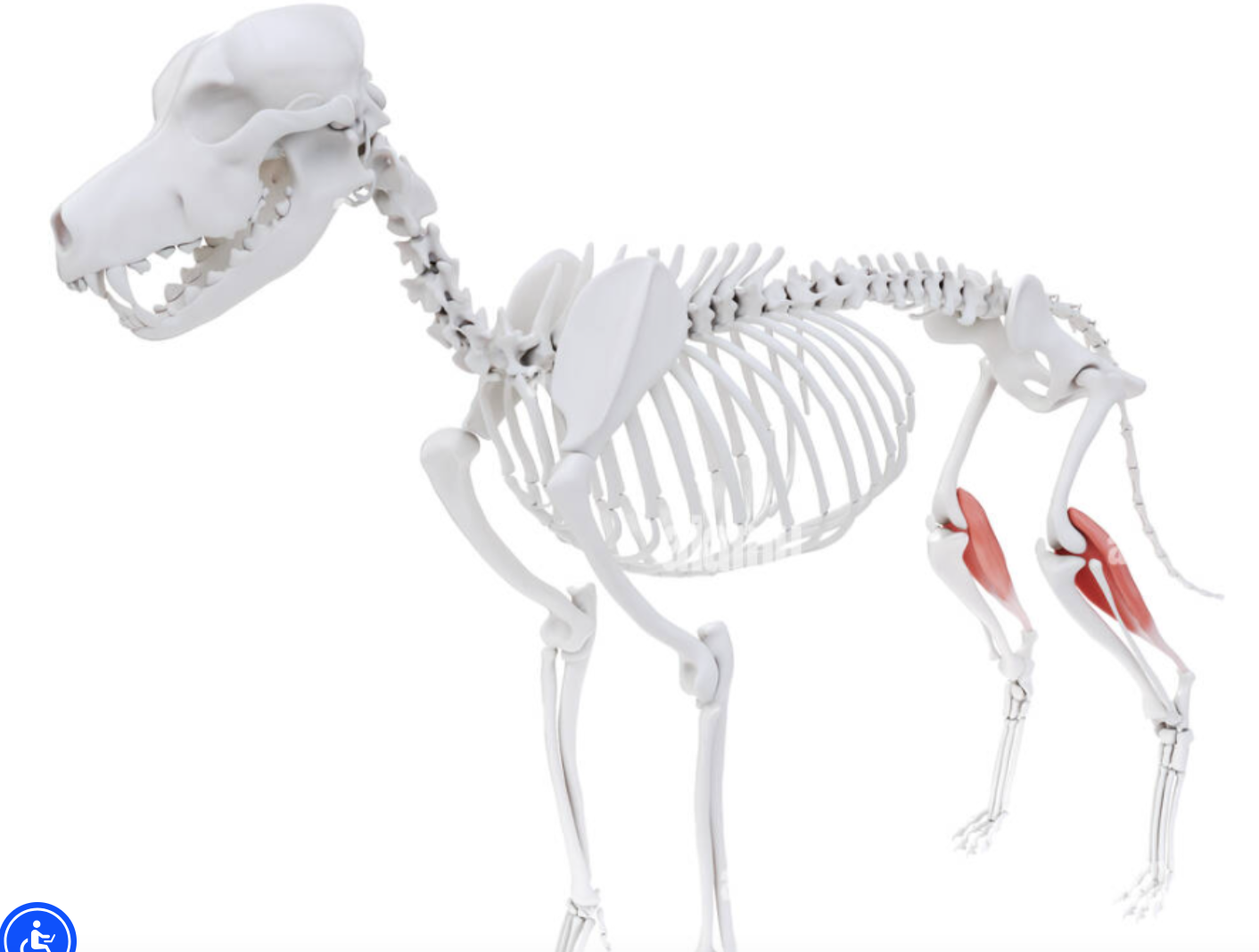
Gastrocnemius Muscle
Chief muscle of the calf of the pelvic limb. Flexes the stifle and hock. Has two heads which originate on the distal femur. The two heads combine and run distally along the caudal aspect of the tibia to form the common calcaneal (Achilles) tendon
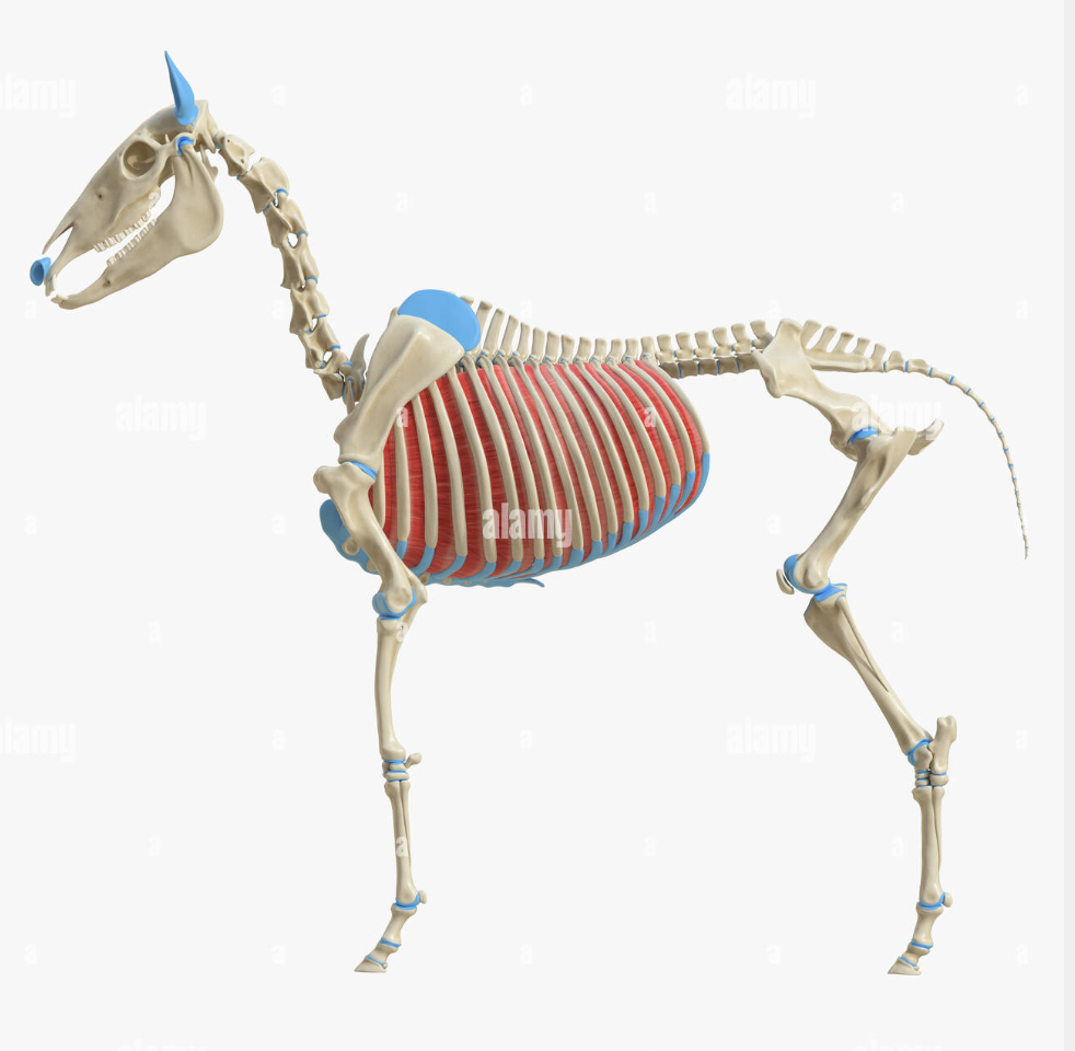
Intercostal Muscles
Internal and External. Connect the caudal edge of each rib to the cranial edge of the rib behind it.
Myofibers
Elongated, multinucleated cells that make up muscle tissue. Typically 2-3 cm long and .05mm in diameter. Fundamental to the excitability and contractivity of muscles
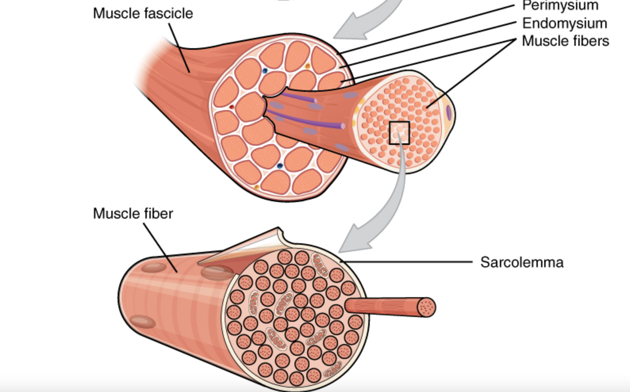
Muscle Fasciles
Bundles of muscle fibers forming a skeletal muscle
Myofibrils
Very fine contractile fibers that run parallel to each other on the long axis of the myocytes. Made up of thick and thin myofilaments (which help give muscle its striped appearance)
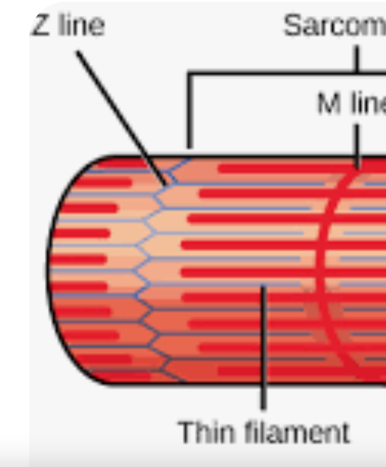
Myofilaments
Protein filaments found within muscle cells. They are responsible for muscle contraction. They are mad up of proteins ACTIN, MYOSIN and TITIN
Actin
The primary component of THIN myofilaments. its a globular (spherical / globe-like) protein that interacts with MYOSIN during contraction
Myosin
The main protein of THICK myofilaments. Its cross-bridges interact with ACTIN to produce muscle contraction
Titin
An elastic protein that forms very thin filaments.
Sarcomeres
Highly organized arrangements of actin and myosin filaments, along with support proteins, that slide past each other during muscle contraction. This sliding action shortens the sarcomere ultimately causing the entire muscle fiber to contract
Z-Line
(Z-Disc , Z-Band) Forms the boundary between adjacent sarcomeres and serves as the anchor point for the thin filaments. Helps maintain the overall structure of the sarcomere
Dense Bodies
Specialized structures, composed of dense aggregates of contractile proteins, within smooth muscle cells that serve as anchoring points for thin filaments. Analogous to Z-Line in skeletal muscles
Neurotransmitters
Substances secreted by involuntary nerve endings that either stimulate or inhibit muscle contraction (depending on the type of receptor on the cell surface) The primary neurotransmitter involved in transmitting nerve impulses to muscles is acetylcholine (ACh)
Tone
State of partial contraction, with low energy consumption, maintained my smooth muscle. Crucial for various physiological functions like maintaining vascular pressure and regulating the diameter of tubes like the digestive tract and urinary bladder. Often maintained by hormones that signal smooth muscle cells to remain in an indefinite state of contraction
Cardiac Muscle
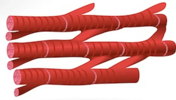
Involuntary, striated muscle. Branched and interconnected by intercalated discs allowing for a synchronized wave of contractions that propels blood. Functions like one large muscle fiber
Intercalated Discs
Connect cardiac muscles together, have a low resistance to electrical current and therefore aid in conduction of electrical currents through heart muscle.