👅 Anatomy + Monogastrics
1/38
There's no tags or description
Looks like no tags are added yet.
Name | Mastery | Learn | Test | Matching | Spaced | Call with Kai | Chat |
|---|
No analytics yet
Send a link to your students to track their progress
39 Terms
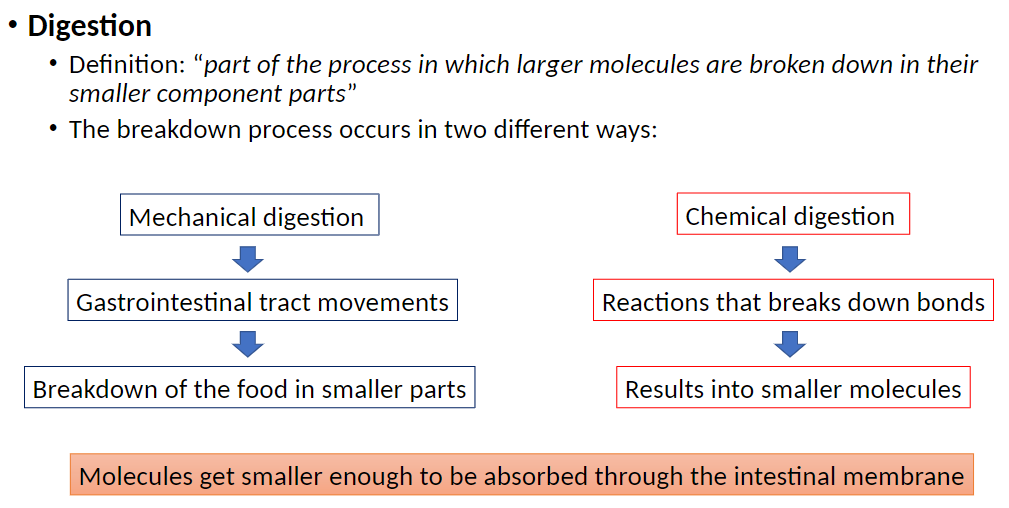
Digestion – Key Points
Definition
Part of the process in which larger molecules are broken down into smaller component parts
Two Types of Digestion
Mechanical Digestion
• Occurs through movements of the gastrointestinal (GI) tract
• Physically breaks food into smaller pieces
Chemical Digestion
• Involves chemical reactions that break molecular bonds
• Produces molecules small enough to be absorbed through the intestinal membrane
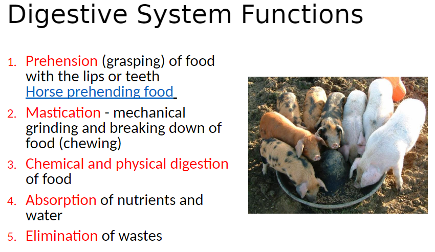
Digestive System – Functions
1. Prehension
• Grasping food using lips or teeth
2. Mastication
• Mechanical grinding and breaking down of food (chewing)
3. Digestion
• Chemical and physical breakdown of food
4. Absorption
• Uptake of nutrients and water into the body
5. Elimination
• Removal of waste products
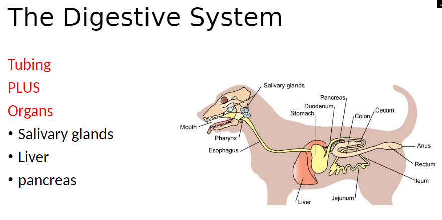
Digestive System – Structure
Tubing
Continuous passage that carries food through the body
Includes mouth, esophagus, stomach, Intestines, and anus
Organs
Salivary Glands: Produce saliva to moisten food and begin starch digestion
Liver: Produces bile to help break down fats and processes nutrients from the blood
Pancreas: Produces digestive enzymes and bicarbonate to aid chemical digestion
Digestive System – Components
Gastrointestinal Tract (GIT): Also known as digestive or alimentary tract
Includes mouth, pharynx, esophagus, stomach, small and large intestine
Considered a long tube from mouth to anus
Accessory Organs: Include tongue, salivary glands, liver, pancreas, gall bladder
Digestive System – Parts
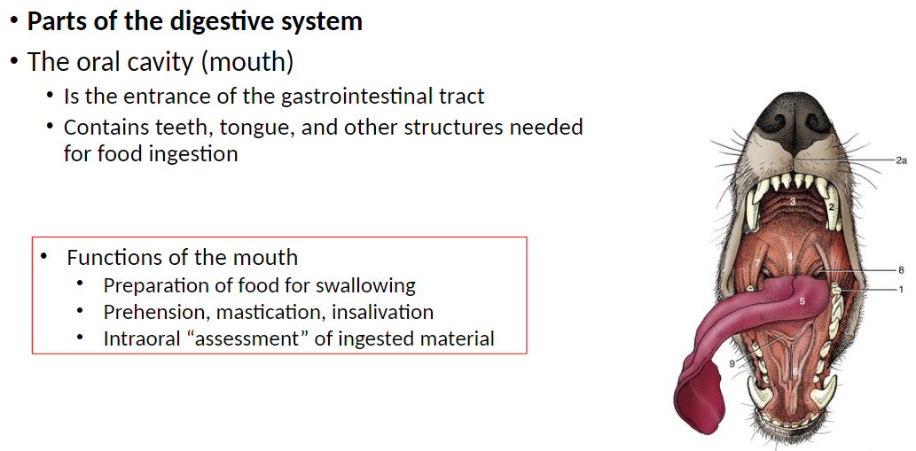
Oral Cavity (Mouth): Is the entrance of the gastrointestinal tract
Contains teeth, tongue, and other structures needed for food ingestion
Functions of the Mouth: Preparation of food for swallowing
Prehension, mastication, insalivation
Intraoral assessment of ingested material
Digestive System – Teeth
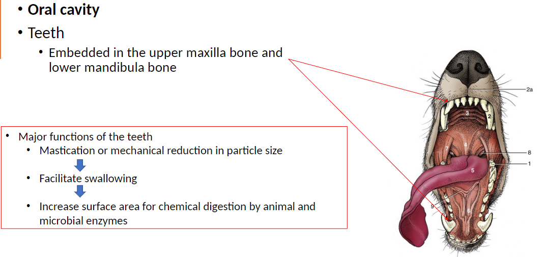
Teeth: Embedded in the upper maxilla bone and lower mandibula bone
Major Functions of the Teeth: Mastication or mechanical reduction in particle size
Facilitate swallowing
Increase surface area for chemical digestion by animal and microbial enzymes
Digestive System – Tongue
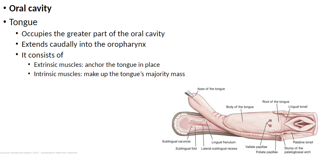
Tongue: Occupies the greater part of the oral cavity
Extends caudally (toward the back of the mouth) into the oropharynx (the part of the throat behind the mouth that connects the oral cavity to the esophagus and larynx)
Structure of the Tongue:
Extrinsic muscles: Anchor the tongue to surrounding structures like the jaw and hyoid bone, allowing movement
Intrinsic muscles: Make up most of the tongue’s mass and allow it to change shape, such as curling, flattening, or lengthening
Digestive System – Tongue Regions (Apex)
Anatomical Regions of the Tongue: Divided into three regions
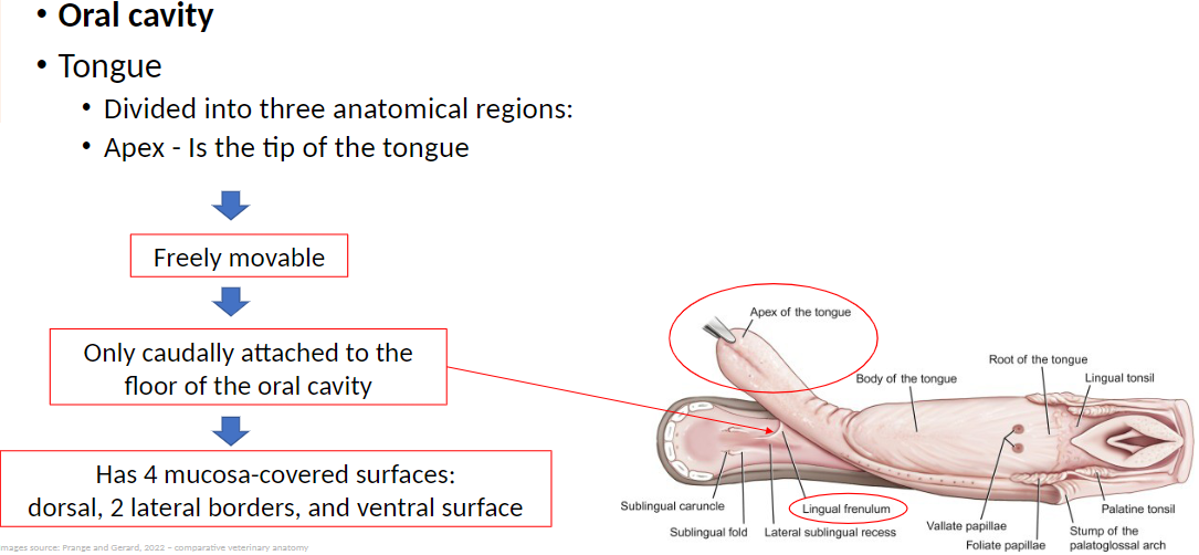
Apex: Is the tip of the tongue
Freely movable to help manipulate food during chewing and swallowing
Only caudally (toward the back) attached to the floor of the oral cavity, allowing flexibility
Has 4 mucosa-covered surfaces: dorsal (top), 2 lateral borders (sides), and ventral surface (bottom)
Digestive System – Tongue Regions (Body)
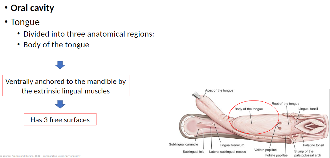
Body of the Tongue: The middle portion of the tongue between the apex (tip) and the root (back)
Ventrally anchored (attached on the underside) to the mandible by extrinsic lingual muscles, allowing movement and support
Has 3 free surfaces that are not attached: dorsal (top) and 2 lateral borders (sides), which help manipulate food during chewing and swallowing
Digestive System – Tongue Regions (Root)
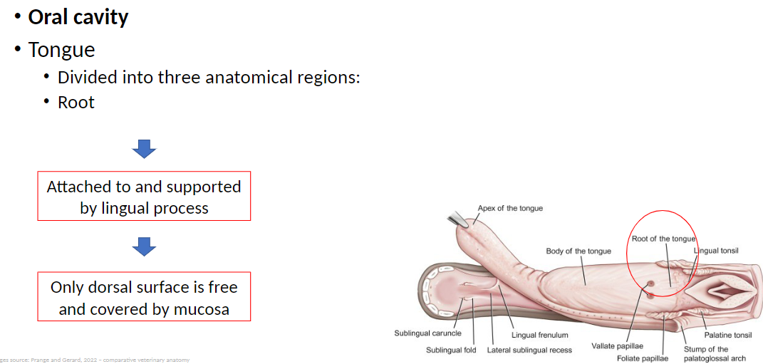
Root: The posterior (back) part of the tongue that connects to the throat
Attached to and supported by the lingual process (a bony structure that anchors the tongue to the skull and hyoid apparatus)
Only the dorsal surface (top) is free and covered by mucosa, while the rest is attached, providing stability during swallowing
Digestive System – Tongue Papillae
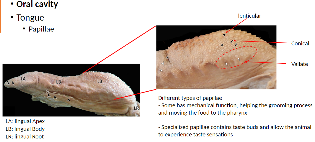
Papillae: Small projections on the surface of the tongue that have different functions depending on type
Located on the Lingual Apex (LA), Lingual Body (LB), and Lingual Root (LR)
Functions of Papillae:
Some have a mechanical function: Help with grooming and moving food toward the pharynx (throat) for swallowing
Specialized papillae contain taste buds: Allow the animal to detect different taste sensations
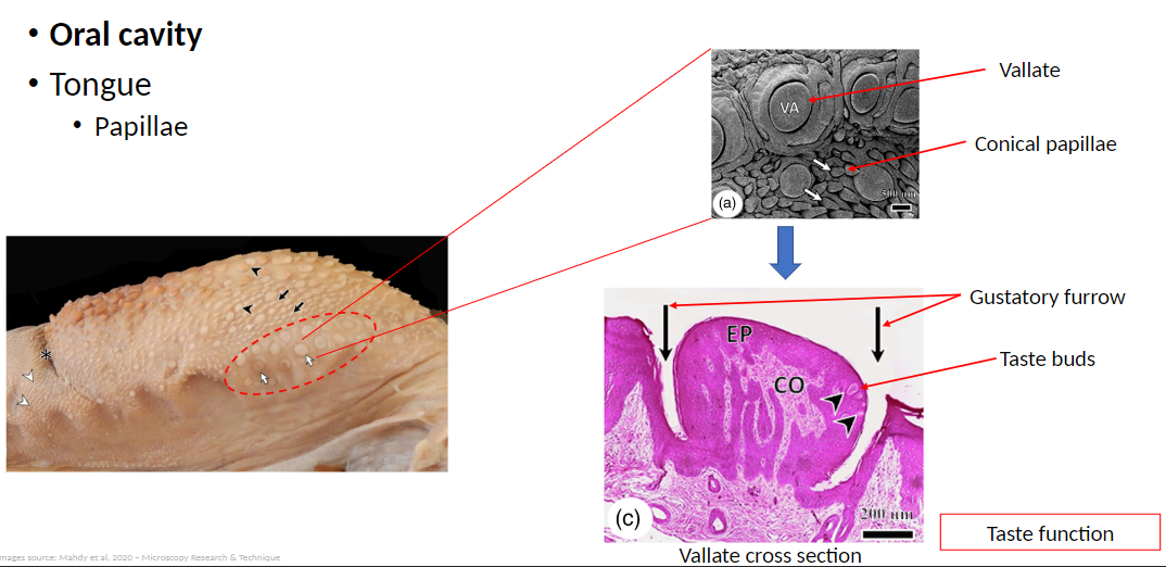
Types of Papillae: Conical, Lenticular, Vallate
Digestive System – Tongue Papillae Details
Vallate Papillae: Large papillae located near the back of the tongue
Often arranged in a V-shape on the back of the tongue
Contain taste buds
Conical Papillae: Cone-shaped papillae mostly involved in mechanical functions
Help move food toward the pharynx and assist in grooming
Gustatory Furrow: Groove surrounding vallate papillae
Houses taste buds and channels dissolved food toward them for taste perception
Taste Buds: Sensory structures within specialized papillae
Detect different taste sensations such as sweet, salty, sour, bitter, and umami
Digestive System – Salivary Glands
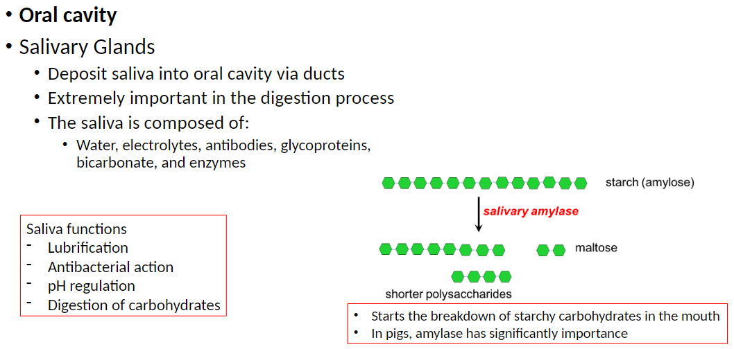
Salivary Glands: Deposit saliva into the oral cavity through ducts
Extremely important in the digestion process
Composition of Saliva: Water, electrolytes, antibodies, glycoproteins, bicarbonate, and enzymes
Functions of Saliva:
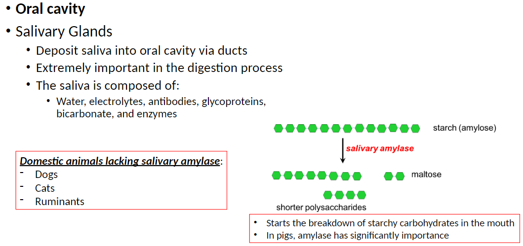
Starts the breakdown of starchy carbohydrates in the mouth (amylase is especially important in pigs)
Lubrication: Moistens food to help swallowing
Antibacterial Action: Helps control oral bacteria
pH Regulation: Maintains optimal acidity for enzymes and oral health
Digestion of Carbohydrates: Begins chemical digestion of starches in the mouth
Digestive System – Animals Lacking Salivary Amylase
Domestic Animals Without Salivary Amylase:
Dogs: Cannot start starch digestion in the mouth
Cats: Cannot start starch digestion in the mouth
Ruminants: Rely on microbial fermentation in the stomach and intestines for starch digestion
Digestive System – Salivary Glands Types
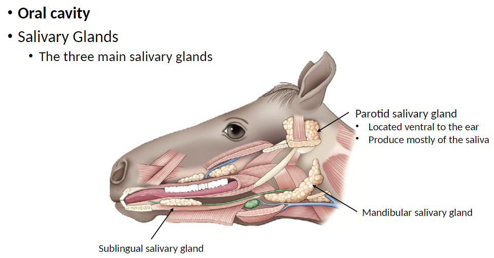
Three Main Salivary Glands:
Mandibular Salivary Gland: Located near the lower jaw (mandible)
Parotid Salivary Gland: Located ventral (below) to the ear
Produces mostly watery saliva and a large part of total saliva
Sublingual Salivary Gland: Located under the tongue
Digestive System – Pharynx (Throat)
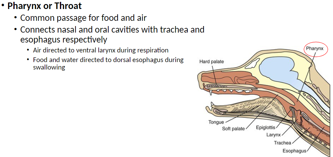
Pharynx: Common passage for food and air
Connects the nasal and oral cavities with the trachea (airway) and esophagus respectively
During respiration, air is directed to the ventral (front) larynx
During swallowing, food and water are directed to the dorsal (back) esophagus
Digestive System – Epiglottis
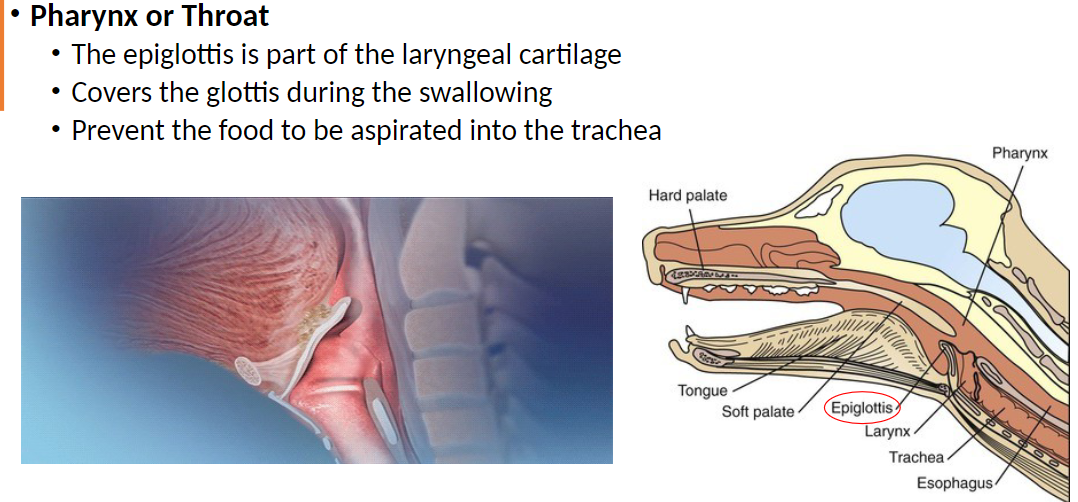
Epiglottis: Part of the laryngeal cartilage
Covers the glottis (opening of the trachea) during swallowing
Prevents food and liquids from being aspirated (inhaled into the lungs) into the trachea
Digestive System – Esophagus
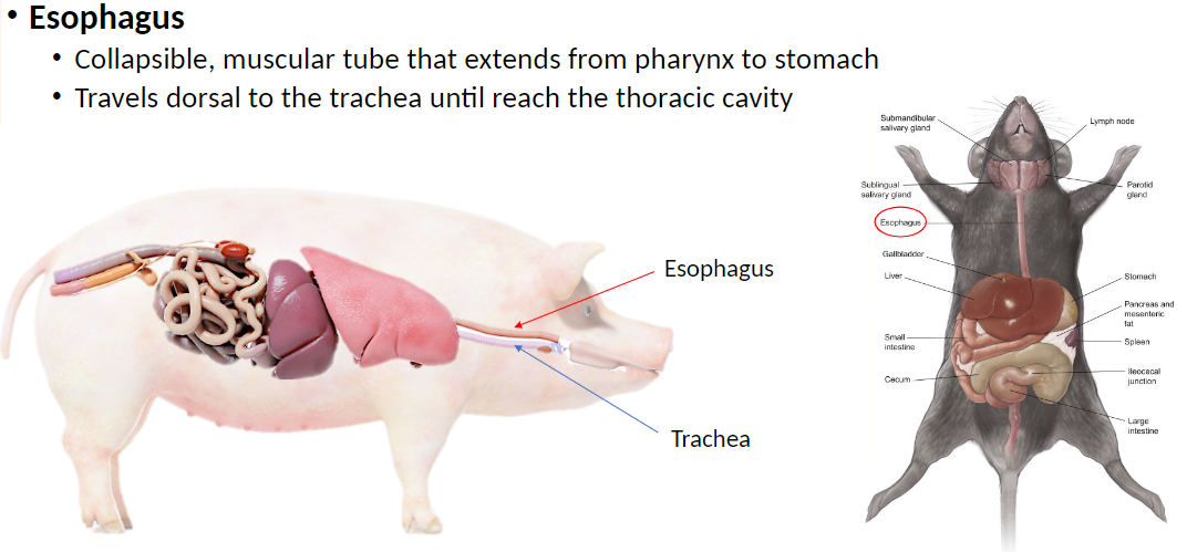
Esophagus: Collapsible muscular tube that extends from the pharynx (throat) to the stomach
Collapsible means the esophagus is normally closed and flattened when empty, but expands when food or liquid passes through
Travels dorsal (behind) to the trachea until it reaches the thoracic cavity
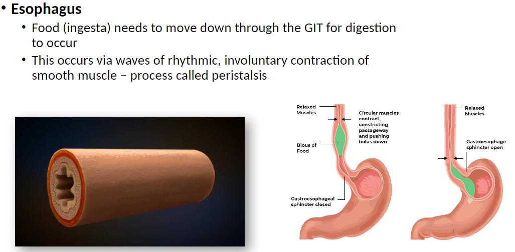
Transports food and liquids from the mouth to the stomach using coordinated muscular contractions called peristalsis
Digestive System – Esophagus Muscle Layers
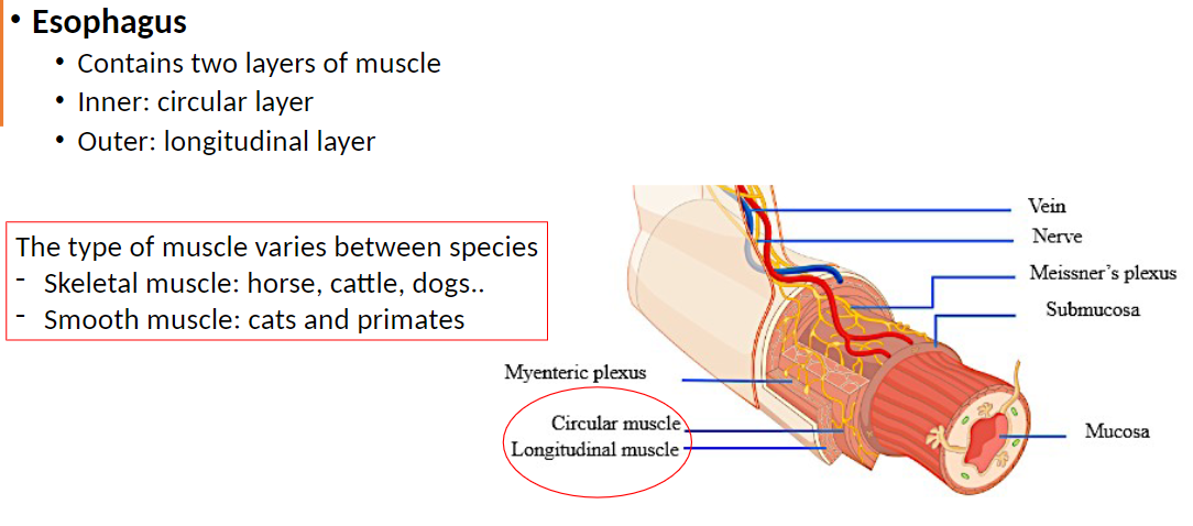
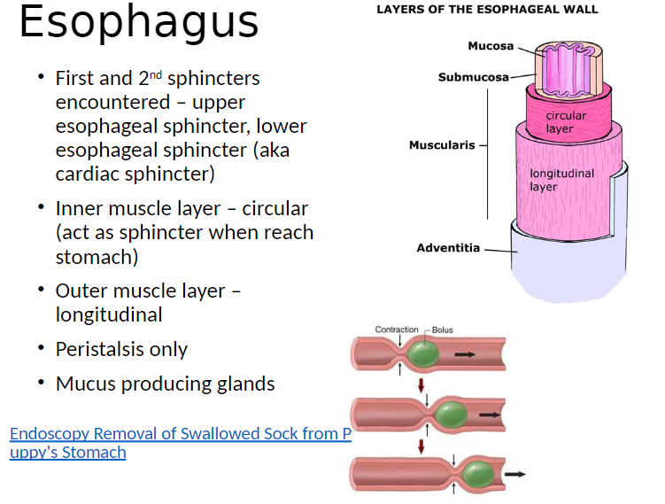
Muscle Layers: Two layers of muscle
Inner: Circular layer, wraps around esophagus to constrict and push food
Outer: Longitudinal layer, runs along esophagus to shorten and move food
Muscle Type by Species:
Skeletal muscle: Horse, cattle, dogs – allows voluntary control of swallowing
Smooth muscle: Cats, primates – involuntary, controlled automatically
Digestive System – Peristalsis
Peristalsis: Rhythmic, involuntary waves of smooth muscle contraction that move food (ingesta) down through GI tract
Allows digestion to occur by transporting food from esophagus to stomach
Digestive System – Esophagus Peristalsis Variations
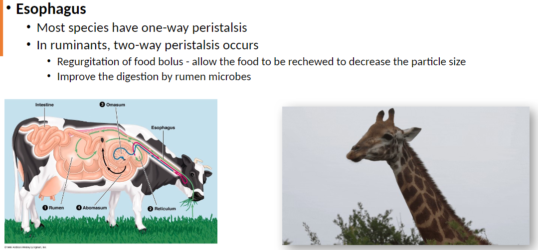
Peristalsis: Most species have one-way peristalsis, moving food toward stomach
Ruminants have two-way peristalsis: Food can move back toward the mouth (regurgitation)
Regurgitation allows rechewing of food bolus to decrease particle size
Improves digestion by rumen microbes, making nutrients easier to break down
Digestive System – Esophagus Sphincters and Layers
Sphincters: First and second sphincters encountered are:
Upper esophageal sphincter – controls entry of food into esophagus
Lower esophageal sphincter (cardiac sphincter) – controls entry of food into stomach
Muscle Layers:
Inner circular layer – acts as sphincter when reaching stomach
Outer longitudinal layer – helps shorten and move esophagus during peristalsis
Other Features:
Peristalsis only moves food in coordinated waves
Mucus-producing glands – lubricate esophagus and ease food passage
Digestive System – Esophagus Species Differences
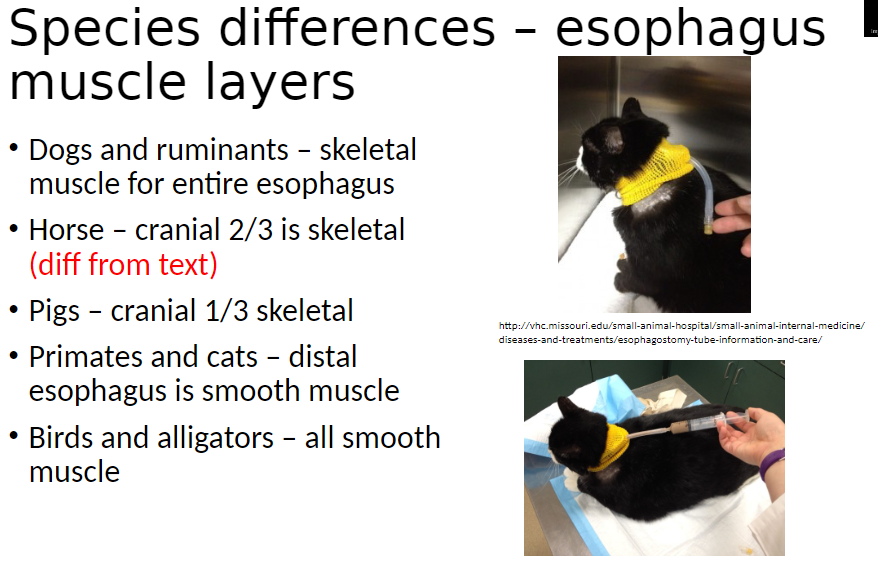
Muscle Type by Species:
Dogs and ruminants – Entire esophagus is skeletal muscle, allowing voluntary control of swallowing
Horse – Cranial 2/3 is skeletal muscle, distal is 1/3 smooth muscle (allows partial voluntary control)
Pigs – Cranial 1/3 is skeletal muscle, rest smooth muscle
Primates and cats – Distal esophagus is smooth muscle, involuntary
Birds and alligators – Entire esophagus is smooth muscle, involuntary
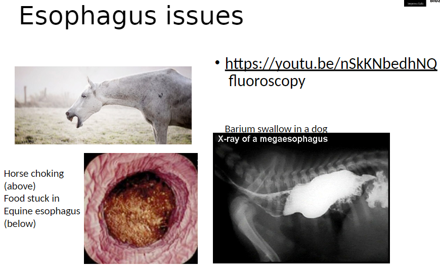
Digestive System – Esophagus Issues
Megesophagus and Obstructions:
Fluoroscopy and barium swallow can be used to visualize esophagus function
Horse choking – food stuck in equine esophagus, can block passage to stomach
Barium swallow in a dog – highlights movement of food and identifies abnormalities like megaesophagus
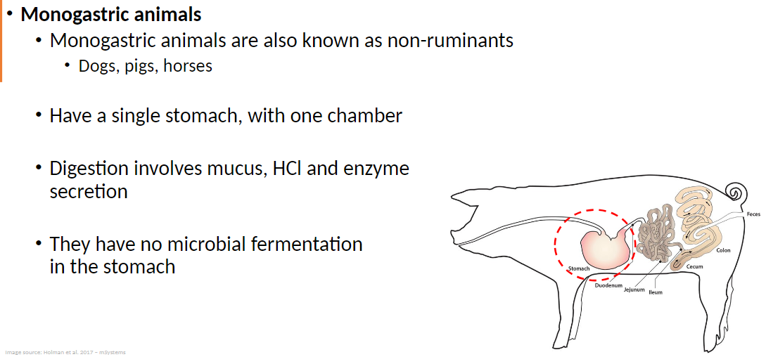
Digestive System – Monogastric Animals
Monogastric Animals: Also known as non-ruminants
Include dogs, pigs, horses
Have a single-chambered stomach
Digestion in Monogastrics:
Involves secretion of mucus, hydrochloric acid (HCl), and digestive enzymes
No microbial fermentation occurs in the stomach
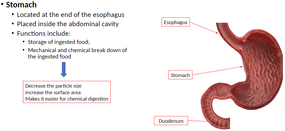
Digestive System – Stomach
Stomach: Located at the end of the esophagus
Placed inside the abdominal cavity
Functions of the Stomach:
Storage of ingested food
Mechanical and chemical breakdown of food
Decreases particle size
Increases surface area for enzymes
Makes chemical digestion easier
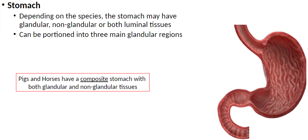
Digestive System – Stomach Regions
Stomach Tissues: Depending on species, stomach may have glandular, non-glandular, or both types of luminal tissues (epithelial layers that line the internal, open space (lumen) of tubular organs, glands, and ducts, such as the mammary ducts, intestines, and blood vessels)
Glandular Regions: Stomach can be portioned into three main glandular regions (fundus, body, pylorus) for secretion of acid and digestive enzymes
Composite Stomach: Pigs and horses have both glandular and non-glandular tissues
Glandular region: Secretes acid and enzymes
Non-glandular region: Mainly storage and mechanical processing of food
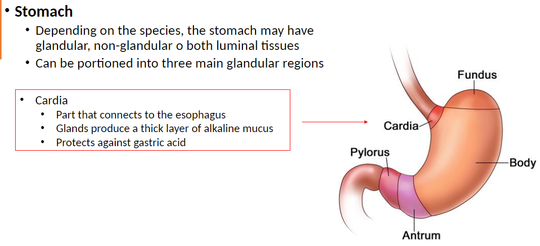
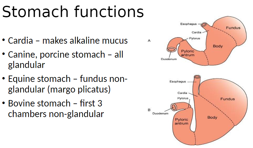
Digestive System – Cardia
Cardia: Part of the stomach that connects to the esophagus
Glands produce thick layer of alkaline mucus
Protects stomach lining from acidic gastric secretions
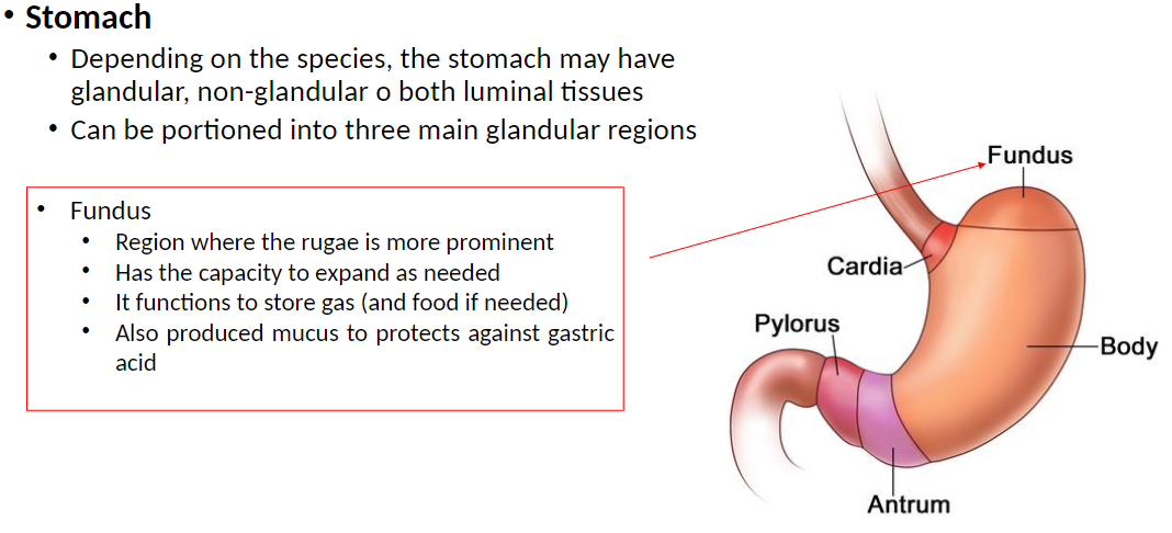
Digestive System – Fundus
Fundus: Region of the stomach where rugae (folds in the stomach lining) are more prominent
Can expand as needed to accommodate food and gas
Functions to store gas and food if needed
Produces mucus to protect stomach lining from gastric acid
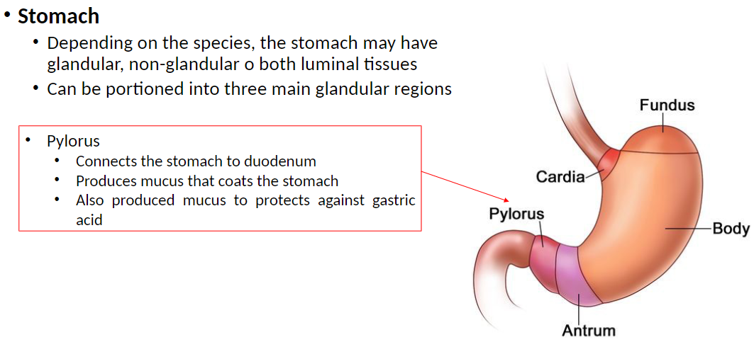
Digestive System – Pylorus
Pylorus: Connects the stomach to the duodenum
Produces mucus that coats the stomach lining
Protects stomach from acidic gastric secretions
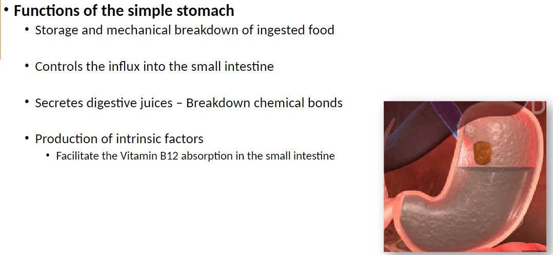
Digestive System – Functions of the Simple Stomach
Functions:
Storage and mechanical breakdown of ingested food
Controls influx of food into the small intestine
Secretes digestive juices to break chemical bonds in food
Produces intrinsic factor, which facilitates Vitamin B12 absorption in the small intestine
Digestive System – Stomach Functions by Region and Species
Cardia: Produces alkaline mucus to protect stomach lining
Species Differences:
Canine, porcine stomach – entirely glandular, secretes digestive acids and enzymes
Equine stomach – fundus non-glandular (margo plicatus separates glandular and non-glandular regions)
Bovine stomach – first three chambers (rumen, reticulum, omasum) non-glandular, primarily for storage and fermentation
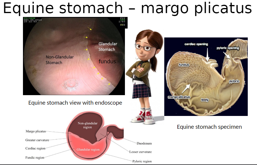
Digestive System – Equine Stomach: Margo Plicatus
Margo Plicatus: Ridge that separates non-glandular fundus from glandular regions of equine stomach
Non-glandular fundus: Mainly storage, no acid or enzyme secretion
Glandular region: Secretes acid and digestive enzymes for chemical digestion
Images: Endoscopic view and stomach specimen showing margo plicatus
From Corrections Post:
Horses
Large non-glandular area near the esophagus (dorsal fundus) → prone to ulcers
Rest of the stomach (cardiac, mostly fundic, pyloric) is glandular
Pigs
Very small non-glandular area near the esophagus
Most of the stomach is glandular
Ruminants
Abomasum is glandular
Forestomachs (rumen, reticulum, omasum) are non-glandular
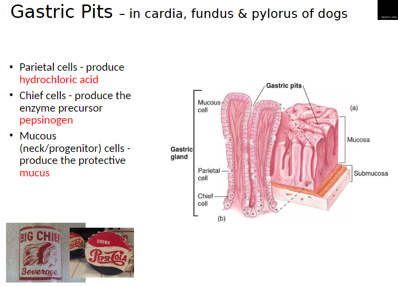
Digestive System – Gastric Pits (Dogs)
Gastric Pits: Found in cardia, fundus, and pylorus regions of the stomach
Cell Types and Functions:
Parietal Cells: Produce hydrochloric acid (HCl) for chemical digestion and killing microbes
Chief Cells: Produce enzyme precursor pepsinogen, which is converted to pepsin to break down proteins
Mucous (Neck/Progenitor) Cells: Produce protective mucus to coat the stomach lining and prevent damage from acid
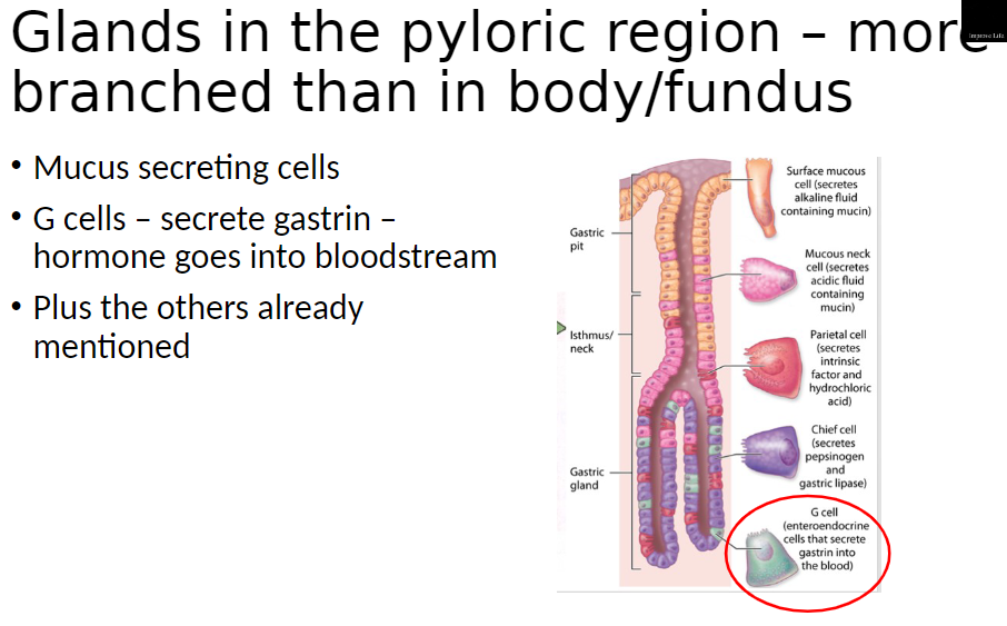
Digestive System – Pyloric Glands
Pyloric Glands: More branched than glands in body or fundus
Cell Types and Functions:
Mucus-Secreting Cells: Produce protective mucus for stomach lining
G Cells: Secrete gastrin, a hormone that enters bloodstream and stimulates acid secretion
Other Cells: Include parietal and chief cells as already described in body/fundus
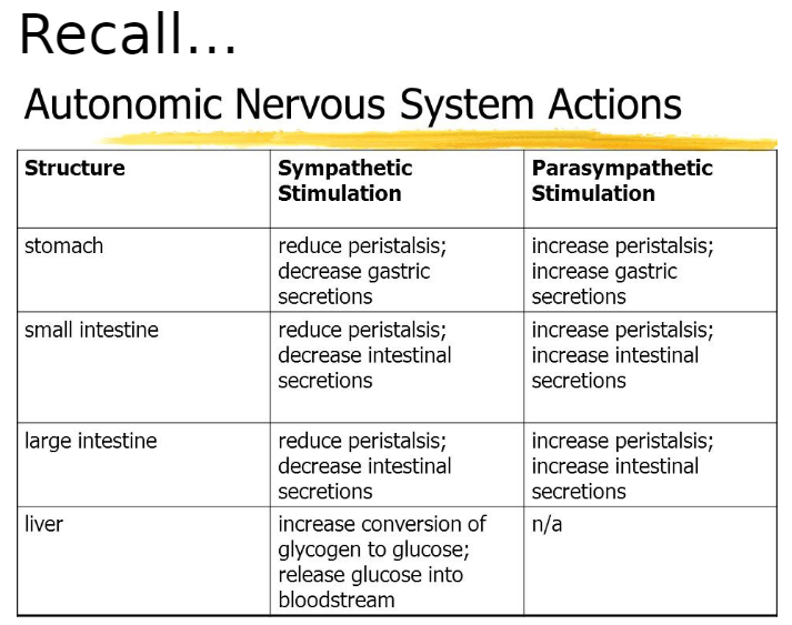
Digestive System – Autonomic Nervous System Actions
Autonomic Nervous System (ANS): Part of the nervous system that controls involuntary body functions
Sympathetic Nervous System: “Fight or flight” branch of ANS
Prepares body for stress or activity
Reduces digestion by slowing peristalsis and decreasing secretions
Parasympathetic Nervous System: “Rest and digest” branch of ANS
Promotes relaxation and maintenance functions
Increases digestion by enhancing peristalsis and stimulating secretions
Stomach:
Sympathetic Stimulation: Reduces peristalsis, decreases gastric secretions
Parasympathetic Stimulation: Increases peristalsis, increases gastric secretions
Small Intestine:
Sympathetic Stimulation: Reduces peristalsis, decreases intestinal secretions
Parasympathetic Stimulation: Increases peristalsis, increases intestinal secretions
Large Intestine:
Sympathetic Stimulation: Reduces peristalsis, decreases intestinal secretions
Parasympathetic Stimulation: Increases peristalsis, increases intestinal secretions
Liver:
Sympathetic Stimulation: Increases conversion of glycogen to glucose, releases glucose into bloodstream
Parasympathetic Stimulation: Not applicable
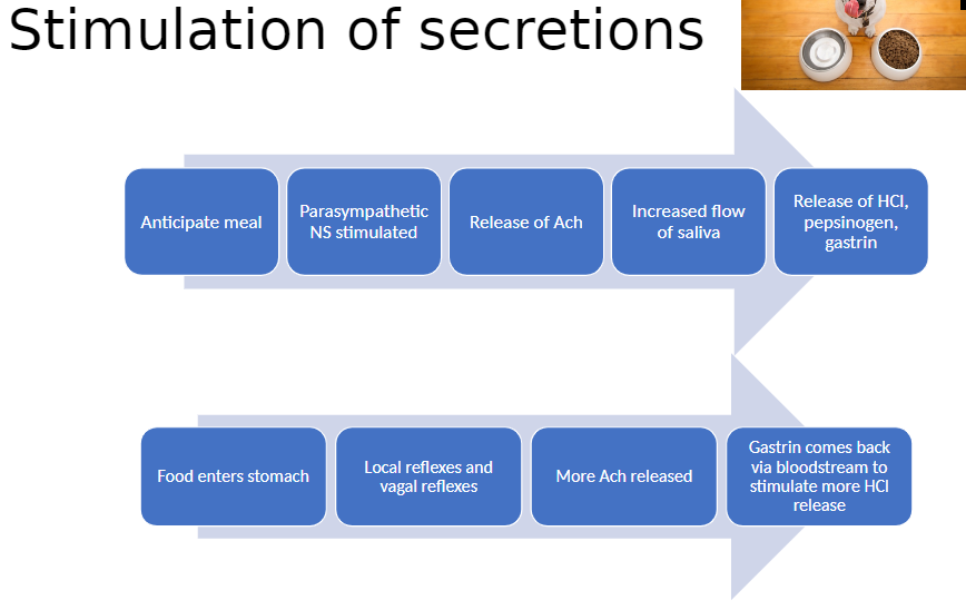
Digestive System – Stimulation of Secretions
Anticipation of Meal:
Parasympathetic nervous system is stimulated
Release of acetylcholine (Ach)
Increases flow of saliva
Triggers release of HCl, pepsinogen, and gastrin in stomach
Food Enters Stomach:
Local reflexes and vagal reflexes stimulate more Ach release
Gastrin returns via bloodstream to stimulate additional HCl release
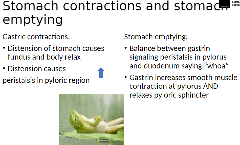
Digestive System – Stomach Contractions and Emptying
Gastric Contractions:
Distension (stretching) of stomach causes fundus and body to relax
Distension also triggers peristalsis in pyloric region to mix and push food toward small intestine
Stomach Emptying:
Controlled by balance between gastrin signaling peristalsis in pylorus and feedback from duodenum slowing emptying
Gastrin increases smooth muscle contraction at pylorus
Gastrin also relaxes pyloric sphincter to allow food to enter duodenum
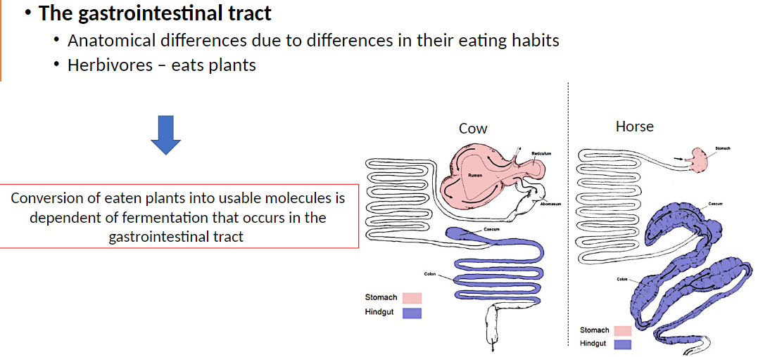
Digestive System – Gastrointestinal Tract (Herbivores)
Gastrointestinal Tract: Anatomical differences exist between species due to eating habits
Herbivores: Eat plants
Conversion of plant material into usable nutrients depends on fermentation in the gastrointestinal tract
Examples: Cow, Horse – specialized stomach and intestines adapted for plant digestion