Bio-Cog Psych
1/207
There's no tags or description
Looks like no tags are added yet.
Name | Mastery | Learn | Test | Matching | Spaced | Call with Kai |
|---|
No analytics yet
Send a link to your students to track their progress
208 Terms
Cognitive psychology
Study of the mind (thinking)
focus on functional explanations; process models
Biological psychology
Study of the biological basis of the mind
focus on brain processes; structural
Noam Chomsky
Linguist; complex cognitive operations (language) can’t be explained in terms of behaviourism
Behaviourism
Study observable behaviour and ignore the mind & mental processes (early 20th century)
Cognitive revolution
From 1960s cognitive psychology argued against behaviourism => mind should not be ignored and can be studied experimentally
→ core: human brain is an information processor
Assumptions of cognitive psychology
mental processes exist
mental processes can be studied in a scientific way
humans are active information processors
A short history of biological psychology
Ablation method (Flourens, 1815)
Discovery of Purkinje cells (neurons in the cerebellum; Purkinje, 1837)
Evolution theory (Darwin, 1859)
Effects of electrical stimulation of the brain (Fritsch & Hitzig, 1870)
Broca & Wernicke
Broca: Patient with left frontal lesion impaired in language production
Wernicke: Patient with left temporal lesion impaired in language comprehension
Henry Molaison (HM)
Epilepsy → removal medial temporal area (bilaterally) / hyppocampus
anterograde amnesia (no new long-term memories)
loss of long-term memory from before the brain surgery
Wilder Penfield
Direct electrical stimulation of cortex
produces "mental" sensations of thinking, perceiving, etc., rather than a sense of the brain being stimulated
Penfield & Jasper (1954) - neural stimulation prior to brain surgery
Motor cortex (motor humunculus)
Neurons project to the spinal cord to activate muscles
Somatosensory cortex (somatosensory humunculus)
Neurons receive activation from the receptors on skin, muscles and joints
Von Helmholtz (1850)
Nerve conduction velocity = 60 m/s (human)
→ Paves the way for mental chronometry
Donder's substruction method
RT (go-nogo) - RT (rimple) = stimulus discrimination time
→ Problems:
depends on assumptions about stages
strong assumption about stages being independent
Integration: Cognitive Neuroscience (since 1990s)
Supported by technological developments: fMRI and PET
Total time hypothesis
The proposal that the amount learned is a simple function of the amount of time spent on learning task
Gladwell (2008): 10.000 hours to become an expert in anything → element of truth, but oversimplification
Distributed vs. Massed practice
Better performance with distributed practice
Baddeley & Longman (1978) tested the effects of training schedule on typing performance → one session of one hour per day is the best
Generation effect
Memory is better if items have been generated by the subject than presented to the subject (Slamecka & Graf, 1978)
retrieval from memory (generation) is better than passive repetition (reading)
testing your knowledge is more effective than additional learning time (Karpicke & Roedigar, 2008 : foreign vocabulary learning
Swahili - English word pairs)
Testing effect
The importance of testing for later remembering
Levels of processing
Processing material in terms of meaning leads to the best retention
Craik & Tulving (1975) : 3 processing tasks, visual (cast), phonological (rhyme), meaning (sentence) → the latter condition does the best on a surprise test
How can we examine cognitive processes?
behavioural methods
reaction time
accuracy
eye movement
How can we determine the brain correlates of the cognitive processes?
brain methods
ERPs
fMRI
PET
lesions
single cell recordings
TMS
Reaction times (RT)
Posner (spatial) cueing task
Press a button as quickly as possible when you see a light, left or right of fixation
A cue indicates the most likely location of the light (80%)
Hold your eyes still in the middle
SO:
Central arrow predicts the location of the target
Subjects attend the cued location (top-down attention!)
Allocation of attention reduces reaction times
→ Reaction times allow us to examine the allocation of attention
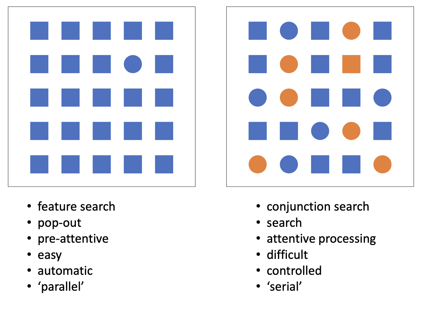
Visual search task
Feature search / Conjunction search
SO:
Reaction times indicate whether attention is required in visual search
Reaction time increase as a function of set size implies attention shifts from item to item to detect the target
steep search slope = attention
flat search slope = no attention
Event-related potentials (ERPs)
electrodes register electrical activity on the scalp, as produced by the brain
ERPs (and single cell recordings) can indicate whether and when items were attended
attentional ERPs effect is typically early (100-400ms)
language ERPs effect is typically late (400-600ms); language ERP effects reflect surprise / error in sentence processing (predictive coding)
syntax & semantics are differentely represented by the brain
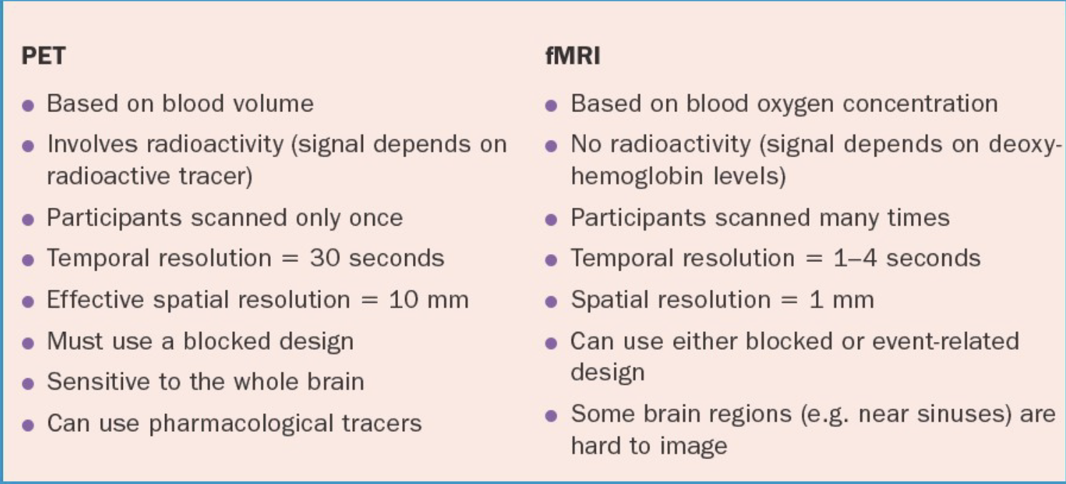
Brain imaging (fMRI, PET)
experimental condition vs. control condition (substruction method)
imagery in the brain (activity)
Imagery
has qualities similar to perception, but perception starts in the outside world, and imagery in our mind; they meet in the middle
SO:
only at the earliest visual projection areas is perception stronger than imagery
but even here the activation in the imagery condition was
nonzero
→ clear brain correlates of imagery
→ visual processes in imagery
fMRI
active brain areas attract blood (need oxygen)
oxygen reduction in hemoglobin -> change in magnetic properties
fMRI detectors pick up the changing magnetic properties
fMRI (like PET) measures neural activity indirectly, via differences in local blood supply
Positron Emission Tomography (PET)
a small amount of a radioactive substance is injected into the blood; active cells absorb more of it
the scanner picks up the gamma rays emitted when positrons from the tracer interact with electrons in the body
the data are reconstructed into detailed 3D images showing metabolic or biochemical activity
PET vs. FMRI
Transcranial Magnetic Stimulation (TMS)
a coil placed on the scalp sends short magnetic pulses through the skull, which induce tiny electrical currents that activate neurons in targeted brain regions
if a certain brain region is needed in a certain task then TMS to this brain region should result in impaired performance => allows an examination of a brain’s causal role
Single-cell recordings
A very fine microelectrode is inserted into brain tissue, positioned near a single neuron.
The electrode detects spikes (action potentials) produced by that neuron.
The timing and rate of these spikes are recorded while the subject is performing a task or perceiving stimuli.
Electroencephalography (EEG)
measures the brain’s continuous electrical activity — all the time, from many overlapping neural processes
ongoing “raw” brain waves (alpha, beta, theta, delta rhythms)
used to study general brain states — like sleep, alertness, or seizure activity
Temporal vs. spatial resolution (EEG vs. fMRI)
EEG: high temporal resolution (accurate in showing when something happens); low spatial resolution (inaccurate in showing where the activation is coming from)
fMRI: low temporal resolution; high spatial resolution
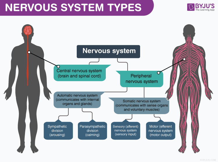
Nervous system types
Diffusion
the flow of dissolved substances from regions with high concentration to regions with low concentration.
Electrostatic pressure / force
the chemical elements with opposite polarity attract, while chemical elements with the same polarity repel each other
Ions
atoms that have an unbalanced amount of protons and electrons, which causes an electrical charge
Cations & Anions
cations are positively charged (cathodes are negative, opposites attract)
anions are negatively charged (anodes are positive, opposites attract)
Types of ions
Ionic bond
a type of chemical bond formed when one atom gives an electron to another atom
the atom that loses an electron becomes a positive ion (because it has more protons than electrons).
the atom that gains an electron becomes a negative ion (because it has more electrons than protons).
these oppositely charged ions attract each other, like magnets — and that attraction is what holds them together
Covalent bond
a type of chemical bond where atoms share electrons instead of transferring them.
In this bond, each atom contributes one or more electrons to be shared in a “shared pair”, i.e. molecule
e.g. glucose, amino acids -> proteins
Peptides
short protein chains
Lipids (fat)
long carbon chains; hydrophobic, because they are connected by an extra phosphate (P) group
Cell membrane is formed by…
a double layer of phospholipids
Transcription
genes are read from the DNA and converted to messenger RNA (mRNA)
mRNA leaves the nucleus through the pores, and is read out by ribosomes (complex of proteins) to form a new protein
Terminal buttons (a.k.a. axon terminals or synaptic boutons)
are tiny structures at the very end of a neuron’s axon; they act like the neuron’s “sending ends.”
Kinesin
anterograde transport from the cell body to the terminal buttons
Dynein
retrograde transport from terminal buttons to soma
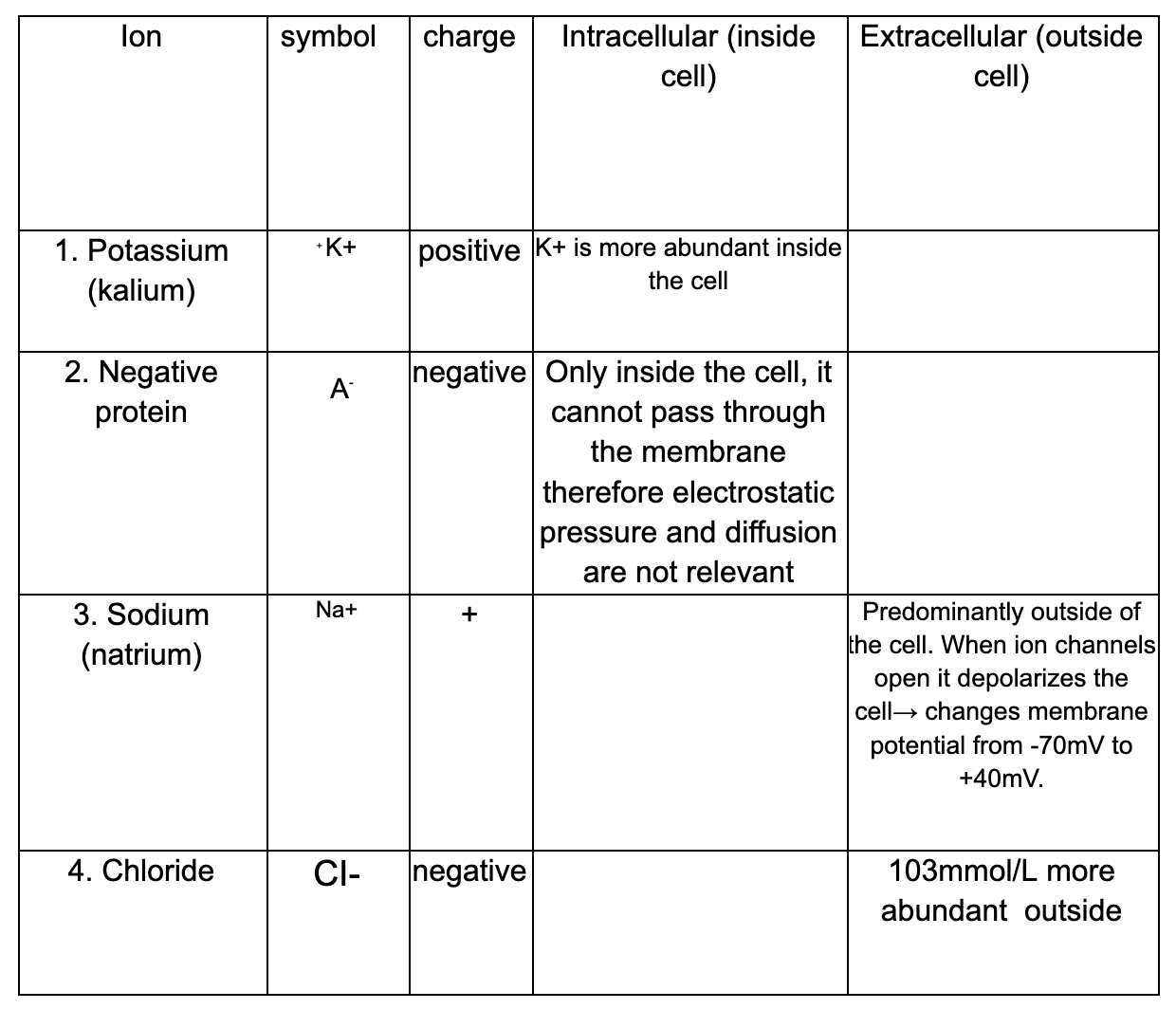
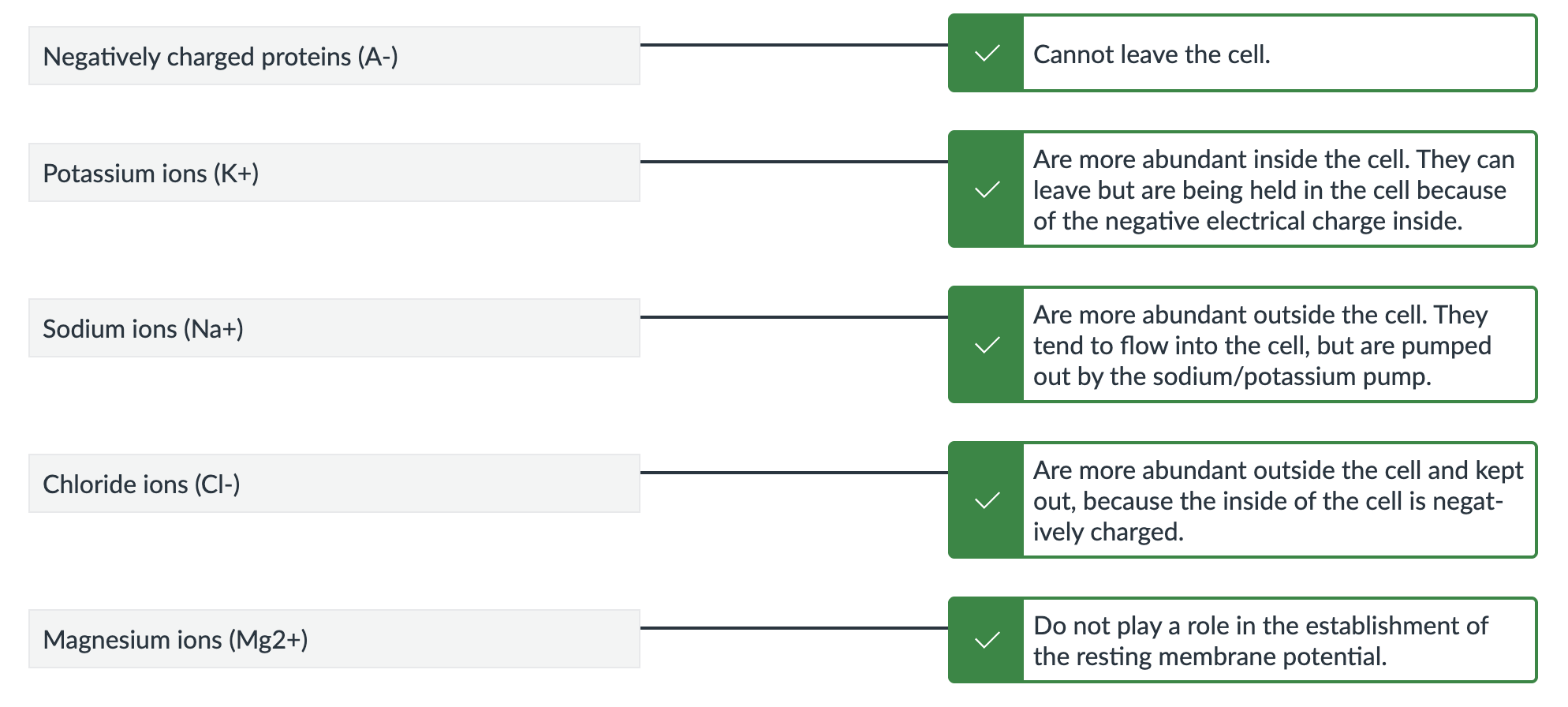
If a neuron is at rest, a stable situation arises, characterised by:
A higher concentration of Sodium (Na+) and Chloride (Cl-) outside the cell, and a higher concentration of Negative proteins (A-) and Potassium (K+) inside the cell
at rest, the negatively charged Chloride (Cl-) ions are more abundant outside the cell; they tend to move into the cell based on the law of diffusion, but are being pushed out based on the law of electrostatic pressure.
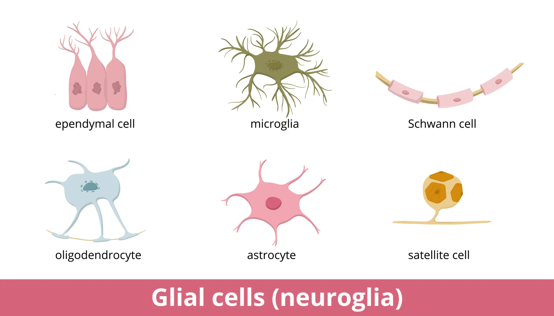
Glia
maintain homeostasis (self-regulation)
form myelin
provide support and protection for neurons
Myelin
An insulating layer that wraps around nerve cell axons, enabling electrical impulses to transmit quickly and efficiently along the nervous system
Microglia
the smallest type of glial cells; the main function of microglia is to support active immune defense in the central nervous system (CNS)
=> they protect the brain in case of possible harm due to foreign substances (for example as a consequence of bacterial or viral infections).
Phagocytosis
breaking down and engulfing of undesirable cells
Astrocytes (macroglia)
physical support for neurons
isolate neuronal contacts
breaking down of neurotransmitters like GABA and glutamate
providing neurons with energy from the blood stream
phagocytosis
Oligodendrocytes (macroglia)
physical support for neurons
producing layers of myelin around the axons of neurons
Schwann cells (macroglia)
producing layers of myelin around the axons of neurons
Oligodendrocytes vs. Schwann cells
oligodendrocytes form myelin around axons in the CNS, while Schwann cells form myelin around axons in the PNS
oligodendrocytes have multiple extensions such that they can form myelin around the axons of multiple neurons, while Schwann cells can only form one layer of myelin around a single axon
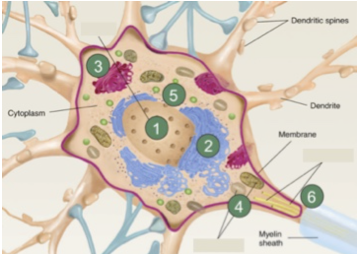
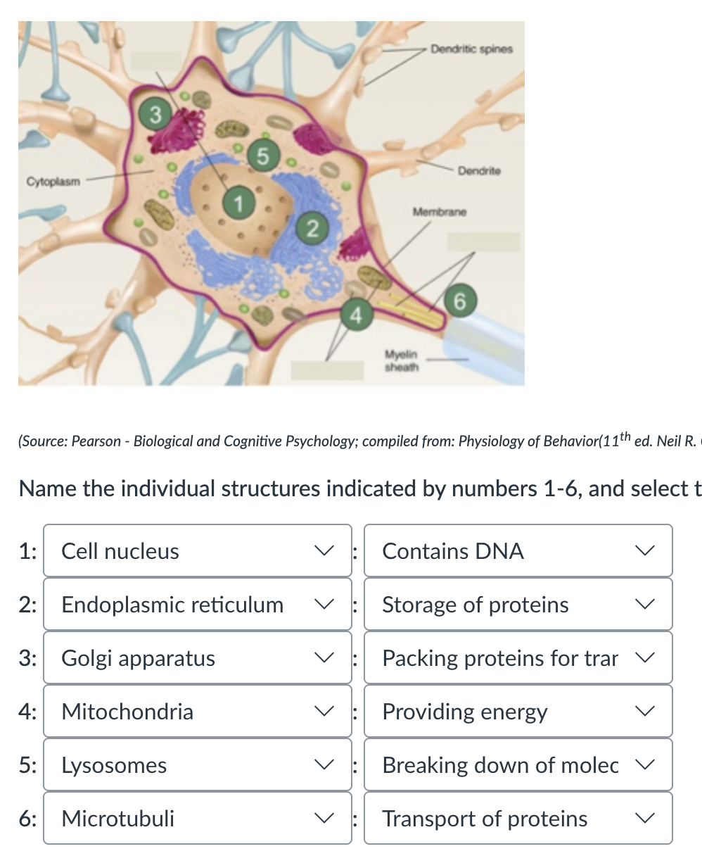
Name the individual structures indicated by numbers 1-6
“All or None Law” / Threshold of excitation
the axon generates an action potential only if the resting potential crosses a threshold → the magnitude of the action potential is always the same
Action potential
the electrical signal that travels along the membrane of a neuron; the main way neurons communicate with each other and send information through the nervous system.
Resting Membrane Potential (= polarisation)
the relative potential difference between the inside and outside of the neuron
When a neuron is not sending a signal, it is polarized
The inside of the neuron is negatively charged relative to the outside
This difference in charge is typically around –70 millivolts (mV)
Voltage-gated ion channels
membrane proteins which open and close in response to changes in membrane potential, allowing certain ions to pass through (sodium, potassium, calcium)
voltage gated sodium channels open first to cause depolarization, then voltage-gated potassium channels open last to cause repolarization
Depolarisation
more Na⁺ channels are opened, Na⁺ flows into the cell, the cell becomes less negative
Repolarisation
K⁺ keeps flowing out, cell inside returns to negative
Hyperpolarisation
Following the massive outflow of K⁺, the membrane temporarily has an extra negative charge
Sodium-potassium pump
a protein transporter in the neuron's membrane that keeps Na+ and K+ balanced inside and outside the cell
the protein pushes 3Na+ out of the cell and 2K+ in the cell, using energy from the AP
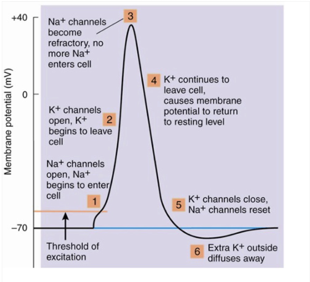
The sequence of events during an action potential
Resting potential around is around -70mV
A stimulus excites the neuron causing depolarization (reaches a threshold of -55mV) triggering an action potential
Voltage gated sodium channels open, positive sodium ions (Na+) flood into the cell making the inside positive (peak around +30mV)
Repolarisation - sodium channels close while potassium channels open, positive potassium ions (K+) flood out the cell causing membrane potential to decrease and become negative again
Hyperpolarisation - potassium channels close causing membrane potential to become temporarily more negative than resting potential (-90mV)
Sodium potassium pump transports ions out to return to resting membrane potential + voltage gated channels reset to prepare for next action potential (3 sodium ions get pushed out and 2 potassium ions go inside) ( around -65 mV)
There is a refractory period (during and after action potential) in which a neuron cannot initiate another one (a one-way transmission of the signal)
Action potential conduction: cons
relatively slow: new action potentials are generated in the neighboring region
energy consuming: resting potential needs to be recovered across the whole axon, by means of the Na⁺ - K⁺ pumps
Myelin passive (w/out new action potentials) conduction
axon covered with pieces of myelin that prevents the generation of action potentials
→ action potential is conducted passively through the myelin
→ a new action potential is generated at the myelin interruptions (nodes of Ranvier); faster, but decays with distance
Myelin passive conduction: pros
saltatory conduction is faster through myelin
saltatory conduction is energy efficient
Nodes of Ranvier
The gaps along the myelin sheath, where action potentials jump from one node to another
Measuring action potential
the size is not measured; it remains consistent → the firing rate is measured
low intensities: slow firing
high intensities: fast firing
How is information represented in the brain?: Coding
specificity coding: representations depend on the activity of individual neurons; one neuron per item
e.g. of specificity coding: Quiroga et al (2005) (Jennifer)
distributed coding (or population coding) : representations depend on the distribution of activity across a whole population of neurons
sparse coding: a small group of neurons represent each item; similar to population coding, even more efficient, but a bit more vulnerable as well
Neural networks (connectionism / distributed networks / parallel distributed processing)
developed during the 80s, biologically inspired
no local representation: concepts have a distributed representation across nodes (distributed coding)
neural networks can learn: on the basis of feedback the strength of connections between nodes is adjusted: (error back propagation (feedback) → adjustment of weights)
allows for powerful computer algorithms: deep learning
"graceful degradation" - if a few nodes or connections are damaged, the network will still work
generalisation is a natural coding
Predictive coding
our brain is continually predicting the immediate future based on:
experience
top-down processing
statistical regularities of the environment
memory
context (spatial, temporal, etc)
Excitatory & Inhibitory neurotransmitters
Axon-hillock
the region of a neuron where the axon joins the cell body
its primary function is to integrate incoming synaptic signals and initiate an action potential, acting as the neuron's "trigger zone”
=> this is because the axon hillock has a high concentration of voltage-gated sodium channels, which are essential for generating the electrical impulse
Exocytosis
a neuron transfers its action potential to other neurons through the synapse
when an action potential arrives, vesicles with neurotransmitter (synaptic vesicles) release their content in the synaptic cleft
How calcium (Ca) triggers exocytosis
depolarisation of the pre-synaptic membrane leads to the opening of “voltage-dependent” Ca channels → Ca will flow into the cell, tearing the vesicles
Effects of neurotransmitter release in post-synaptic neuron
the neurotransmitter diffuses into the post-synaptic membrane, BECAUSE
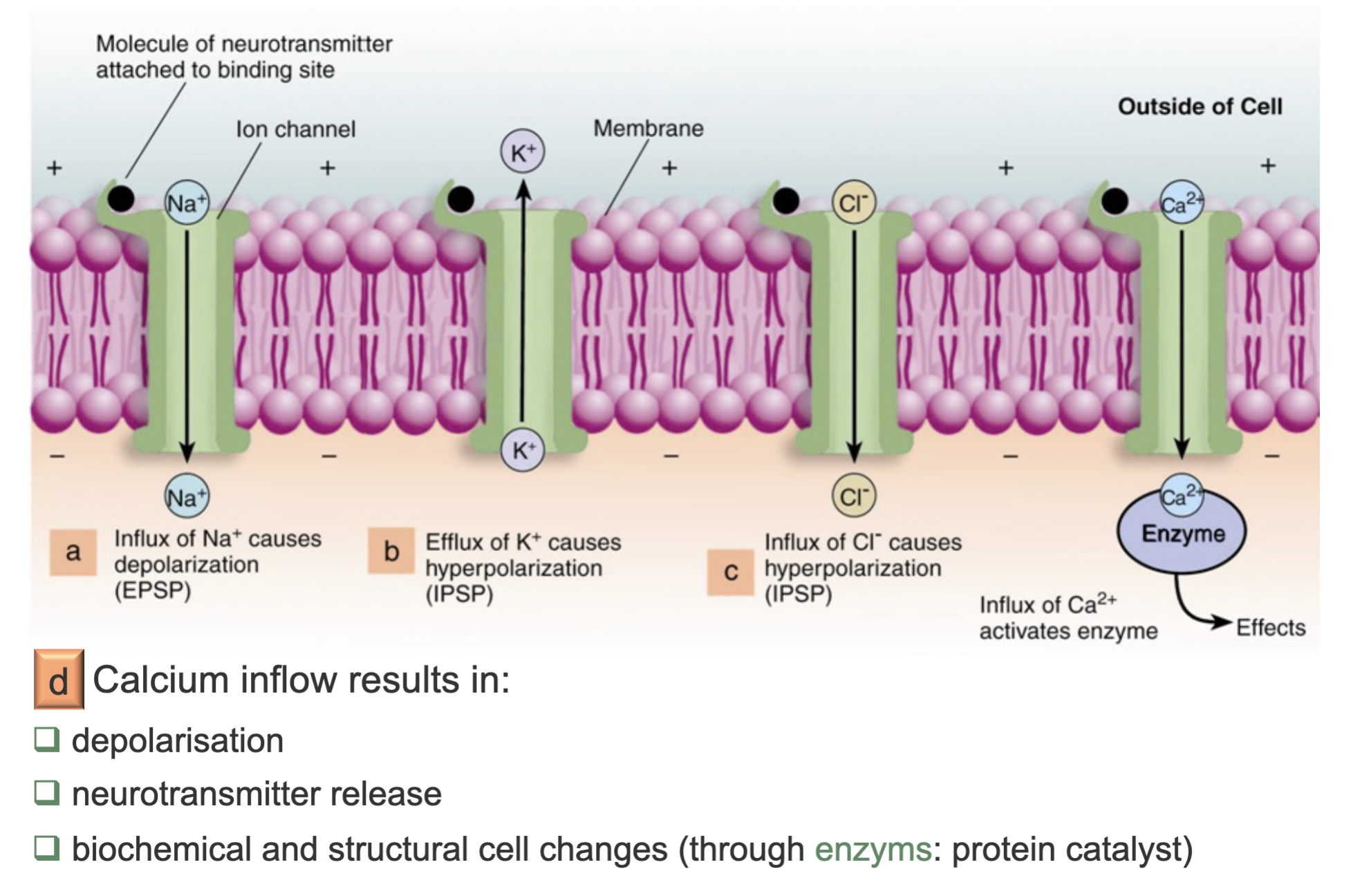
the post-synaptic membrane has transmitter-dependent ion channels (ionotropic receptors)
Ionotropic receptors
group of proteins which open to allow ions (e.g. Na+, K+, Ca2+, and / or Cl−) to pass through the membrane in response to the binding of a chemical messenger (i.e. a ligand)
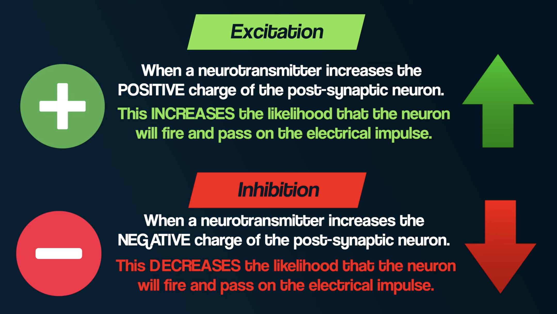
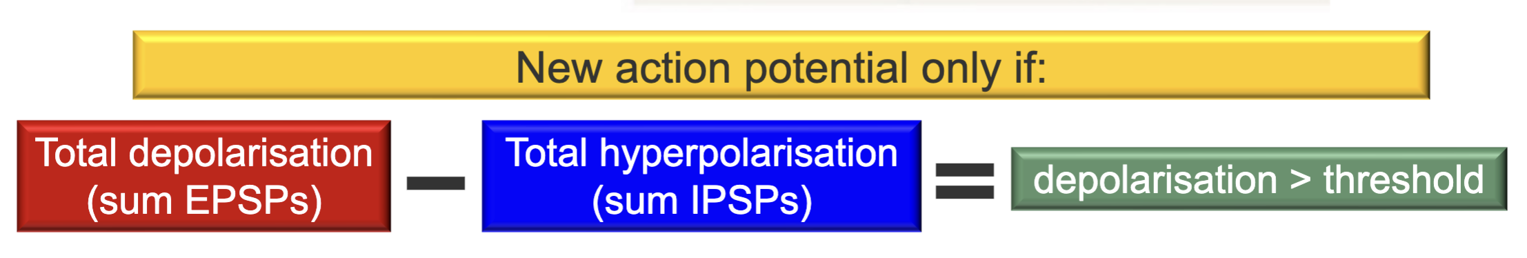
Excitatory Post-Synaptic Potential (EPSP)
depolarisation of the post-synaptic neuron will occur when the neurotransmitter opens Na+ channels
Inhibitory Post-Synaptic Potential (IPSP)
hyperpolarisation of the post-synaptic neuron will occur when the neurotransmitter opens Cl- channels
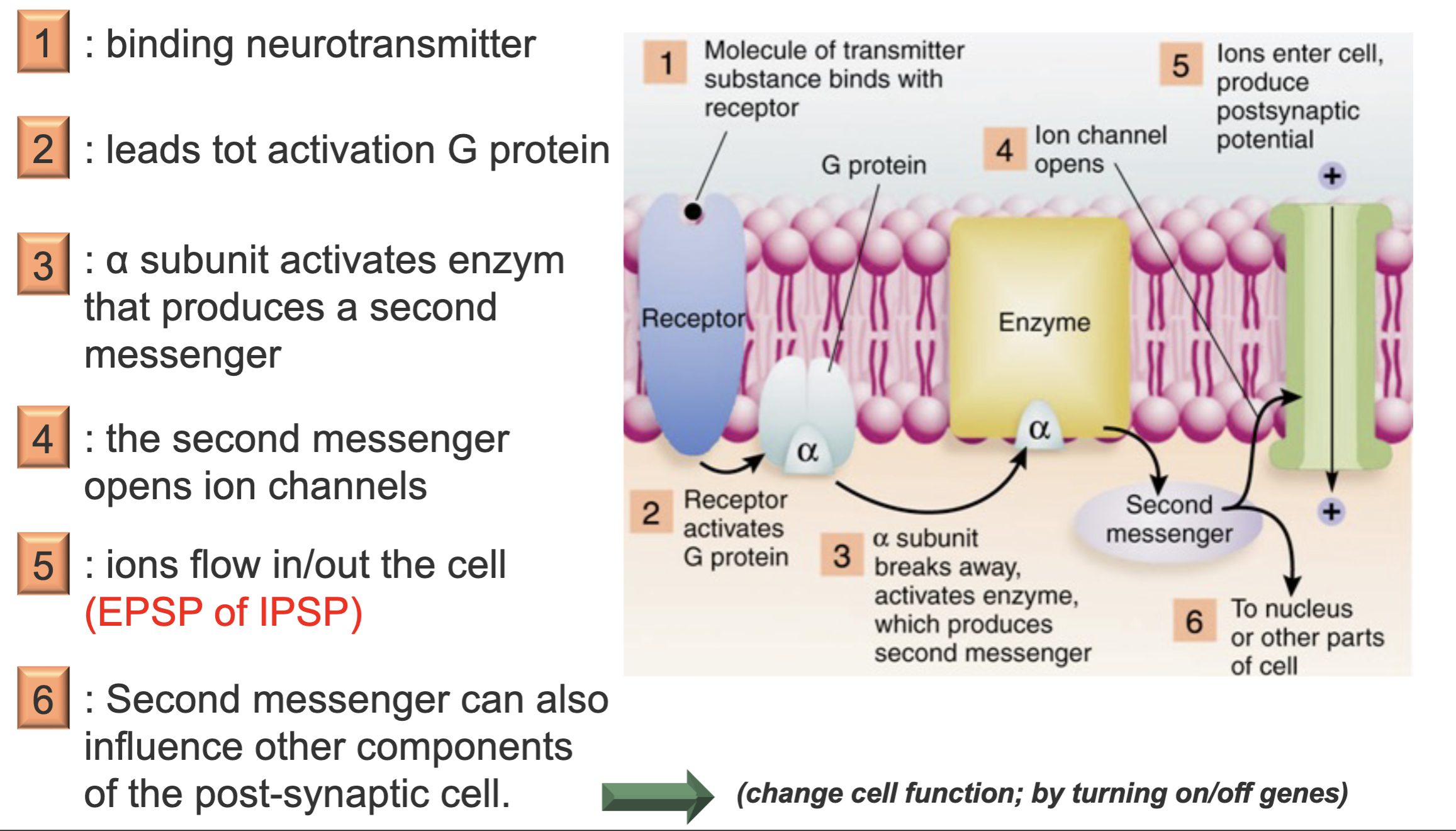
Metabotropic receptors
these receptors are indirectly linked with ion channels through signal transduction mechanisms, such as G protein
Summary of possible ion flows
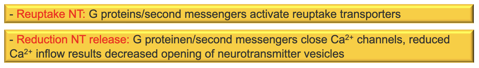
Regulation of the neurotransmitter concentration
neurotransmitter cleared from synaptic cleft through:
diffusion
re-uptake
enzymatic degradation: the enzyme Acetylcholine-esterase (AChE) splits ACh into Choline and Acetate
Autoreceptors
regulate the production and release of neurotransmitter by the neuron
metabotropic
generally inhibitory
New action potential only if:
Neuromodulators
wander through the brain and sensitize / desensitize complete neural networks (through subthreshold depolarisations or hyperpolarisations)
Hormones
excreted by glands: kidneys, pituitary, thyroid, etc
e.g.: Vasopressin (agression), Oxytocin (bonding)
Nucleosides
part of a DNA base pair bound to sugar
e.g.: Adenosine accumulates during the day, making one sleepy -> Caffeine blocks Adenosine receptors
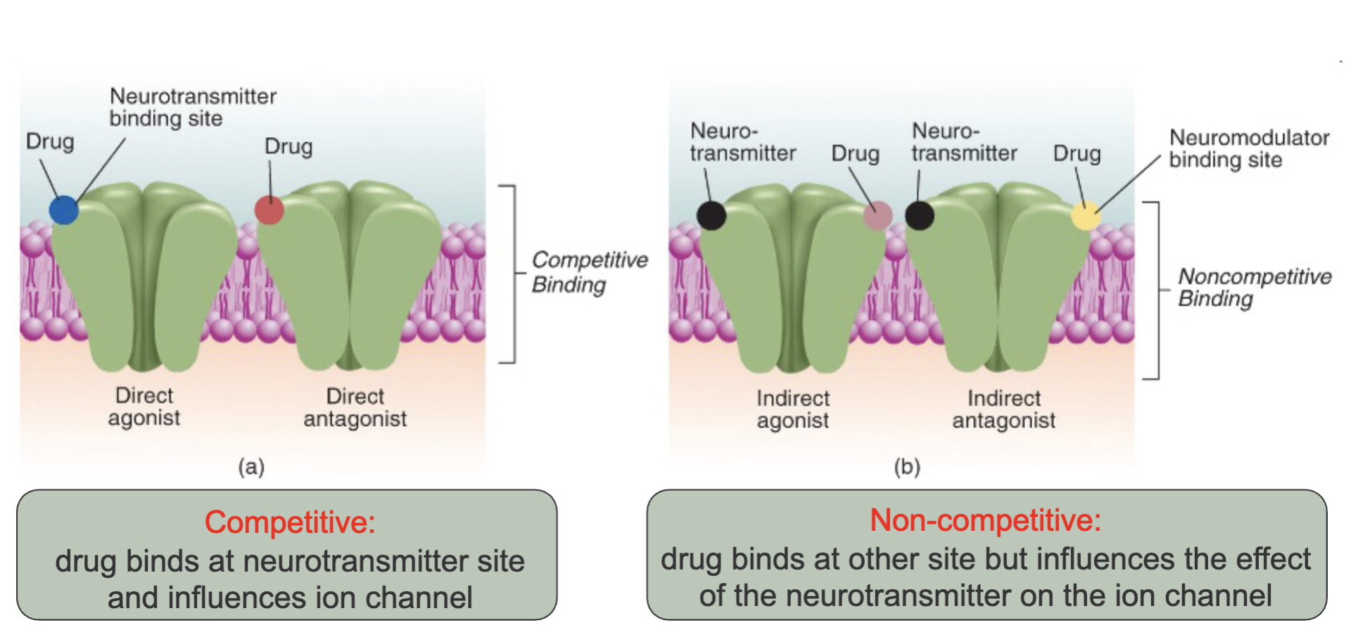
Ways of operation of drugs
drugs exert their effect mainly by binding to pre- and post-synaptic receptors
Ligand
a chemical that binds to a receptor
Competitive vs. non-competitive binding
General overview cerebral cognitive functions
overall: posterior = perception; frontal = processing
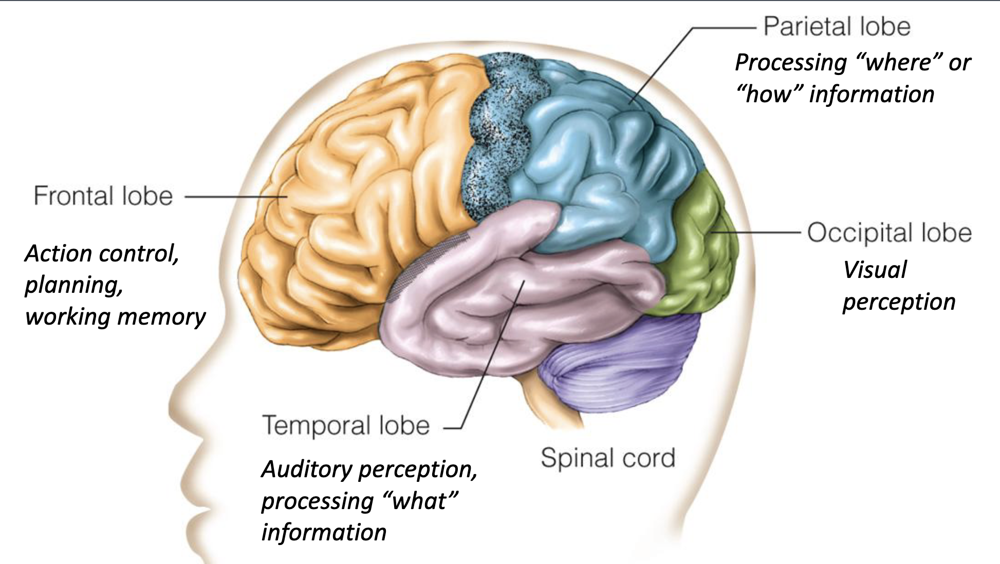
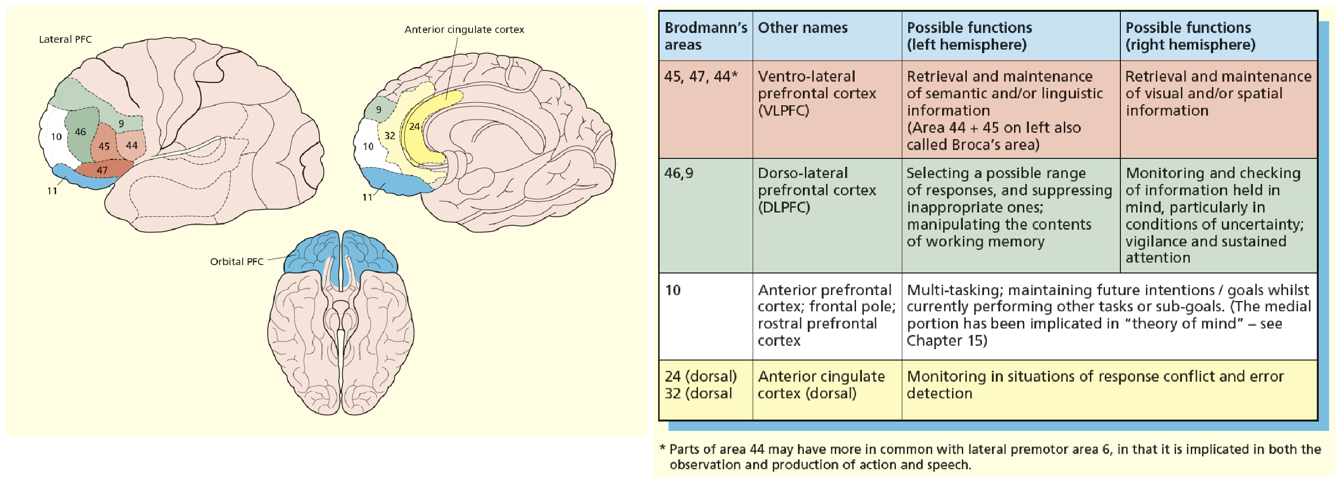
Prefrontal cortex
involved when tasks get more complex/require some form of “control”
particularly large in humans, and matures relatively late during development
may hold the key to our ability to thrive as humans in complex societies (both from social and technological perspective)
Three surfaces of the prefrontal cortex
lateral, medial and orbital
lateral surface implicated in cognitive aspects of executive functions
orbital and medial more implicated in emotional / social regulation of behavior
Curtis and D’esposito (2003)
prefrontal cortex is involved in the top-down selection of stimulus representations in the parietal cortex
representations stored in posterior regions; representations controlled from prefrontal regions
Working memory
maintaining information needed for ongoing cognitive processing
best-known model by Baddeley and Hitch (1974)
“slave systems” controlled by a central executive
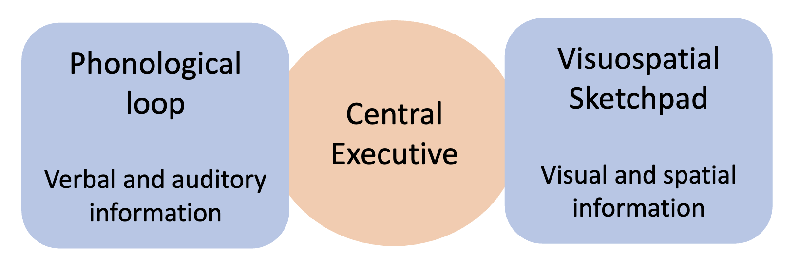
Working memory: Central executive
recruits relevant “slave systems”
attentional controller (distributes attention across tasks)
coordination of complex cognitive operations