Radiology Lab 9 - Neuroradiology, head + neck
1/20
There's no tags or description
Looks like no tags are added yet.
Name | Mastery | Learn | Test | Matching | Spaced | Call with Kai |
|---|
No analytics yet
Send a link to your students to track their progress
21 Terms
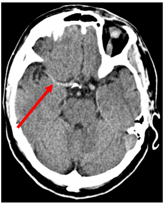
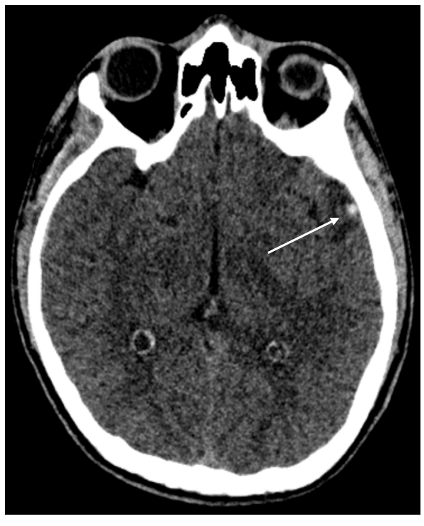
What is shown in this native CT aspect image?
Hyperintense right middle cerebral artery - indirect sign of supraacute ischemic stroke
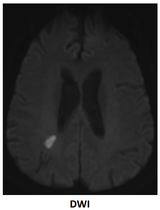
What is shown in the image here?
Supraacute ischemic stroke
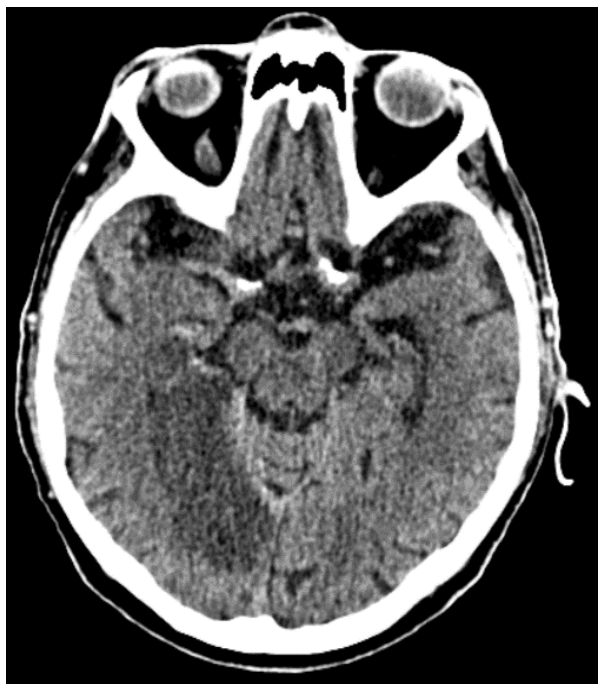
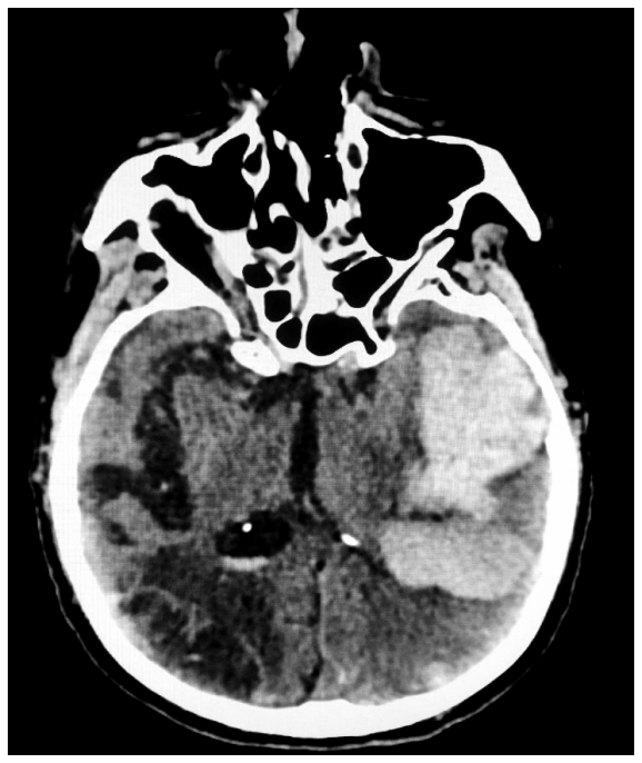
What does this CT image show?
Acute Ischemic Stroke: hypointense diffuse area on the right + gyral effacment
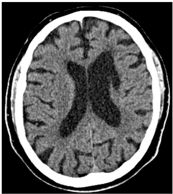
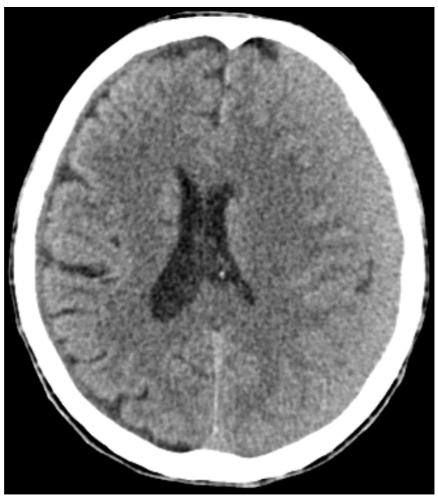
What does this CT image show?
Chronic Ischemic Stroke (sequalae); tractioned frontal horn of the left ventricle
What is shown in this imaging?
Acute Hemorrhagic Stroke - with right ventricular flooding and mass effect generated over the ventricular system
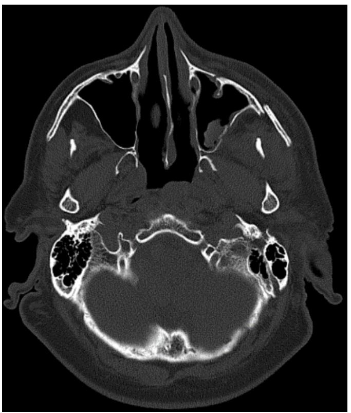
What is shown in this CT bone window?
Bone fractures affecting:
Zygomatic bone
Posterior wall of the left maxillary sinus + Left maxillary hemosinus
What is shown in this CT image?
Brain Hemorrhagic Contusion + Surrounding edema
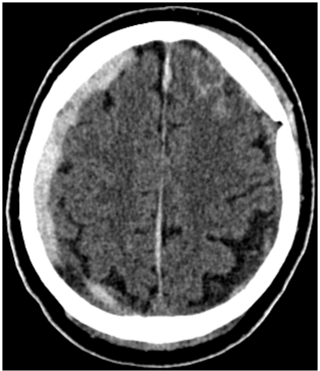
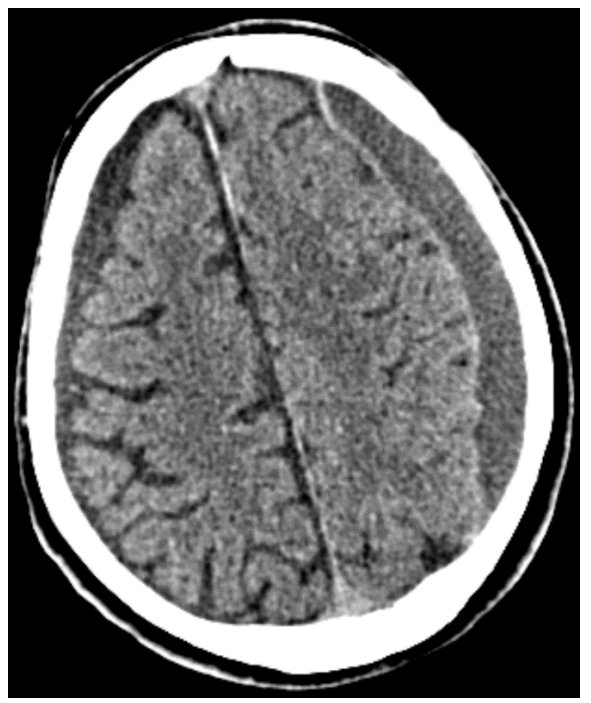
What can be seen in this CT image?
Right fronto-parietal acute subdural hematoma
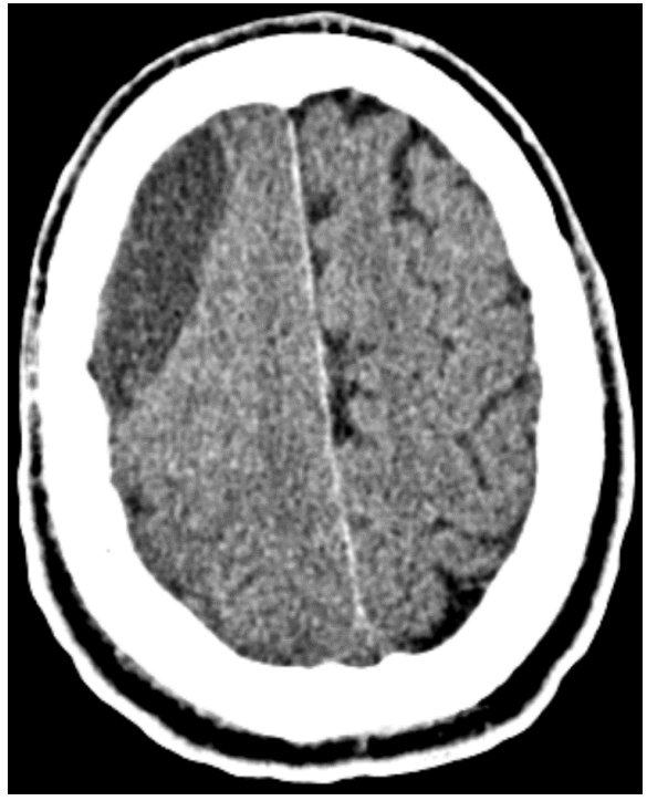
What is this CT image showing?
Left fronto-parietal subacute subdural hematoma - mass effect over brain parenchyma + ventricuar system
What is shown in this CT image?
Left fronto-parietal chronic subdural hematoma
What does this CT image show?
Chronic epidural hematoma
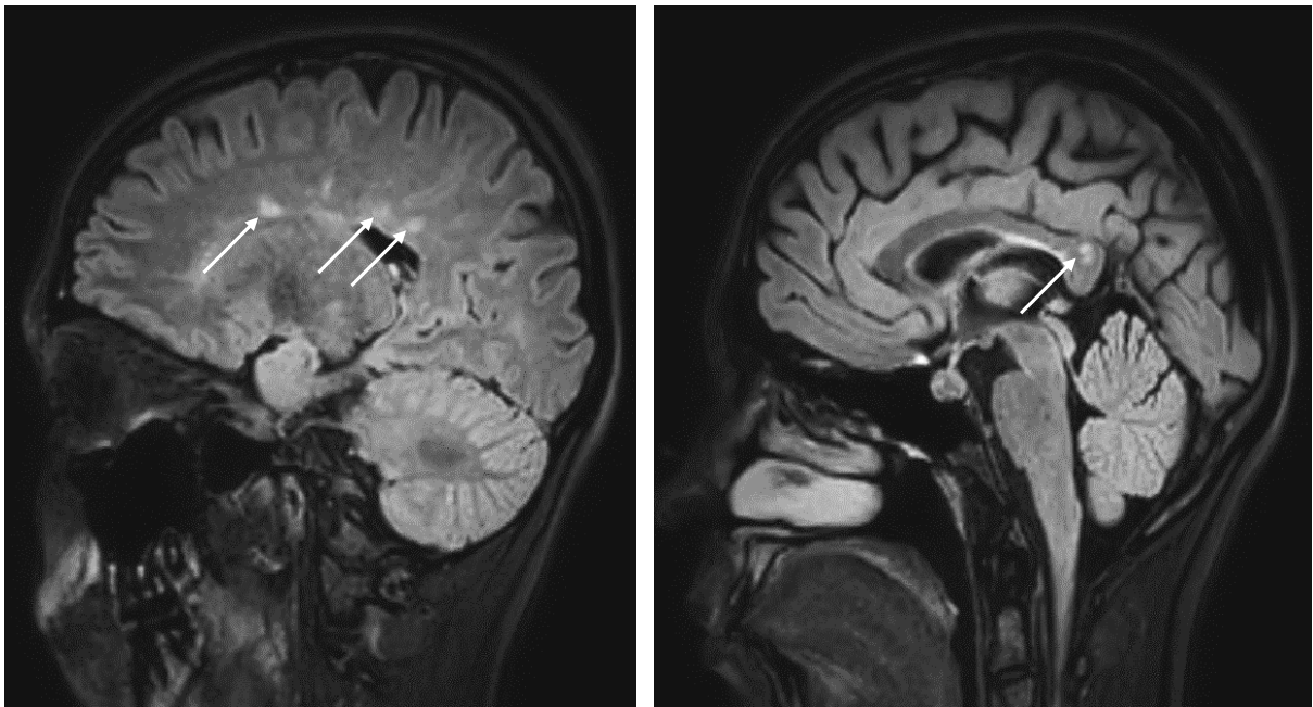
This MRI, FLAIR sequence, imaging shows?
Demyelinating lesions - Multiple sclerosis
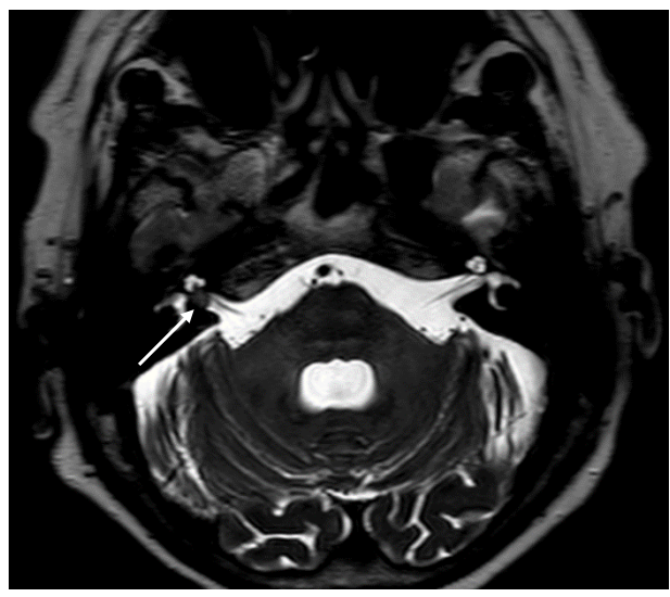
What does this MRI image show, and what phase?
Acoustic neuroma - T2
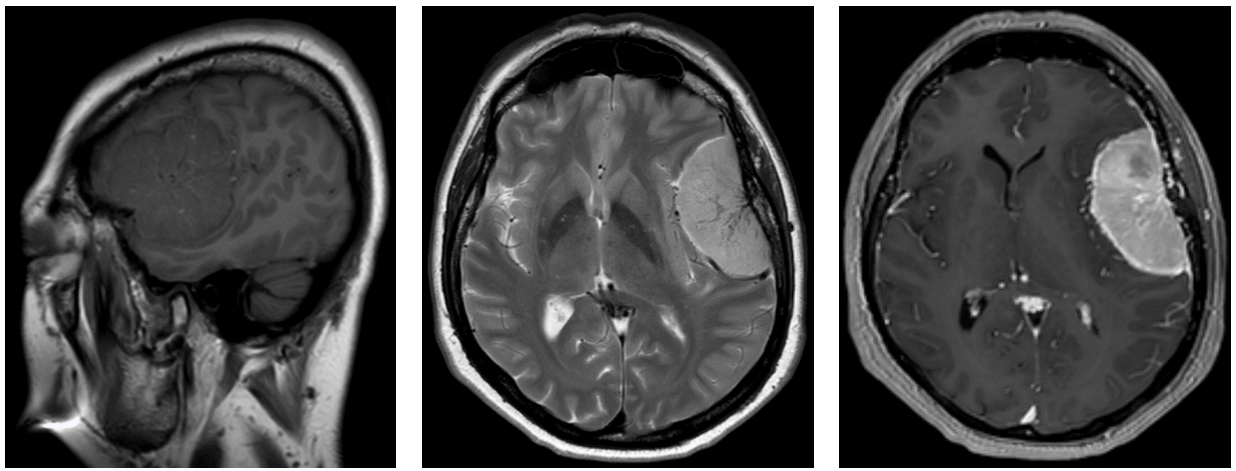
what does this MRI image show, and what phases are each of these?
Meningioma: T1, T2, postcontrast T1 sequences
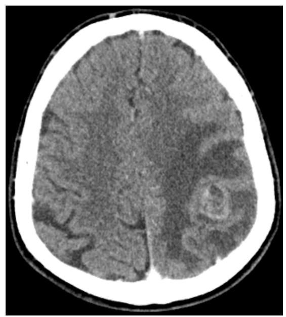
What can be seen in this CT image?
Malignant brain tumor + perilesional edema
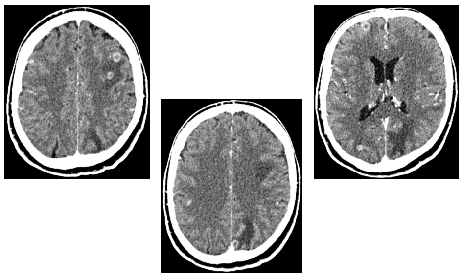
What does this postcontrast CT image show?
Multiple brain metastases, perilesional edema
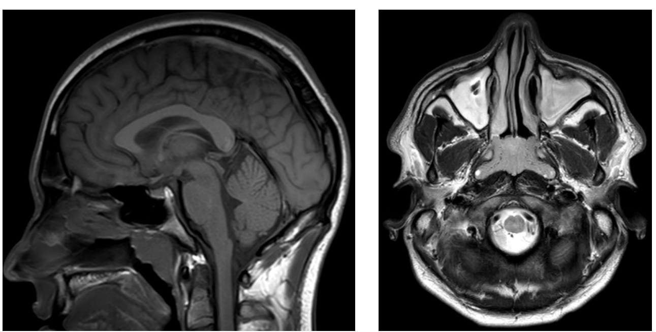
What does this MRI image show, and which phase is this?
High amount of fluid inside the right maxillary sinus - T2
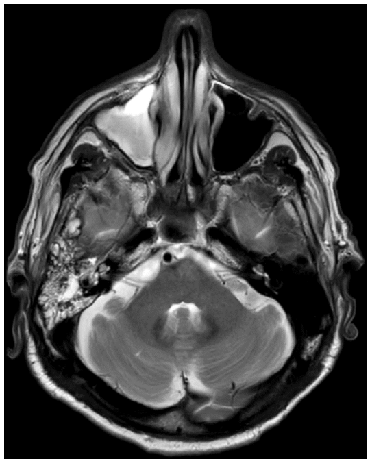
What can be seen in this MRI image?
Adenoid hypertrophy + fluid inside both maxillary sinuses
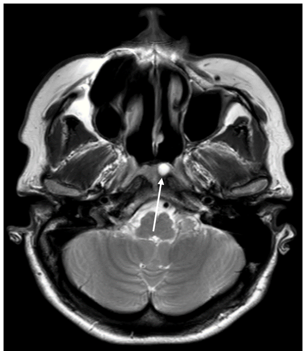
What does this MRI image show, and which phase?
Thornwaldt cyst - T2
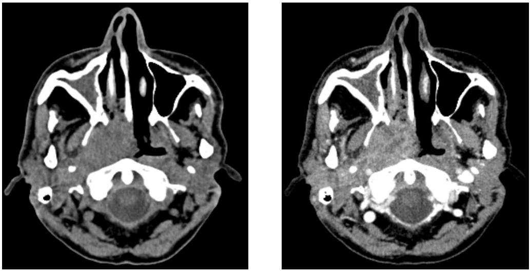
What does this CT image show?
Right nasopharyngeal tumor + fluid inside the right maxillary sinus
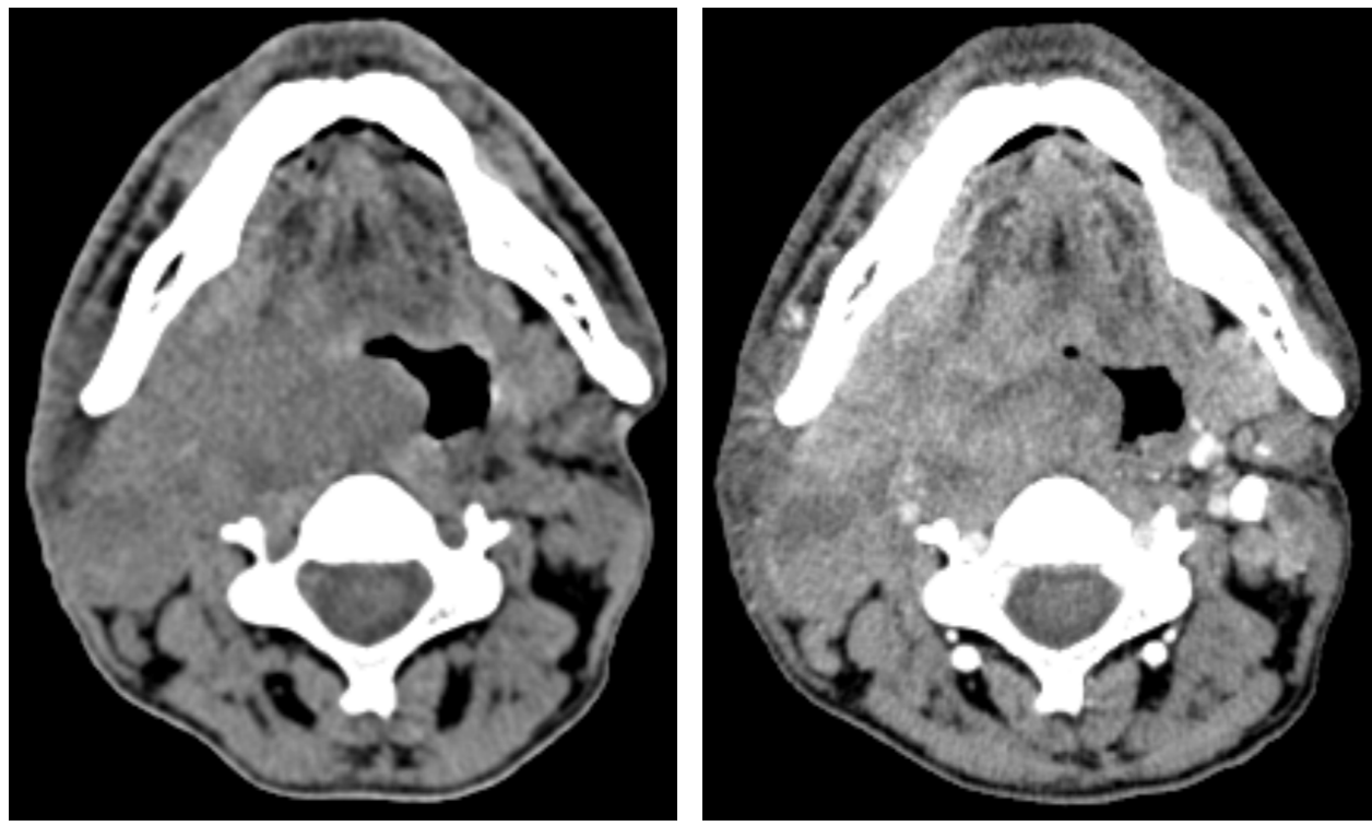
What does this CT image show?
Right oropharyngeal tumor