FULL Meninges & the Cerebral Hemispheres
1/309
There's no tags or description
Looks like no tags are added yet.
Name | Mastery | Learn | Test | Matching | Spaced |
|---|
No study sessions yet.
310 Terms
What is the medullary center?
The deep white matter located beneath the cerebral cortex, consisting of axons transmitting sensory input to cortex and motor output away from cortex.
What types of neural information travel in the medullary center?
Ascending sensory information and descending motor commands.
Why does the right hemisphere control the left body and vice versa?
Because projection fibers decussate (cross the midline) in the brainstem/spinal cord.
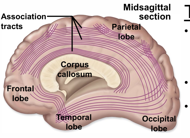
What do association fibers connect?
Different cortical areas within the SAME cerebral hemisphere.
Example of long association fibers?
Fibers connecting frontal lobe to occipital lobe within the same hemisphere.
Example of short association fibers?
Fibers connecting adjacent gyri within the same hemisphere.
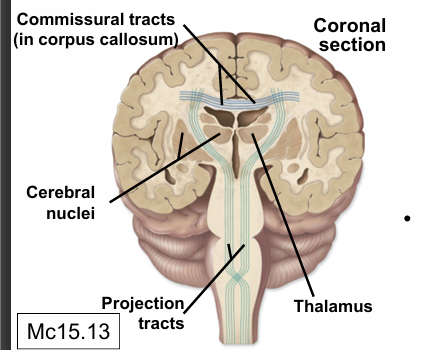
What do commissural fibers connect?
Corresponding areas of the LEFT and RIGHT cerebral hemispheres across the midline.
What is the largest commissural fiber bundle?
The corpus callosum.
Where do you see the corpus callosum in brain sections?
In any mid-sagittal section and many coronal sections because it is a massive structure.
What is the function of the corpus callosum?
Allows communication and integration between left and right hemispheres.
What is the anterior commissure?
A smaller commissural fiber bundle connecting the two temporal lobes across the midline.
What do projection fibers connect?
The cerebral cortex with lower CNS structures (brainstem and spinal cord).
What do descending projection fibers carry?
Motor commands from cortex to brainstem and spinal cord.
What do ascending projection fibers carry?
Sensory information from body → spinal cord → brainstem → cortex.
Where do descending motor projection fibers decussate?
In the brainstem (pyramidal decussation).
Where do ascending sensory fibers decussate?
In the spinal cord or brainstem, depending on the sensory pathway.
What is the clinical importance of projection fiber decussation?
Lesions above the decussation cause contralateral deficits
What gray matter structures are embedded in the white matter of the medullary center?
Basal nuclei (caudate, putamen, globus pallidus) and parts of the diencephalon (thalamus, hypothalamus, epithalamus).
What is the masa intermedia?
A small midline connection joining the right and left thalami in most people.
What does the masa intermedia do to the third ventricle?
It interrupts the third ventricle, creating a circular bump in ventricular casts.
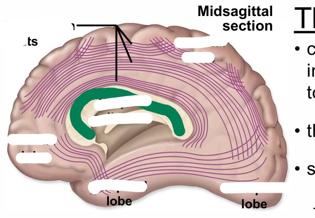
LABEL THIS
association tracts
parietal, temporal, occipital, frontal lobes
corupus callosum
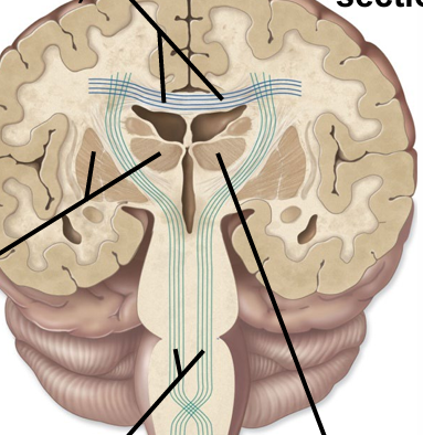
Label this
commusiural tracts
cerebral nuclei
projection tracts
thalamus
What are the basal nuclei?
Deep gray matter clusters involved in refining voluntary movement.
What older term is sometimes used for basal nuclei?
Basal ganglia (incorrect because ganglia = PNS).
What three major nuclei make up the basal nuclei?
Caudate nucleus, putamen, globus pallidus.
What is the globus pallidus also called?
The pallidum (“pale body”).
What two additional structures functionally associate with the basal nuclei?
Subthalamic nucleus and substantia nigra.
What is the function of the basal nuclei?
Refine and improve voluntary movement (not initiate it).
Do basal nuclei directly control the spinal cord?
No — they influence movement indirectly via motor cortex loops.
What type of feedback loop do basal nuclei participate in?
Cortex → basal nuclei → thalamus → back to cortex (modulating motor output).
What are two major categories of movement disorders caused by basal nuclei dysfunction?
Hypokinesia (too little/slowed movement) and hyperkinesia (excess/involuntary movement).
Which basal nuclei–related structure degenerates in Parkinson’s?
Substantia nigra (loss of dopaminergic neurons).
What classic tremor occurs in Parkinson’s?
Pill-rolling tremor (thumb and finger rolling motion).
What gait abnormality occurs in Parkinson’s?
Shuffling gait with reduced stride length.
What difficulty with movement initiation is seen in Parkinson’s?
Trouble taking the first step — patients may lean forward to “fall into” walking.
What facial expression change is seen in Parkinson’s?
Masked facies (reduced facial expression).
What posture issue occurs in Parkinson’s?
Stooped posture and difficulty adjusting postural reflexes.
Do Parkinson’s patients develop cognitive issues?
Yes — cognitive decline may occur in later stages.
What is hyperkinesis?
Excess, involuntary, or abnormal movements caused by basal nuclei dysfunction.
What is the thalamus?
A pair of large relay nuclei located superior to the hypothalamus
Which sensory modality does NOT relay in the thalamus?
Olfaction.
What does the lateral geniculate nucleus (LGN) process?
Vision (relays visual input to primary visual cortex).
What does the medial geniculate nucleus (MGN) process?
Auditory input (relays to primary auditory cortex).
What does the ventral posterior nucleus relay?
Somatosensation (touch, pain, temperature, vibration) and taste.
What is conscious proprioception?
Awareness of body position in space
Why is it strange that the thalamus has a “ventral posterior” nucleus?
Because the neuraxis bends—so ventral ≠ anterior and posterior ≠ dorsal anymore.
What is the main function of the thalamus?
Relay and modulate sensory, motor, limbic, and arousal signals to the cortex.
Which thalamic nuclei are involved in arousal and wakefulness?
Diffuse projecting nuclei.
Which thalamic nuclei contribute to emotion and memory?
Anterior nuclear group (limbic functions).
What is the masa intermedia?
A small midline connection between the right and left thalami present in most people.
How does the masa intermedia affect the third ventricle?
It interrupts the third ventricle with a circular “bump” in the midline.
Where is the hypothalamus located?
Below the thalamus, forming part of the walls of the third ventricle.
What is the main role of the hypothalamus?
Maintain homeostasis through neural and hormonal control.
How does the hypothalamus influence the autonomic nervous system?
Regulates sympathetic and parasympathetic output.
What reproductive functions does the hypothalamus regulate?
Controls hormonal regulation of reproduction via pituitary hormones.
What behaviors or drives does the hypothalamus regulate?
Feeding, water balance, circadian rhythms, emotional responses.
What gland does the hypothalamus control directly?
The pituitary gland.
What structure is included in the epithalamus?
The pineal gland.
What hormone does the pineal gland secrete?
Melatonin.
What is the function of melatonin?
Regulates circadian rhythms and contributes to puberty onset.
Which sensory modality does NOT relay through the thalamus?
Olfaction (smell).
Where do all other sensory modalities synapse before reaching cortex?
A specific relay nucleus in the thalamus.
What does “relay” mean in sensory pathways?
One neuron synapses on another inside the thalamus before projecting to cortex.
What is a primary sensory cortex?
The cortical region where a sensory modality is first consciously perceived.
Examples of primary sensory cortices
Primary somatosensory (postcentral gyrus), primary visual (calcarine sulcus), primary auditory (temporal lobe/insular region), primary olfactory (temporal lobe).
What is a sensory association cortex?
A cortical region that interprets and gives meaning to sensory input after it is first perceived.
Why is association cortex needed?
Perception is not comprehension — interpretation requires additional processing.
Where are association cortices typically located?
Adjacent to their corresponding primary cortices.
What is a multimodal association cortex?
A region where multiple sensory modalities combine and integrate (e.g., sight + sound + touch).
Why are multimodal cortices important?
Allows holistic perception — e.g., seeing a cookie + smelling it + remembering past experiences.
Where is the major multimodal association region?
Inferior parietal lobe at the intersection of somatosensory, visual, and auditory areas.
What functions is the prefrontal multimodal association cortex responsible for?
Reasoning, prediction, judgment, personality, emotional regulation.
What deficits occur with prefrontal cortex lesions?
Impulsivity, poor judgment, personality changes, inability to predict consequences.
Which famous case demonstrated prefrontal cortex injury?
Phineas Gage (railroad spike → personality changes).
What does “somatotopic organization” mean?
Adjacent body parts are represented in adjacent cortical regions.
Where is the motor homunculus located?
Primary motor cortex (precentral gyrus).
What determines the size of a body part in the motor homunculus?
Precision of movement required, NOT size of the body part.
Which body areas have the largest representation in the motor homunculus?
Hands, face, and tongue (high precision movements).
Which body areas have the smallest representation in the motor homunculus?
Trunk and proximal limbs.
What side of the body does each motor cortex control?
The contralateral (opposite) side.
Where is the sensory homunculus located?
Primary somatosensory cortex (postcentral gyrus).
What determines the size of a body part in the sensory homunculus?
Density of sensory receptors and precision of sensation.
Which areas have the largest sensory representation?
Fingertips, lips, tongue (dense receptors).
Why do fingertips have such precise sensation?
Tightly packed sensory receptors with very small receptive fields.
Which areas have the smallest sensory representation?
Trunk and limbs (large receptive fields).
How do dermatomes relate to the sensory homunculus?
Body regions with lower spinal dermatomes (S2–S5) are represented deeper on the medial postcentral gyrus (e.g., genitals/perineum).
Example: Where is L5 dermatome (top of foot) represented in the homunculus?
More superior on the medial surface than S2–S5 (genital region).
What is two-point discrimination?
Testing how close two stimuli can be before they are perceived as one.
Why can the arm confuse two close points?
Large receptive fields & lower receptor density.
Why can fingertips distinguish two very close points?
Small receptive fields & high receptor density.
Why is homunculus knowledge vital for stroke localization?
Different vascular territories supply different body parts on the map — deficits reveal which artery is occluded.
Which artery supplies medial pre/postcentral gyrus (leg area)?
Anterior cerebral artery (ACA).
Which artery supplies lateral pre/postcentral gyrus (face/arm area)?
Middle cerebral artery (MCA).
What is Broca’s (motor speech) area located anterior to?
The precentral gyrus in the dominant frontal lobe (usually left).
What is the primary function of Broca’s area?
Motor aspects of speech production: sequencing, rate, and putting words in order.
What is Wernicke’s (sensory speech) area primarily responsible for?
Understanding/interpreting language (sensory language comprehension) in the dominant temporal lobe.
What is the typical hemisphere of language dominance?
Left hemisphere in the majority of people.
What characterizes expressive (Broca’s) aphasia?
Non-fluent speech, impaired speech production (labored, broken), preserved comprehension (usually), patient aware and frustrated.
What characterizes receptive (Wernicke’s) aphasia?
Fluent but meaningless speech, poor comprehension, patient usually unaware and content (no insight).
What is global aphasia?
Severe impairment of both comprehension and expression (both Broca’s and Wernicke’s areas affected).