Posterior Thigh and Popliteal Region
 Call Kai
 Learn
Learn Practice Test
 Spaced Repetition
 Match
Match Flashcards
 Knowt Play
1/32

There's no tags or description
Looks like no tags are added yet.
Name | Mastery | Learn | Test | Matching | Spaced | Call with Kai | Chat |
|---|
No analytics yet
Send a link to your students to track their progress
33 Terms
What are the compartments of the thigh muscles?
Anterior, medial, and posterior separated by 3 fascial intermuscular septa
What are the muscles of the posterior thigh?
Semitendinosus, semimembranosus, biceps femoris
What are the attachments of the semitendinosus?
Ischial tuberosity to medial surface of superior part of tibia
(1/2 tendinosus from its attahcment)
What is the action of the semitendinosus?
Extend thigh, flex leg, medially rotate leg when knee is flexed; can extend trunk when thighs and legs are flexed
What innervates the semitendinosus?
Tibial division of sciatic nerve L5,S1/2
What are the attachments of the semimembranosus?
Ischial tuberosity to posterior part of medial condyle of tibia
(1/2 membranous from its attahcment)
What is the action of the semimembranosus?
Same as semitendinosus: extend thigh, flex leg, medially rotate leg when knee is flexed; can extend trunk when thighs and legs are flexed
What innervates the semimembranosus?
Tibial division of sciatic nerve L5, S1/2
What are the attachments of the long head of the biceps femoris?
Ischial tuberosity to lateral side of head of fibula
What are the attachments of the short head of the biceps femoris?
Linea aspera and lateral supracondylar line of femur to lateral side of head of fibula
What is the action of the biceps femoris?
Flex leg, rotate it medially when flexed
*Short head of biceps femoris only flexes the knee
What innervates the long head of biceps femoris?
Tibial division of sciatic nerve L5, S1/2
What innervates the short head of biceps femoris?
Common fibular division of sciatic nerve
How do the posterior thigh muscles work synergistically?
Except short head of biceps femoris, they all attach to iliotibial tract and act on 2 joints which cause extension of the hip and flexion of the knee
What are the borders of the popliteal fossa?
Diamond shaped:
Superolateral: biceps femoris
Superomedial: semimembranosus and semitendinosus
Inferolateral and inferomedial: gastrocnemius
What are the contents of the popliteal fossa?
Fat, popliteal vessels, tibial and common fibular nerves, small saphenous vein, articular branch of obturator nerve, 4-6 popliteal lymph nodes, popliteus bursa
What is the path of the popliteal artery?
The deepest structure in the popliteal fossa; continuation of femoral artery as it emerges from adductor hiatus, ends by dividing into anterior and posterior tibial artery at the inferior border of popliteus muscle
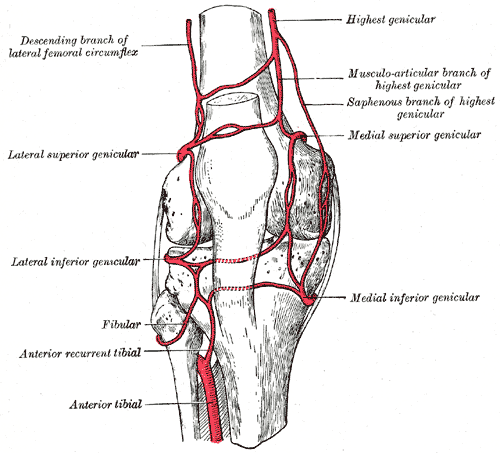
What is the genicular anastomosis?
5 genicular branches (lateral superior and inferior, medial superior and inferior, and middle) of popliteal artery supply capsule and ligaments of knee joint; like a highway around the knee
What is the path of the popliteal vein?
Formed at distal border of popliteus muscle by union of venae comitantes of anterior and posterior tibial veins; lies in same fibrous sheath as popliteal artery, ends at adductor hiatus where it becomes femoral vein
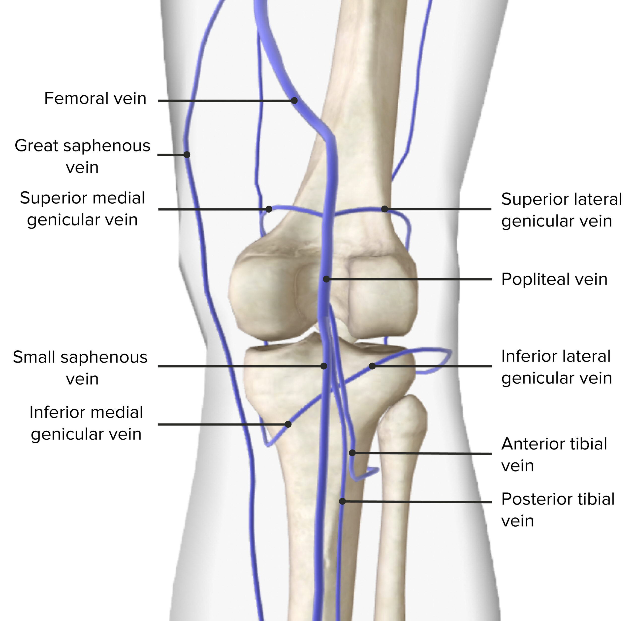
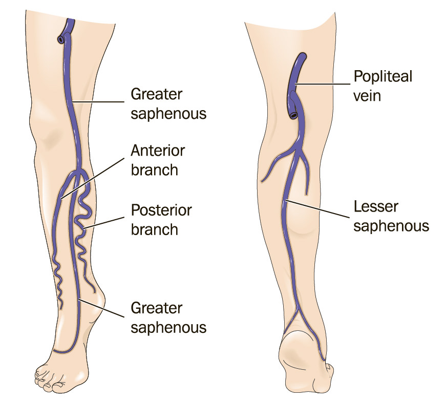
Where does the small saphenous vein drain into?
Popliteal vein
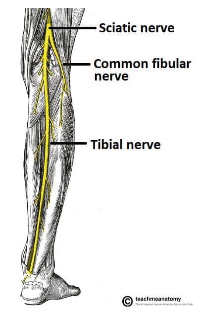
What is the path of the tibial nerve?
After breaking from sciatic nerve, the genicular branches innervate knee joint, muscular branches innervate soleus, gastroc, popliteus, and plantaris; medial sural cutaneous joins lateral (from common fibular) to make sural cutaneous to supply lateral side of leg and ankle
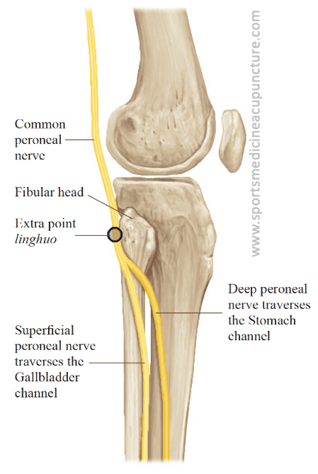
What is the path of the common fibular nerve?
Leaves popliteal fossa by passing superior to lateral head of gastroc; passes over posterior aspect of head/neck of fibula, superficially divides at neck to fibula into superficial and deep fibular nerves; within popliteal fossa it gives articular branches to knee and proximal tibiofibular joint, lateral sural cutaneous (joins with medial to make sural), peroneal communicating branch
What is the acetabular labrum?
The fibrocartilaginous rim attached to margin of acetabulum; increases acetabular articular area nearly by 10%
(more than ½ of femoral head is contained within the acetabulum)
What is the structure of the acetabulum?
Forms ¾ of a circle; the missing inferior segment is the acetabular notch and is bridged by the transverse acetabular ligament
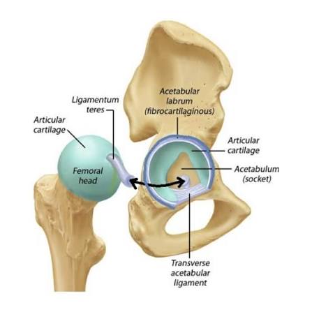
What is the transverse acetabular ligament?
A continuation of the acetabular labrum
What ligaments strengthen and thicken the articular cartilage of the acetabular capsule?
Iliofemoral, pubofemoral, and ischiofemoral ligament
What is the body’s strongest ligament?
Iliofemoral ligament
Which is the weakest hip joint ligaments?
Pubofemoral
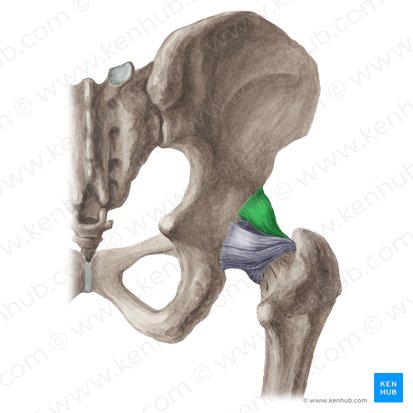
What is the role of the iliofemoral ligament?
Prevents hyperextension of hip during standing (helps maintain erect position); lies superior and anteriorly
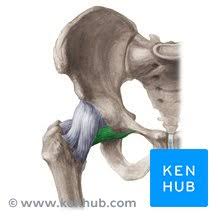
What is the role of the pubofemoral ligament?
Strengthens inferior and anterior parts of capsule; lies anterior and inferiorly
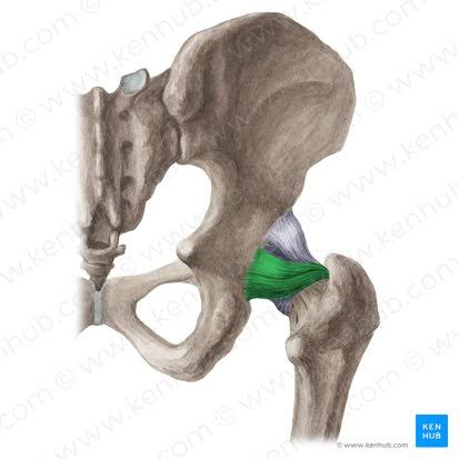
What is the role of the ischiofemoral ligament?
Strengthens posterior part of fibrous capsule and prevents hyperextension; lies posteriorly
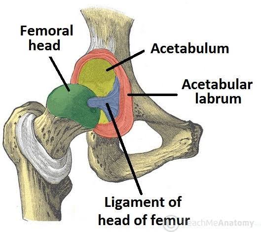
What is the ligamentum capitis femoris and what does it do?
AKA ligamentum teres: a weak intracapsular ligament surrounded by a sleeve of synovial membrane; contains a small artery to head of femur; is stretched when flexed thigh is adducted or lateral rotated
What supplies blood to the hip joint?
Branches of the medial and lateral cutaneous circumflex artery, deep divisions of the superior and inferior gluteal arteries, and the artery to head of femur (runs though ligamentum teres)