GI and Biliary
 Call Kai
 Learn
Learn Practice Test
 Spaced Repetition
 Match
Match Flashcards
 Knowt Play
1/197

Earn XP
Description and Tags
EXAM 7/30
Name | Mastery | Learn | Test | Matching | Spaced | Call with Kai | Chat |
|---|
No analytics yet
Send a link to your students to track their progress
198 Terms
Liver (3-4 pounds)
What is the largest solid organ in the body?
RUQ
Where is the liver located in the abdomen?
Over 100 different functions
- Synthesis of substances concerned with blood clotting
- Storage of vitamins
- Detoxification & excretion of substances
- Production of bile (1 qrt a day)
- Metabolism of substances delivered via portal circulation
How many functions does the liver have?
about 1 quart
How much bile is produced by the liver each day?
Emulsifying fat (contains cholesterol)
What is biles function?
Majors: Right (larger & lateral) & Left (smaller & medial)
Minor (both associated with the right lobe): Quadrate (inferior) & Caudate (superior)
What are the loves of the liver? Major & Minor?
Falciform ligament
What separates the major lobes of the liver?
ducts that drain bile into the duodenum
What is the biliary tree?
Triggered by cholecystokinin (CCK) by the presence of fatty food
What triggers the release of bile?
In the lobules of the liver
Where is bile formed?
Right and Left Hepatic duct --> Common hepatic duct --> Common bile duct --> Pancreatic duct (or Duct of Wirsung) --> hepatopancreatic ampulla (or Ampulla of Vater) --> Hepatopancreatic sphincter (or Sphincter of Oddi) -->
Duodenum
Or
Right and Left Hepatic duct --> Common Hepatic duct --> Cystic Duct --> Gallbladder
Explain the biliary tree to the duodenum and to the gallbladder.
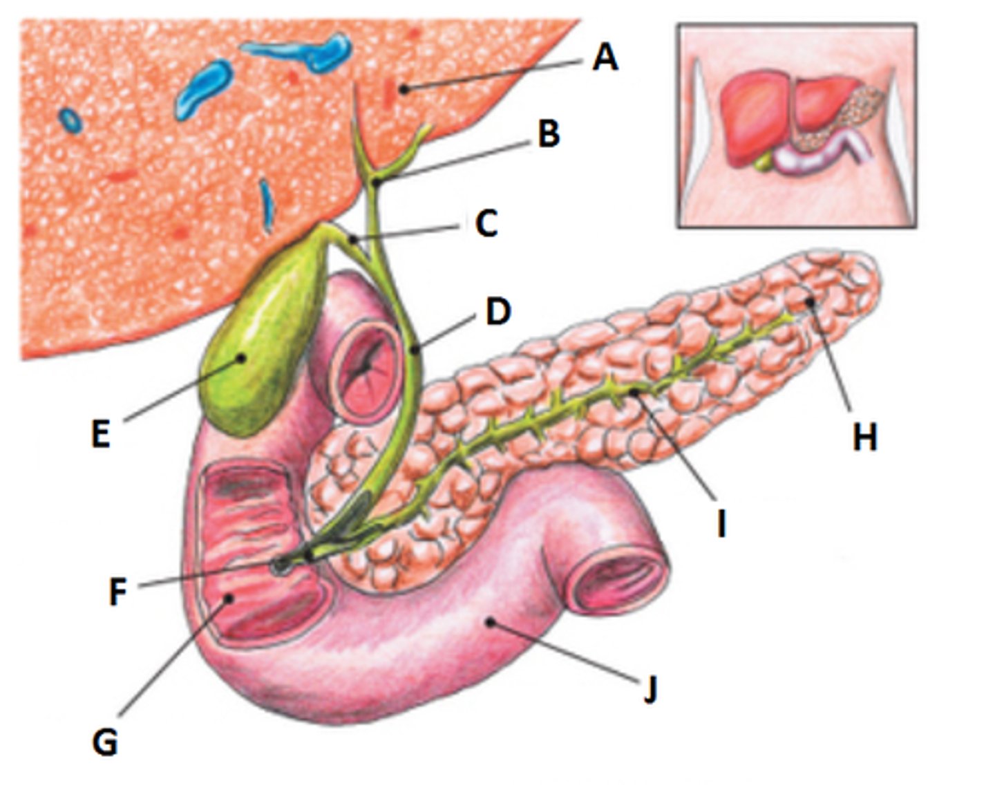
Hepatopancreatic Ampulla
Part of the biliary tree where the common bile duct & the Pancreatic duct merge
What is the alternate name for the Ampulla of Vater? What does connect?
Hepatopancreatic Sphincter
What is the alternate name for the Sphincter of Oddi|?
Duct of Wirsung
What is the alternate name for the pancreatic duct?
Cystic duct
(connects off of the common hepatic duct)
What is the name of the duct that takes bile to the gallbladder?
Pear-shaped sac found under the surface of the liver
Describe the shape of the gallbladder.
Store of bile
Concentrate bile,
Contraction when triggered
What are the three sole functions of the gallbladder?
Ultrasound
What is the primary modality of imaging the Gallbladder?
Hydrolysis (removal of water)
How dos the Gallbladder concentrate bile?
Cholelithiasis
What is the condition of having gallstones?
Choleliths - too much water removal forms stones
What is the medical term for gallstones?
Sphincter of Oddi
What is a common site of choleliths?
Immediate Cholangiogram
Surgical Cholangiogram
What are the alternate names for an OR Cholangiogram?
Immediately following a cholecystectomy
In the OR after Surgery
When and Where is an OR Cholangiogram performed?
laparoscopic cholecystectomy
- less hospital times and cost
- Less invasive (outpatient)
- endoscope is inserted through umbilicus
- GB removed & cystic duct cauterized
- Cholangiogram can be performed
How are cholecystectomies usually performed? What are the advantages of this method?
T-tube cholangiography
What is the alternate name for Post-Operative Cholangiography?
Done in radiology dept (IR) by a radiologist
Performed about 24 hours post cholecystectomy
Where & when is T-tube cholangiography performed?
- T-tube catheter is inserted into the common bile duct and extends to outside of the body
- T tube clamped
- scout films
- injection of contrast
- can show residual choleliths undetected during surgery
- May remove stones through a basket catheter passed over a guide wire
Explain T Tube Cholangiography.
Both
Is a T Tube cholangiography diagnostic or therapeutic?
can mimic stones
What do air bubbles mimic in cholangiography?
Percutaneous Transhepatic Cholangiography or PTC
What is a PTHC?
- Direct puncture of biliary ducts using a 22g chiba needle
- done in IR
- needle passes through the skin through the liver
- PT and PTT need to be checked for blood clotting
- can be diagnostic or therapeutic
- most invasive
Explain a PTHC.
1) Liver Hemorrhage
2) Pneumothorax
3) Escape of bile
Risks of PTHC
Endoscopic Retrograde Cholangiopancreatography
ERCP
- Gastroenterologist uses Fiber optic endoscope (duodenoscope)
- Can be dones in OR GI suite, or in fluoro
- Catheter through sphincter of oddi to common bile duct & main pancreatic duct
- diagnostic or therapeutic
Explain an ERCP
Gastroenterologist - using a fiber optic endoscope (duodenoscope)
Who performs ERCP?
Radiologist in IR
Who performs PTHC?
Radiologists in IR
Who performs T Tube Cholangiography?
- no ionizing radiation
- detection of small stones
- no contrast
- less pt prep
4 Reasons why Ultrasound is preferred
Oral cavity (mouth) --> Pharynx -> Esophagus -->Stomach--> Small Intestines --> Large Intestines --> Anus
List the parts of the alimentary canal.
Salivary glands
Pancreas
Liver
Gallbladder
List the accessory organs of the digestive system.
- Intake & digestion of food, water, vitamins, and minerals
- Absorbs food, water, vitamins, and mineral into the blood & lymphatic capillaries
- Eliminate any unused material
Functions of the Digestive System
- Modified Barium Swallow
- Esophagogram
- Upper GI series (UGI)
What are some common radiographic procedures of the upper digestive system?
Oral cavity (mouth)
Where does the alimentary canal begin?
Upper/lower teeth, hard & soft palates, tongue, uvula
What forms the oral cavity?
Salivary glands
- secrete saliva = water, salts, and enzymes
What are the accessory glands of the mouth and what is their function?
Parotid - near the ear
Submandibular (submaxillary) - below maxilla
Sublingual - below the tongue
List the pairs of Salivary glands
act of swallowing the bolus
What is deglutition?
teeth and tongue cooperate in chewing motions to mx with saliva - initiate the mechanical part of digestion
What is mastication?
Nasopharynx
Oropharynx
Laryngopharynx
What are the three parts of the pharynx?
oropharynx
Which part of the pharynx is epiglottis attached to?
depresses over the laryngeal opening - respiration is inhibited
What does the epiglottis do during swallowing?
about 10 inches and 3/4 inches in diameter
How long is the esophagus?
Begins posterior to the level of the lower border of the cricoid cartilage of the larynx (C5-C6)
Terminates at the connection to the stomach around T11
At what level does the esophagus start and end?
T10
At what level does the esophagus pass through the diaphragm?
True
T/F: The esophagus is posterior to the trachea.
Esophagus
IVC
Aorta
Three diaphragmatic opening
Cardiac antrum
What is the abdominal segments of the esophagus called?
Esophagogastric junction, Gastroesophageal junction or cardiac orifice
What is the opening between the esophagus and stomach called?
wavelike series of involuntary muscular contractions propelling solids and semisolid materials through the alimentary canal
Define peristalsis
stomach
What is the most dilated portion of the alimentary canal?
serves as reservoir for swallowed food and fluid - can expand
What is the function of the stomach?
longitudinal folds in the internal lining of the stomach
- assist with mechanical digestion of food within the stomach
What are rugae?
formed by rugae along the lesser curvature that funnels fluid directly from the body of the stomach to the pylorus
What is the gastric canal?
fundus
What is the most posterior aspect of the stomach?
Pylorus
What is the most posterior portion of the stomach?
- fundus (highest portion) filled with air
- pylorus filled with barium
If a patient is prone describe where the barium and air would be within the stomach.
- fundus is the filled with barium
- pylorus if filled with air
If a patient is supine describe where the barium and air would be within the stomach.
- air in the fundus and barium in the pyloric portion (air/barium line is straight)
If a patient is erect describe where the barium and air would be within the stomach.
duodenum
What is the first part of the small intestine?
head of pancreas sits in the C loop of duodenum
What is the romance of the abdomen?
T
T/F: Duodenum is a retroperitoneal part of the small intestine.
Shortest, widest, and most fixed portion of the small bowel.
Describe the duodenum.
Superior (first)
Descending (second)
Horizontal (third)
Ascending (fourth)
Duodenojejunal flexure
List the parts of the duodenum
4-8 seconds - solids
1 second - liquids
How long does it take for solids and liquids to pass from mouth to stomach?
semifluid mass in the stomach
What is chyme?
2-6 hours
- protein/fat move slower than carbs
- movement by mixing and peristalsis
About how long does chyme remain in the stomach?
3-5 hours
Rhythmic segmentation (churning) and peristalsis
How long for the small intestine?
- 5% of population (massive body build)
- abdomen broad and deep, diaphragm high, transverse colon high
- GB high and transverse
- stomach is high and almsot transverse (T9-T12)
- center of stomach is about 1 inch distal to xiphoid tip
- duodenal bulb about T11-T12
Describe the abdomen, stomach, and GB in a hypersthenic patient.
- Almost opposite
- More slender, narrower, low diaphragm
- Large intestine very low
- Stomach is J shaped (T11- to iliac crests or L5)
- Duodenal bulb (L3-L4)
- GB - L3-L4
Describe the abdomen, stomach, and GB in a hyposthenic/Asthenic patient.
- Average body build
- Stomach is somewhat J shaped (T10 or T11 down to L2)
- Duodenal bulb - L1, L2
- GB - less transverse
- Splenic flexure quite high
Describe the abdomen, stomach, and GB in a Sthenic patient.
1-2 inches
How far do the parts of the abdomen drop when in the erect position?
False - Barium is a colloidal suspension and never dissolves in water
T/F: Barium dissolves in water.
Perforated hallow viscus
Presurgical Procedure
What are the contraindications of barium?
Perforated hallow viscus or presurgical procedure
What are the clinical indications for water soluble iodinated contrast media?
hypersensitivity to iodine
Contraindications of water soluble iodinated contrast media
T
T/F: Isovue moves through the alimentary canal faster than barium.
- Anatomical anomalies - congenital or disease caused like a stroke
- Esophageal reflux (GERD)
- Barretts Esophagus - mid to distal esophagus erodes away, precursor for cancer
- Esophageal varices - cirrhosis of liver, causes enlargement of veins
- Foreign body obstruction - fish bones
- Dysphagia - Impaired swallowing mechanism
- Carcinoma of esophagus
- Zenker Diverticulum - outpouching of esophageal lining due to weakening
List clinical indications of a Esophagogram
- Breathing Exercises (two types)
o Valsalva Maneuver – deep breathing bear down like moving bowels – forces air against closed glottis
o Mueller Maneuver – exhales then tries to inhale against closed glottis
o Increase of intraabdominal pressure may produce reflux
- Water Test
o Esophagogastric junction regurgitation
o Supine – LPO position – fundus fill with barium – significant barium regurgitates into the esophagus from the stomach
- Compression Technique
o Esophageal reflux – pt prone, compression paddle inflated against stomach region
- Toe-Touch Maneuver
o Investigate cardiac orifice, hiatal hernias, reflux as pt bends over touches toes
What are the 4 techniques in diagnosing esophageal reflux?
- Peptic ulcer - erosions of the stomach or duodenal mucosa
- Hiatal hernia -portion of stomach herniates through diaphragm sliding hernia (Schatzki's ring)
- HPS (hypertrophic pyloric stenosis - most common gastric obstruction in infants
- Diverticula - outward pouching
- Gastritis - inflammation of the lining of the stomach
- Tumor -Gastric adenomas adenomcarcinomas - 95%
- Bezoar - masses that are not digested and built up - hair, fibers, seeds,
- Emesis (hematoemeis)
Clinical indications of an UGI series
- determine pregnancy
- NPO for 8 hours
What is the patient prep for an UGI series?
1) Clinical history - review clinical history with patient and documentation
2) Body habitus - affects positioning
3) Fluoroscopy - identifying positioning landmarks
4) High kVp - 100-125 single or 90-100 for double - short exposure time
What are some imaging considerations for UGI.
90-100 kVp
What kVp would we use for a double contrast UGI?
RAO, Lateral, AP (PA)
Routine projections for an Esophagogram post-fluoro
LAO & Soft tissue lateral
Special projections for an esophagogram post-fluoro
Puts Esophagus between vertebral column and the heart
Why is an RAO esophagogram preferred over an LAO?
35-40 degrees
How much should the patient be obliqued for an RAO esophagogram?
2 inches above the shoulders
What is the IR for an RAO esophagogram?
CR to T5-T6 (2-3 inches inferior to jugular notch or 1 inch inferior to the sternal angle)
What is the CR for an RAO esophagogram?
3 swallows - thick barium bolus or continuous thin barium
How many swallows of barium should be taken before an projection?