b223 special senses 16
1/54
There's no tags or description
Looks like no tags are added yet.
Name | Mastery | Learn | Test | Matching | Spaced |
|---|
No study sessions yet.
55 Terms
olfaction and gustation
located in specific sense organs
chemoreceptors
exteroceptors - respond to chemical stimuli from external environment
receptor areas have chemically gated ion channels
receptors (cells) deteriorate rapidly and are continuously replaced with new cells
exteroreceptors
respond to chemical stimuli from external environment
olfactory organs
olfactory (bowman) gland, regenerative basal cell (divides to replace worn out olfactory receptor cells), cribriform plate, lamina propria, olfactory epithelium, olfactory nerve fibers, olfactory bulb, olfactory receptor cell/developing cell, supporting cell, mucous layer, knob, olfactory cilia (surfaces contain receptor proteins)
olfaction
sense of smell
olfactory epithelium - located in superior portion of nasal cavity
olfactory receptors
basal stem cells
supporting cells, glands, blood vessels, etc
epithelium surface coated with mucus secretions from olfactory glands
to be detectable as a smell, chemical must be volatile and soluble in mucus
mucus cleans olfactory epithelium, removing chemicals
voluntary cell must diffuse through mucus
olfactory receptors (cilia) are:
g-protein coupled receptors located on the dendrites of bipolar neurons
large area of membrane on which chemically gated channels are located
humans have approximately 390 functional olfactory receptors
each neuron type is sensitive to only one chemical
olfactory epithelium
located in superior portion of nasal cavity
olfactory receptors
basal stem cells
supporting cells, glands, blood vessels, etc.
olfactory mucus
epithelium surface coated with mucus secretions from olfactory glands
to be detectable as a smell, chemical must be volatile and soluble in mucus
mucus cleans olfactory epithelium, removing chemicals
voluntary cell must diffuse through mucus
cilia (olfactory receptors)
g-protein coupled receptors located on the dendrites of bipolar neurons
large area of membrane on which chemically gated channels are located
humans have approximately 390 functional olfactory receptors
each neuron type is sensitive to only one chemical
olfactory receptors
1) the binding of an odorant to its receptor protein leads to the activation of adenylate cyclase, the enzyme that converts ATP to cyclic AMP (cAMP)
2) the cAMP opens sodium ion channels in the plasma membrane, which then begins to depolarize
3) if sufficient depolarization occurs, an action potential is triggered in the axon, and the information is relayed to the CNS
olfactory discrimination
humans can discriminate at least 1 trillion different “smells”
CNS interprets different smells on the basis of the overall pattern of activity in the 390 different receptor/neuron types
example: smell A (popcorn) activates primary smell neurons 1, 3, 4; smell B (mint) activates neurons 4, 27, 32, 41
very sensitive - only a few molecules needed to open enough gates to reach AP threshold in some olfactory neurons
1st order neuron (olfactory pathways)
receptor cell in olfactory epithelium - axon extends into CNS as cranial nerve I
approximately 20 small bundles of axons (nerves) go through cribriform plate
instead of one single peripheral nerve
2nd order neuron (olfactory pathways)
located in olfactory bulb
much processing, including central adaptation
2nd order axons form olfactory tract that diverges to multiple sites
3rd order neurons (olfactory pathways)
at various locations
olfactory cortex - medial surface temporal lobe
hypothalamus
limbic system
gustatory discrimination
humans can detect 4 to 6 “primary tastes”
sweet, salty, sour, bitter, umami, water
CNS interprets different tastes on the basis of the overall pattern of activity in the 4 to 6 different receptor types
to be detectable as a taste, chemical must be dissolved
much less sensitive than olfactory receptors
least sensitive to sweet and salty (requires more to trigger taste)
1000 times more sensitive to acids (sour) than sweet and salty
most sensitive to bitter (plant secondary metabolites)
taste sensitivity shows significant individual differences, some of which are inherited
phenylthiocarbamide (PTC) - bitter or tasteless
gustatory reception
involves: transitional cell, gustatory cell (not a nerve), basal cell, taste hairs (microvilli), taste pore
taste buds
contain gustatory receptor cells and basal stem cells
recessed into surface of surrounding epithelium of lingual papillae
gustatory receptor cells
four to six different types of gustatory receptor cells, each sensitive to only one type of chemical
most taste buds have all 4-6 types
salt and sour channels
the diffusion of sodium ions from salt solutions or hydrogen ions from acids or sour solutions into the gustatory epithelial cells lead to depolarization
sweet, bitter, and umami receptors
receptors responding to stimuli that produce sweet, bitter, and umami that produce sweet, bitter, and umami sensations are linked to g-proteins called gustducins—protein complexes that use second messengers to produce their effects
gustatory pathways
receptor cell in taste bud - gated ion channels open, producing receptor potential
1st order neuron
dendrite receive synapse from receptor cell
amount of NT released by receptor cell determines AP frequency in 1st order neuron
axon extends into CNS in cranial nerves
CN VII (facial) - anterior 2/3 of tongue
CN IX (glossopharyngeal - posterior 1/3
olfactory and gustation parallels
level of stimulation of olfactory receptors has major role in taste perception
olfactory receptors much more sensitive than gustatory receptors
aging reduces olfactory and gustatory sensitivity
number of receptors declines with age as fewer new cells are produced
receptor sensitivity declines
inner ear
bony labyrinth
surrounds and protects membranous labyrinth
contain perilymph
membranous labyrinth
contains endolymph
labyrinths are divided into 3 areas
vestibule with utricle and saccule
semicochlear canals with semicircular ducts
cochlea with cochlear duct
bony labyrinth
surrounds and protects membranous labyrinth
contains perilymph
membranous labyrinth
contains endolymph
3 labyrinth areas
vestibule with utricle and saccule
semicircular canals with semicircular ducts
cochlea with cochlear duct
equilibrium and hearing
sensory organs located in the inner ear
specialized receptor cells termed hair cells are mechanoreceptors
hair cells respond to physical distortion of their cilia
amount of neurotransmitter released by hair cells controls AP frequency in 1st order neurons
cranial nerve VIII - vestibulocochlear
contain 1st order neurons that conduct APs into CNS
organization of sensory pathways (ear)
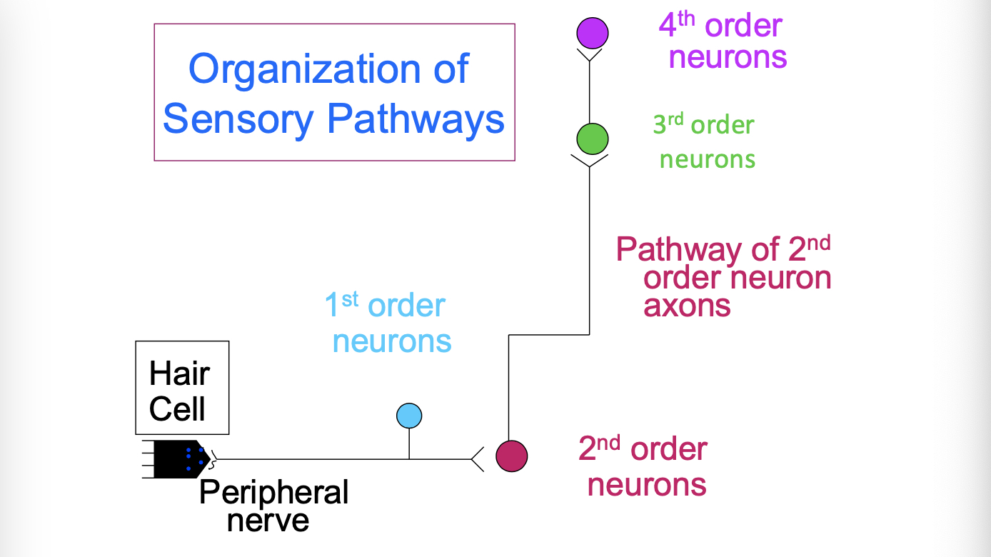
hair cell components
kinocilium, gelatinous material, stereocilia, hair cell, sensory nerve ending, supporting cell
location of hair cells
semicircular ducts
in cristae - small patch of hair cells in ampullarf region at one end of each of the 3 ducts
utricle and saccule
in maculae - small patch of hair cells - one inside utricle, one inside saccule
cochlear duct
in organ of corti on basilar membrane - long strip of hair cells extending the length of the coiled duct
equilibrium (vestibular sensation)
semicircular ducts - cristae hair cells
sense rotational movement
activated only during movement
cupula - floating gelatinous mass in which “hairs” are embedded
utricle and saccule - maculae hair cells
sense head position and change in movement
active at all times
otolith - “hairs” embedded in gelatinous mass containing calcium carbonate crystals
vestibular neural pathway
hair cells synapse onto 1st order sensory neurons, which enter CNS as vestibular branch of CN VIII
2nd order neurons located in vestibular nuclei of the brainstem
integrate input from left and right inner ears
send information to the somatosensory cortex
send information to the cerebellum
send information to motor nuclei in BS and SC for reflex control of eye, head, neck
motion sickness
disconnect between equilibrium and vision
nystagmus
involuntary eye movement
postrotatory and opticokinetic (induced) (spinning in chair)
pathological from damage to the vestibular system
hearing - auditory sense
external and middle ear assist in getting stimulus (sound waves) to receptors in the cochlea of the inner ear
external ear (auricle, external acoustic canal)
collects and directs sound waves toward middle ear
middle ear (tympanic membrane and auditory ossicles)
conducts and applies vibrations from tympanic membrane to inner ear
pathway of sound
1) sound waves arriving at tympanic membrane cause it to vibrate
2) auditory ossicles conduct and amplify the vibration onto the oval window of the inner ear
tensor tympani and stapedius muscles contract to reduce the amount of movement when loud sounds arrive
3) in/out movement at the oval window creates pressure wave in the perilymph of the cochlea
4) pressure waves vibrate the basilar membrane area of the cochlear duct
5) hair cells of the organ of corti are pushed against the tectorial membrane
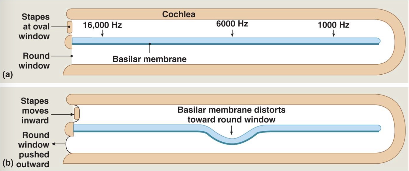
sound frequency and intensity discrimination
basilar membrane flexibility varies along length of cochlear duct
highest frequency sounds (20000 hertz) vibrates areas closest to oval window
lowest frequency sounds (20 hertz) vibrates areas furthest from oval window
higher intensity (louder) sounds cause larger vibration and stimulate more hair cells (intensity is measured in decibels)
frequency discrimination
auditory neural pathway
hair cells synapse onto 1st order sensory neurons, which enter CNS as cochlear branch of CN VIII
2nd order neurons located in cochlear nuclei of the brainstem
2nd order axons decussate and ascend to 3rd order neurons in inferior colliculi of mesencephalon
inferior colliculus coordinates reflex movement of head and neck to sounds
3rd order axons ascend to thalamus
conscious awareness and interpretation of sound
primary auditory cortex - superior surface of temporal lobe
auditory association cortex in surrounding areas of temporal lobe
left hemisphere interprets language
eye
sensory organ that collects and focuses light onto the photoreceptors (rods and cones)
cornea and lens focus light rays onto retina located on the inner surface of eye ball
light passes through layers of retinal neurons to reach photoreceptors
retina
outer pigmented portion
absorbs excess light
transports nutrients to neural part
inner neural part
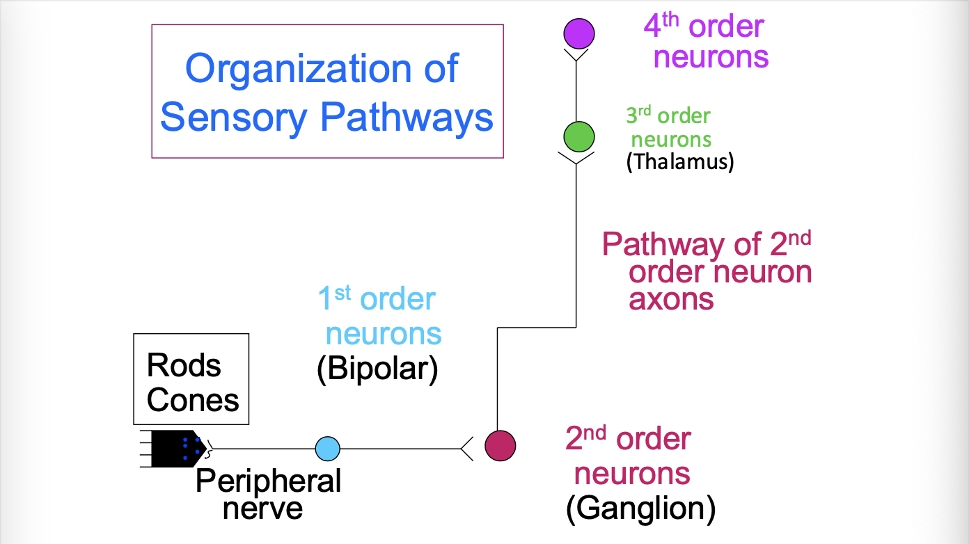
photoreceptors: rods and cones
1st order neurons: bipolar cells
2nd order neurons: ganglion cells, axons form the optic nerve and tract
embryologic origin - outgrowth from brain
organization of sensory pathways (eye)
cones
provide color vision
require more light to be activated than rods
located in macula lutea portion of retina
in highest concentration in fovea portion of macula
re
rods
do not distinguish colors
highly sensitive to light, allow for vision in dim light conditions
found in retinal areas peripheral to macula
inner segment (rods and cones)
the inner segment contains the photoreceptor’s major organelles and is responsible for all cell functions other than photoreceptor, it also releases neurotransmitters
outer segment (rods and cones)
the outer segment of a photoreceptor contains flattened membranous plates, or discs, that contain the visual pigments
pigmented epithelium (rods and cones)
the pigmented epithelium absorbs photons that are not absorbed by visual pigments. it also phagocytizes old discs shed from the tip o the outer segment
photoreceptors (rods and cones)
convert light signals into electrical signals
gated ion channels
respond to amount of light absorbed by pigment molecules in the photoreceptor
current flowing through gated ion channels changes membrane potential, controls amount of neurotransmitter released onto 1st order neurons
photoreceptor physiology
gated ion channels are indirectly controlled by light
light absorption activates enzyme pathway that controls 2nd messenger (cGMP) levels that open gated channels
light is absorbed by retinal molecule embedded in an opsin protein molecule
retinal (photo reactive molecule) - synthesized from vitamin A
opsin differ in which light frequencies can reach the retinal molecule
rods (color vision)
retinal and rhodopsin
respond to wide range of light wavelengths, responds to all the different wavelengths of the different colors
cones (color vision)
retinal and one of the 3 different opsin (red, blue, and green sensitive cones)
respond to specific, narrower ranges of wavelengths, ranges overlap
color discrimination
integration of information from red, blue and green cones
white perceived when all cones equally stimulated
yellow perceived when green cones stimulated strongly, red cones are stimulated moderately, and blue cones are not stimulated
variable stimulation of the three cones leads to the ability to discriminate >10 million colors
colorblindness
inability to detect certain colors due to lack of one or more color opsin
inherited genetic trait - genes for red and green opsin are located on X chromosome
10% of males, 0.67% of females
visual pathway
each ganglion cell receives input from a specific receptive field of the retina, a group of photoreceptors
image of retina surface is mapped onto visual cortex
receptive fields are very small in fovea area of retina
highest visual acuity
lowest area in visual cortex
photoreceptors to bipolar cells to ganglion cells
axons of ganglion cells form the optic nerve and optic tract
synapse in superior colliculus for visual reflex pathways
synapse on 3rd order neurons in the thalamus for conscious processing pathway
conscious processing of visual input (visual pathway)
primary visual cortex and visual association cortex are in occipital lobe of brain
right visual field projects to left visual cortex
left half of both retinas view right visual field
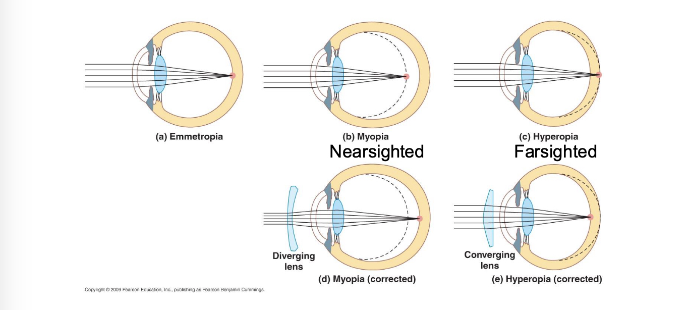
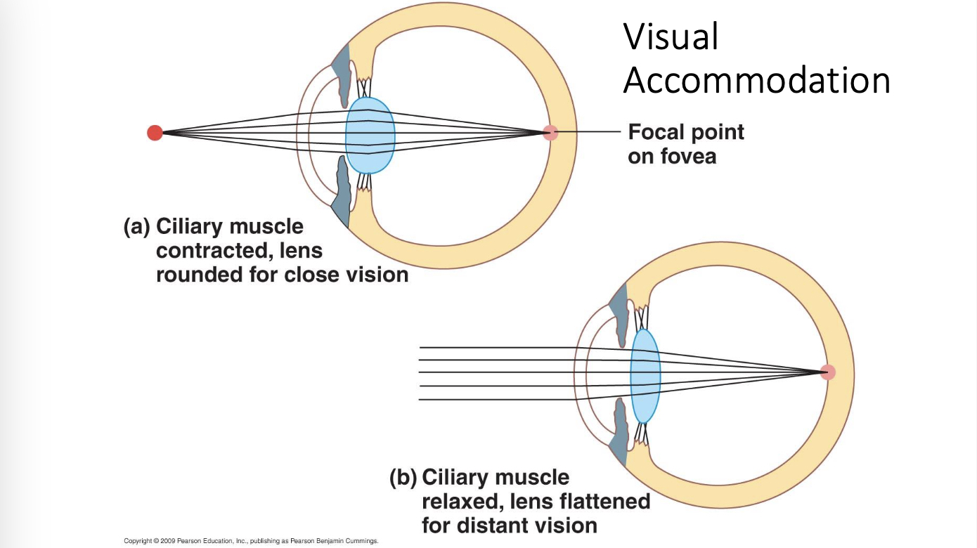
visual accommodation
nearsighted and farsighted