common sports injuries presenting to FM
 Call Kai
 Learn
Learn Practice Test
 Spaced Repetition
 Match
Match Flashcards
 Knowt Play
1/66

There's no tags or description
Looks like no tags are added yet.
Name | Mastery | Learn | Test | Matching | Spaced | Call with Kai | Chat |
|---|
No analytics yet
Send a link to your students to track their progress
67 Terms
Family history of sudden cardiac death
red flags in family history to not in sports physial exam
Hypertrophic cardiomyopathy (HCM), arrhythmogenic right ventricular cardiomyopathy (ARVC), congenital coronary anomalies.
cardiac findings to look for in athletes
Hypertrophic cardiomyopathy (HCM)
leading cause of sudden cardiac death in young athletes
Commotio Cordis
When the heart stops due to a sudden blow to the chest
Spirometry before/after exercise → ≥10-15% fall in FEV1 confirms diagnosis.
spirometry finding of exercise induced asthma
Pre-exercise SABA (albuterol)
first line management of exercise induced asthma
Increased iron loss (sweating, GI blood loss, hematuria, heavy menses).
Increased demand (endurance training).
Poor intake (vegetarian/vegan diets).
main causes of iron deficiency in athletes
Low hemoglobin/hematocrit.
Low ferritin (
lab findings indicative of iron deficiency anemia
sports anemia
False anemia or pseudo anemia caused by extensive aerobic training, which increases the plasma volume, producing a relative decrease in hematocrit
-normal ferritin and iron stores
heme iron
the iron in foods that is bound to the hemoglobin and myoglobin proteins; found only in meat, fish, and poultry.
vitamin C
supplement to hellp absorb iron
1. Low energy availability (with or without eating disorder).
2. Menstrual dysfunction (amenorrhea, oligomenorrhea).
3. Low bone mineral density (osteopenia/osteoporosis, stress fractures).
name the components of the female athlete triad
Amenorrhea >3 months.
History of stress fractures.
Restrictive eating or eating disorder.
red flags of the female athlete triad
ACL injuries
pts with female athlete triad have a higher risk for these injuries due to anatomic and hormonal factors
ensure adequate calcium/vitamin D intake
first line management for female athlete triad
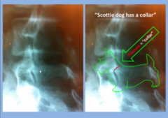
spondylolysis
stress fracture of pars interarticularis — common in adolescent athletes, e.g., gymnasts, football linemen
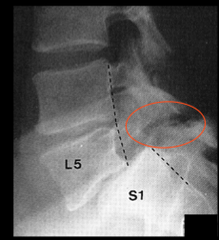
spondylolisthesis
forward slipping of one vertebra over another
new fracture at same or other sites.
prior fracture history increases risk for
X-ray = initial.
MRI = best for stress fractures (early detection).
Bone scan = sensitive, less specific.
best forms of imaging for fractures
RED-S
Relative Energy Deficiency in Sport affecting health, new name for female athlete triad
Overuse, periostitis at tibial insertion
cause of medial tibial stress syndrome
medial tibial stress syndrome
-diffuse pain along medial tibia
-gradual onset, related to training errors, poor footwear
-xray normal
Rest, ice, stretching, footwear correction
treatment for mdeial tibial stress syndrome
chronic exertional compartment syndrome
-Increased pressure in fascial compartment with exercise
-Tight, burning pain with exertion, resolves with rest
Predictable: after certain distance/time of exercise
describe the onset of chronic exertional compartment syndrome
pressure testing
gold standard testing for chronic exertional compartment syndrome
surgical fasciotomy if severe
tx for chronic exertional compartment syndrome
stress fracture
Repetitive loading → microfracture
Localized point tenderness, worsens with activity; focal bony tenderness, possible swelling
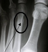
MRI best for early detection
imaging for stress fracture
Rest, gradual return, activity modification; surgical fixation if severe
management of stress fracture
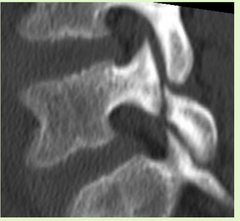
Spondyloysis
for low back pain in young athletes, think
Either:
Bone tenderness at the posterior edge or tip of the lateral malleolus, OR
Bone tenderness at the posterior edge or tip of the medial malleolus, OR
Inability to bear weight immediately after injury and for 4 steps in the emergency department/clinic.
an ankle X-ray is required if there is pain in the malleolar zone AND
Either:
Bone tenderness at the base of the 5th metatarsal, OR
Bone tenderness at the navicular bone, OR
Inability to bear weight immediately and for 4 steps in the ED/clinic.
a foot X-ray is required if there is pain in the midfoot zone AND
oblique
angled xray view for overlapping bones
Multiple views (≥2 perpendicular)
how many xray views are needed for an accurate diagnosis
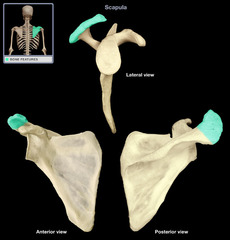
acromion
an extension of the scapula that forms the high point of the shoulder
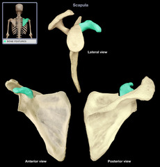
coracoid process
process above the glenoid cavity that permits muscle attachment
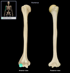
capitulum
Name this specific part of the humerus.
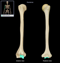
trochlea
Name this specific part of the humerus.
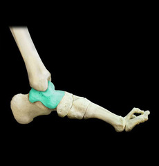
talus
Name this specific bone of the foot.
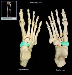
navicular
Name this specific bone of the foot.
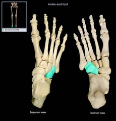
cuboid
Name this specific bone of the foot.
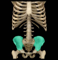
ilium
Name this specific region of the pelvic bone.
ischium
the lower, posterior portions of the pelvis
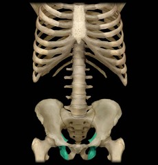
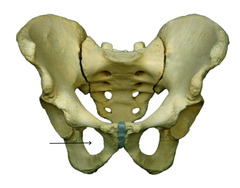
obturator foramen
opening in hip bone formed by the pubic and ischial rami
fracture through the physis only
S= straight across
define a salter harris I fracture
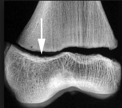
fracture through the physis and metaphysis
A= above
define a salter harris II fracture
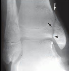
fracture through the physis and epiphysis
L=Lower
may involve joint surface--> moderate risk
define a salter harris type III fracture
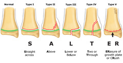
Fracture through metaphysis, physis, and epiphysis
T= through
high risk for growth disturbance
define a salter harris type IV fracture
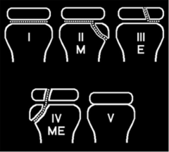
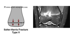
Crush injury to the physis
-high risk of premature growth plate closure
define a salter harris type V fracture
SALTR → Straight, Above, Lower, Through, Ramme
mnemonic to remember the salter harris fractures
types III-V
salter harris fracture types requiring orthopedic referral and possibly open reduction to restore growth plate alignment
tibia, metatarsals (2nd-3rd), navicular, femoral neck, fibula.
most common sites of stress fractures
anterior fat pad sign
indicates hemarthrosis in radial head fx
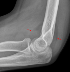
posterior fat pad sign
always pathologic; visible only when effusion displacees it out of the olecranon fossa
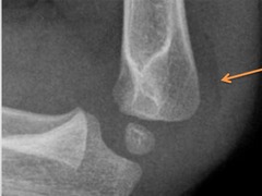
lateral xr
best view for elbow effusions
occult radial head fracture (adults) or supracondylar fracture (children).
common causes of elbow effusions
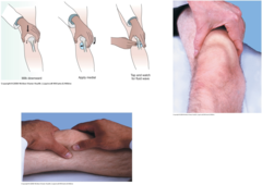
bulge sign
milk fluid toward the medial side; small wave on lateral side suggests effusion.
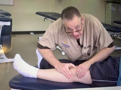
ballottement sign
patella "floats" and rebounds when pushed down.
-indicates pleural effusion
trauma, meniscal tear, ACL injury, infection, gout, inflammatory arthritis.
common causes of knee effusion
CT
imaging best for Bony detail, complex fractures (esp. pelvis, spine, facial bones, intra-articular)
MRI
imaging best for Soft tissue structures (ligaments, tendons, cartilage, marrow, menisci, discs)
Expensive, time-consuming, motion artifacts, contraindicated with metal
limitations of MRI
CT
what imaging should be used to evaluate comminuted fractures, subtle cortical breaks, preoperative planning.
MRI
what imaging should be used to Assess ACL/PCL tears, meniscal injury, rotator cuff tears, stress fractures not seen on X-ray.
inversion injury, which causes the fibular head to glide posteriorly at the proximal tibiofibular joint and the lateral malleolus to glide anteriorly at the distal fibula.
most common ankle sprain MOA
1. Patient PositionPatient seated or supine, knee flexed to 90°.
2. Physician PositionAt foot of table, stabilizing distal tibia with one hand; grasp lateral malleolus with the other.
3. Engage Restriction BarrierGently apply posterior pressure on the lateral malleolus to take up slack (toward correction).
4. Patient EffortAsk patient to evert the foot (activating peroneus longus/brevis) against physician's counterforce for 3-5 seconds
muscle energy for anterior medial mallolus