Lab 4
1/129
There's no tags or description
Looks like no tags are added yet.
Name | Mastery | Learn | Test | Matching | Spaced | Call with Kai | Chat |
|---|
No analytics yet
Send a link to your students to track their progress
130 Terms
Diaphragm
big sheet of muscle that separates thoracic & abdominal cavity

During inhalation, the diaphragm…
contracts DOWNWARD


During inhalation, the ribs…
expand
The thoracic cavity gets larger.
Volume inside lungs increases
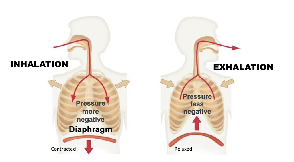

During inhalation pressure is…
more negative!
(B/c the same amount of air is temporarily occupying a larger space, the pressure inside the lungs decreases)
air driven in by negative pressure


During exhalation the diaphragm…
relaxes & moves UPWARD
pushed up against


During exhalation the ribcage
moves inward again
The thoracic cavity gets smaller.
lung volume decreases

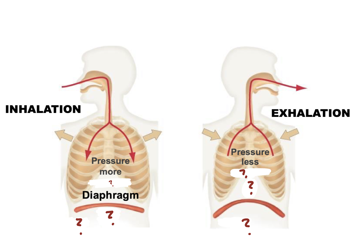
In exhalation, pressure is
less negative!
Since the same amount of air is now compressed into a smaller space, the pressure inside the lungs increases
air flows out of the lungs.

Inhale = Increase volume
→ Decrease pressure (Negative Pressure) → Air In
Exhale = Decrease volume
→ Increase pressure (Less Negative) → Air Out
Pathway of Oxygen
Lungs (Trachea into Bronchi into Bronchioles) → Alveoli → Blood → Heart → Body

What is number 1?
Trachea
What is the function of the trachea?
Conducts air from the upper respiratory tract to the bronchi.

What is number 2?
Lung

What is number 3?
Bronchi
What is the function of the bronchi?
Carry air into each lung and branch into smaller bronchioles.

What is number 4?
Bronchioles
What is the function of the bronchioles?
Conduct air to the alveoli and regulate airflow by constricting or dilating.

What is number 5?
Smallest Bronchiole
air goes into alveoli (multiple sacs) → into singular alveolus (single, individual air sac in the lungs)

What is number 6?
Alveolus
single, individual air sac in the lungs
What are alveoli?
Tiny air sacs where gas exchange occurs between air and blood.

What is number 7?
Capillaries
smallest blood vessels
What surrounds each alveolus?
A network of capillaries that allows gases to diffuse between the alveoli and blood.

What does number 8 showcase?
oxygen poor blood going in to get oxygen from air brought into alveoli from smallest bronchiole

What does number 9 showcase?
oxygen rich blood going out after alveolar gas exchange
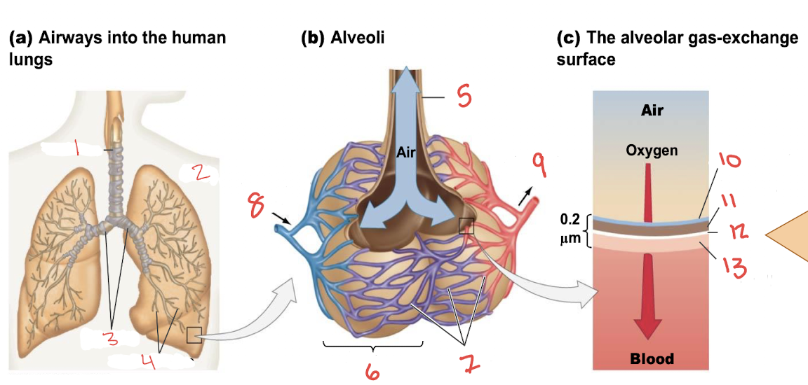
Alveolar Gas Exchange: What is number 10?
aqueous film

What is number 11?
Epithelium of alveolus

What is number 12?
extracellular matrix (ECM)
thin layer between the alveolar epithelium & capillary wall, consisting mainly of fused basement membranes that minimize diffusion distance.

What is number 13?
Wall of Capillary
Alveolar Gas Exchange: How does oxygen cross the respiratory membrane?
By passive diffusion to oxygen poor blood
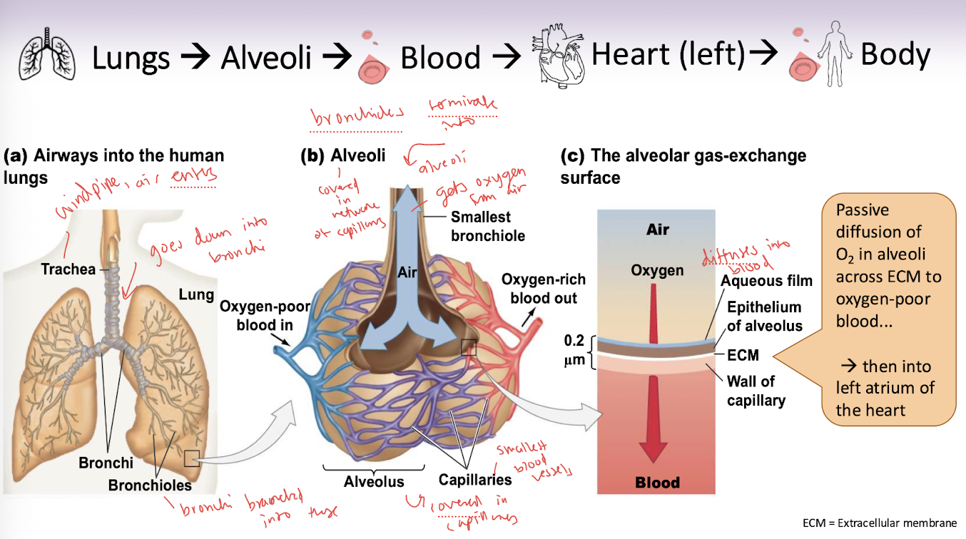
Why is oxygen diffusion passive?
Because oxygen moves down its partial pressure (PO₂) gradient and does not require ATP.
What drives oxygen to diffuse from the alveoli into the blood?
The PO₂ is higher in the alveoli than in the oxygen-poor blood, so oxygen diffuses down its partial pressure gradient.
What happens to oxygen after it enters the blood?
Most oxygen binds to hemoglobin in red blood cells for transport.
Where does oxygen-rich blood go after leaving the pulmonary capillaries?
Through the pulmonary veins to the left atrium of the heart.
What happens to carbon dioxide during alveolar gas exchange?
Carbon dioxide diffuses from the blood into the alveoli down its partial pressure (PCO₂) gradient and is exhaled.
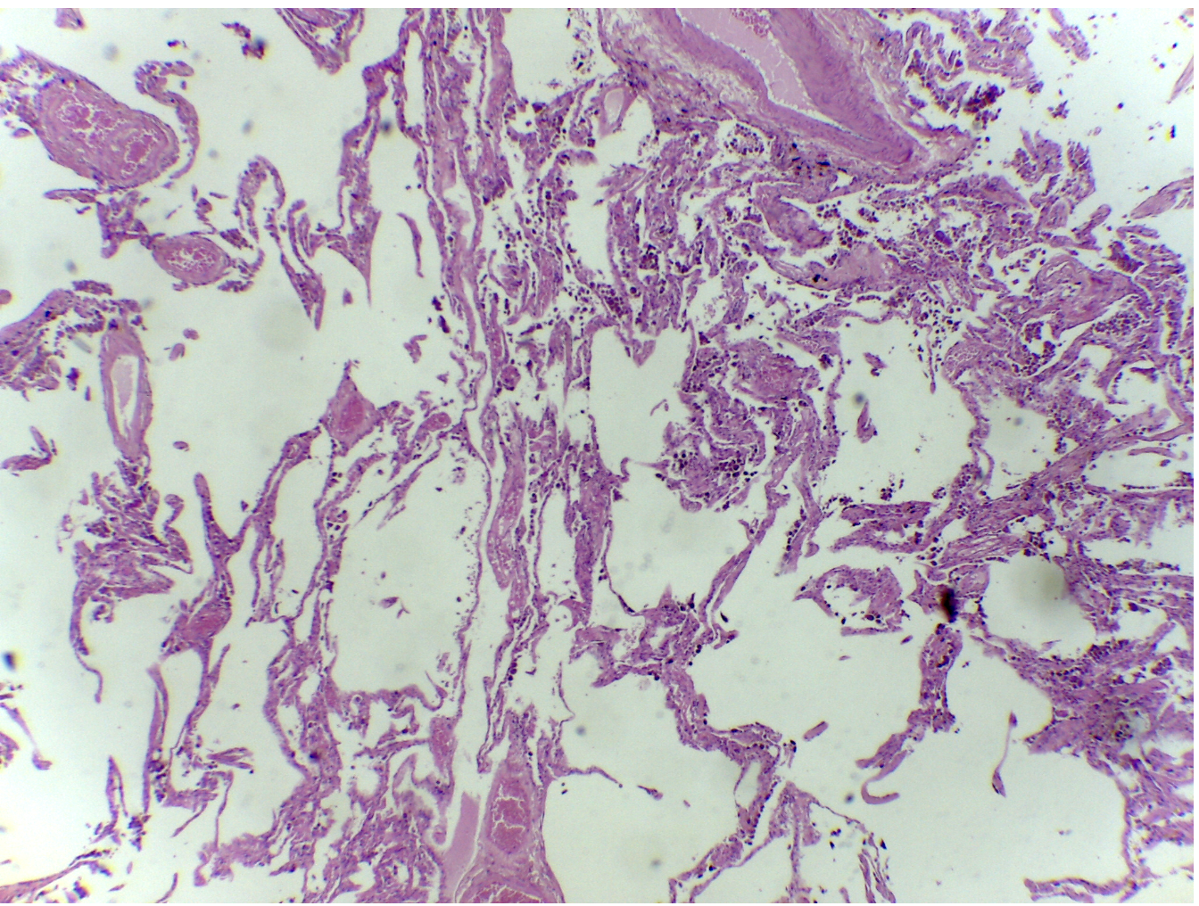
What is Emphysema (COPD)?
causes progressive damage to alveoli = tiny air sacs in your lungs
alveoli compressed/collapsed, limiting O2 absorption BC: reduction in the surface area available for gas exchange
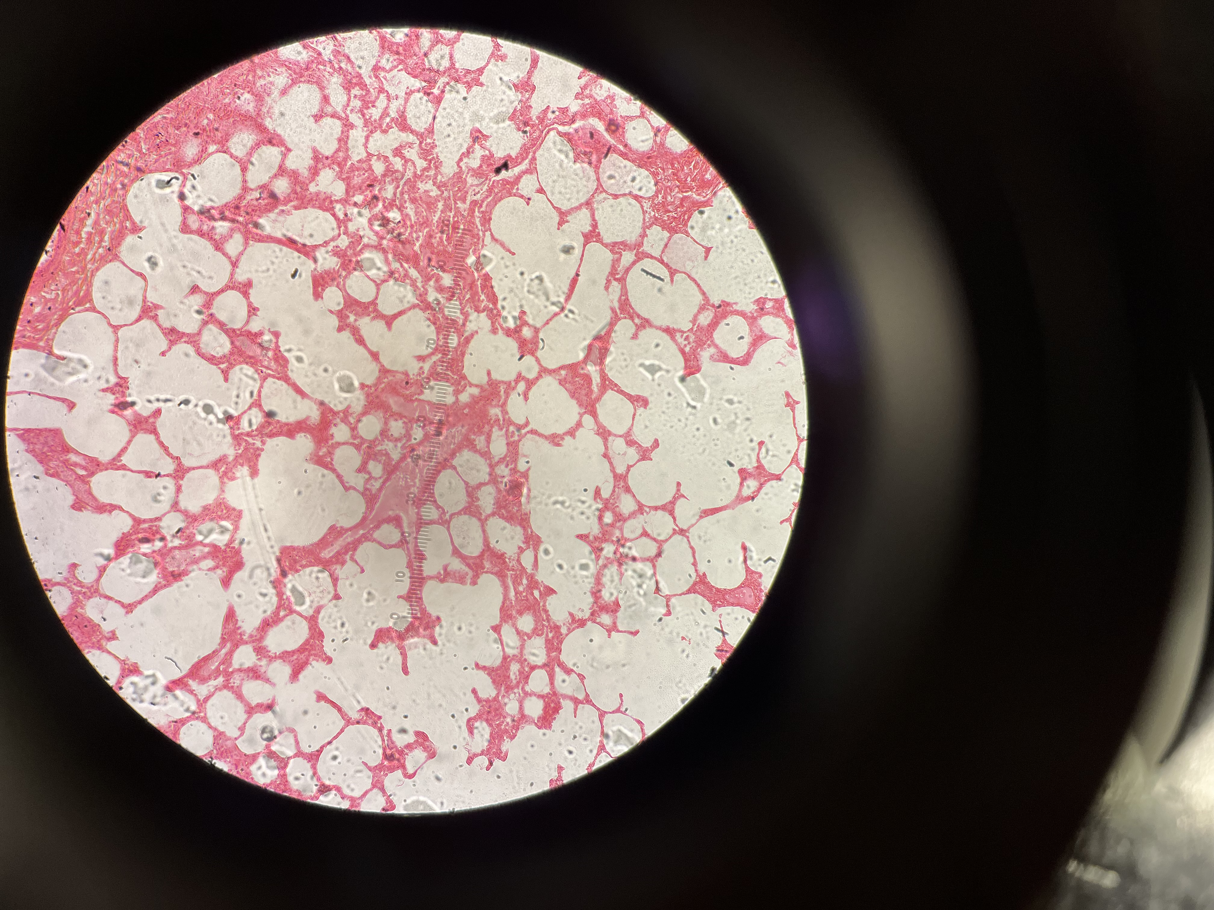
What is this an image of?
A healthy lung
Features a tight, uniform, and sponge-like array of small, distinct air sacs (alveoli) (2nd image shows alveoli better)
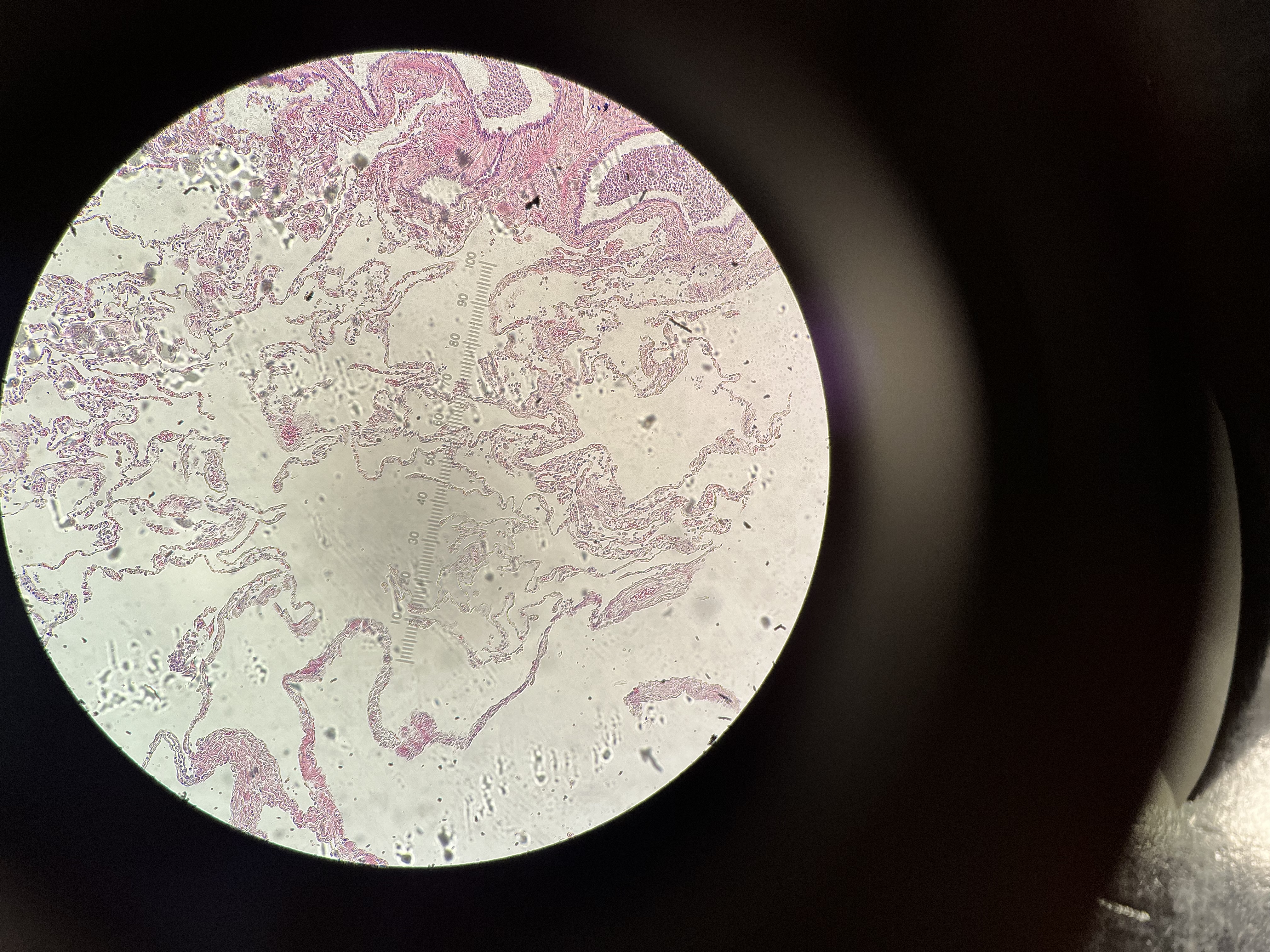
What is this an Image of?
Emphysema Lung (COPD: Chronic Obstructive Pulmonary Disease)
alveoli compressed/collapsed, merged air spaces
Artery….
has rigid/thick outer wall and a lumen
structurally reinforced → to withstand the intense, high-pressure surges of blood pumped directly from the heart
Arteries usually….
carry oxygenated AWAY from the heart
Artery exception?
Pulmonary artery carries deoxygenated blood from heart to lungs!
Vein….
flimsier outer wall than in arteries (looks less circular)
has lumen
thinner walls, only hold shape when liquid flows through
Veins usually…
carry deoxygenated blood TO the heart
Vein exception
pulmonary veins carry oxygenated blood from lungs to heart!
Nerves….
solid fiber bundles
conduct electrical signals
no lumen!!
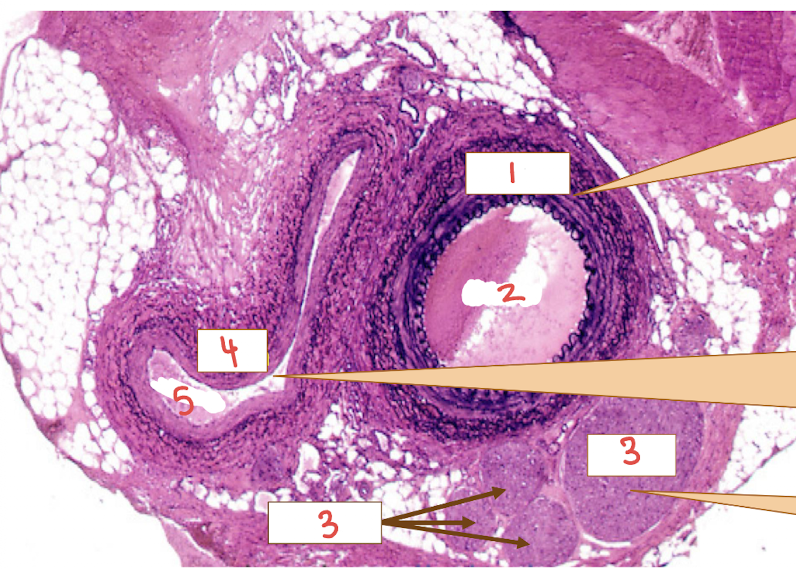
what is number 1?
Artery

What is number 2?
Lumen of artery

What is number 3?
Nerves!
no lumen!!!!

What is number 4?
Vein

What is number 5?
Lumen of vein
Why are artery walls thicker?
high pressure
Arteries receive blood directly pumped from the heart and need thick, muscular, and elastic walls to withstand that high-force impact
Why are vein walls thinner?
because they carry blood at a much lower pressure to heart (instead of receiving blood from heart)
What is arteriosclerosis?
a broad, general term for the hardening and thickening of artery walls
hardening of artery from age
What is atherosclerosis?
a specific, very common type of arteriosclerosis caused specifically by the buildup of fatty plaque
hardening of artery from plaque buildup
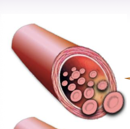
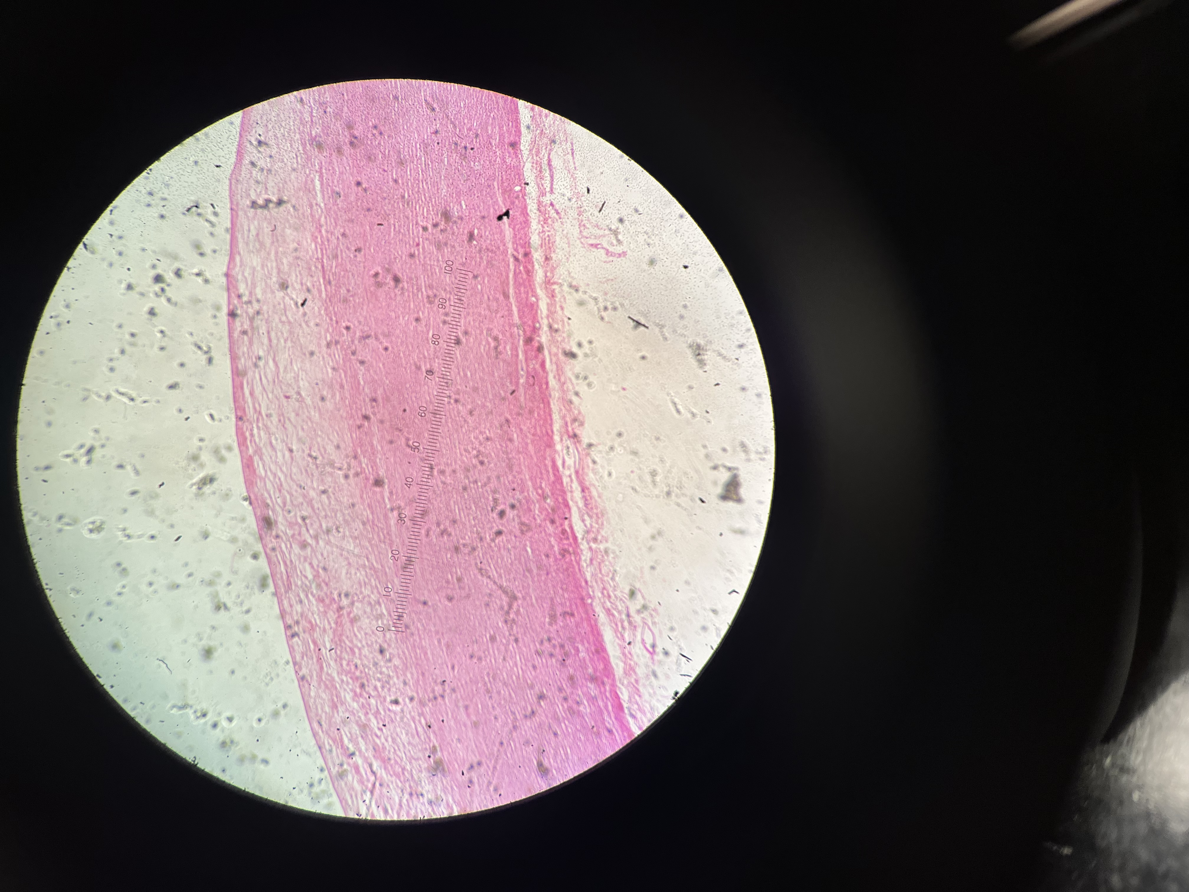
This is a…
normal artery
thicker wall than vein but not abnormally
allow consistent flow of blood through
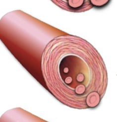
This is an image of
Arteriosclerosis
hardening of artery from age
restriction of bloodflow
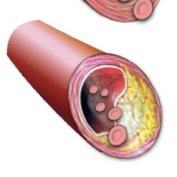
This is an image of
atherosclerosis
buildup of plaque
restriction of blood flow

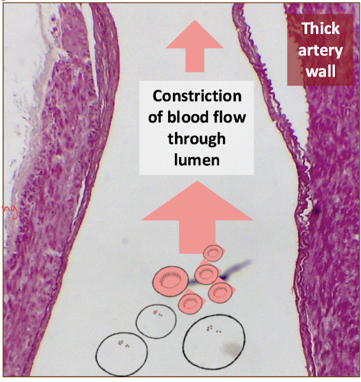
Longitudinal section of diseased artery: What does the star showcase?
thick artery wall
outgrowth of artery
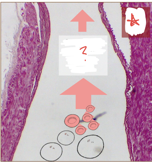
Longitudinal section of diseased artery: What does the thicker artery well result in?
constriction of blood flow through lumen
Human Atherosclerosis aorta section
plaque (fat, cholesterol, and calcium) builds up on the inner walls of the aorta
More red blood cells =
higher capacity to store oxygen in body
Mammals are endotherms
rely on metabolic heat production
need more oxygen to keep metabolic rates high to keep body temperature consistent → need more binding affinity……
Reptiles are ectotherms
rely on surrounding to keep warm, so they require less O2
having nucleated red blood allows their cells to undergo mitosis!
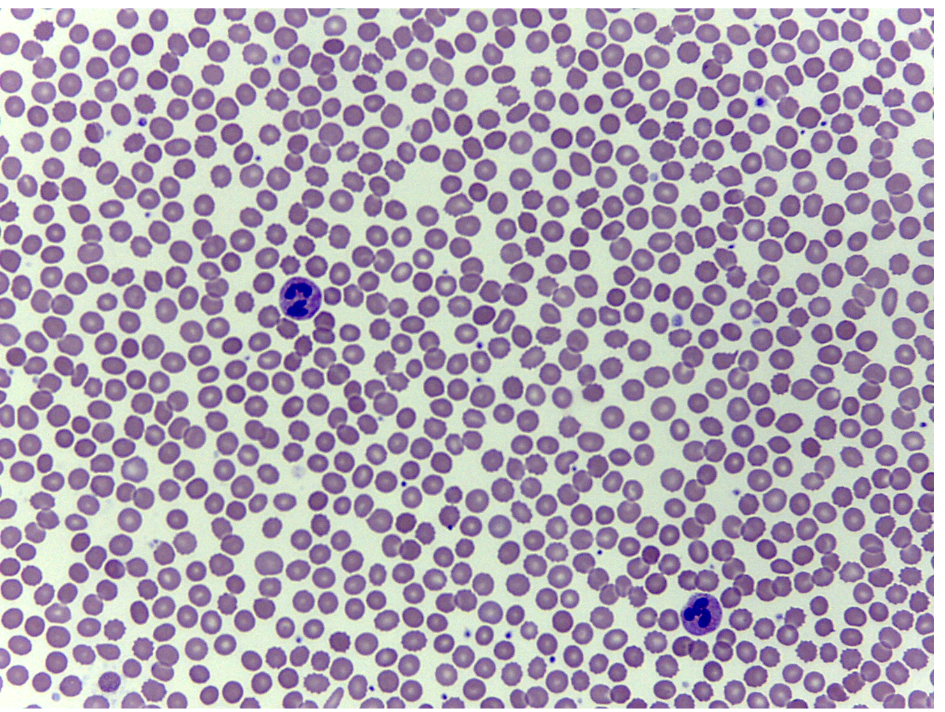
This is an image of…
Healthy Human Blood Smear
lacking nuclei
Why is lacking nuclei in red blood cells advantageous ?
since endotherms need more oxygen to keep up w/metabolic rate & keep temperature more consistent
losing the nucleus in our red blood cells allow for a higher surface area for hemoglobin & O2, boosting our metabolism

What is this an image of?
Reptile Blood Smear
Why do reptiles have nucleated red blood cells?
retained nucleated red blood cells because they have lower metabolic rates than mammals (bc they are ectotherms)
so they don't require the extreme oxygen-efficiency of enucleated (nucleus-free) cells
require less O2 + cells undergo mitosis
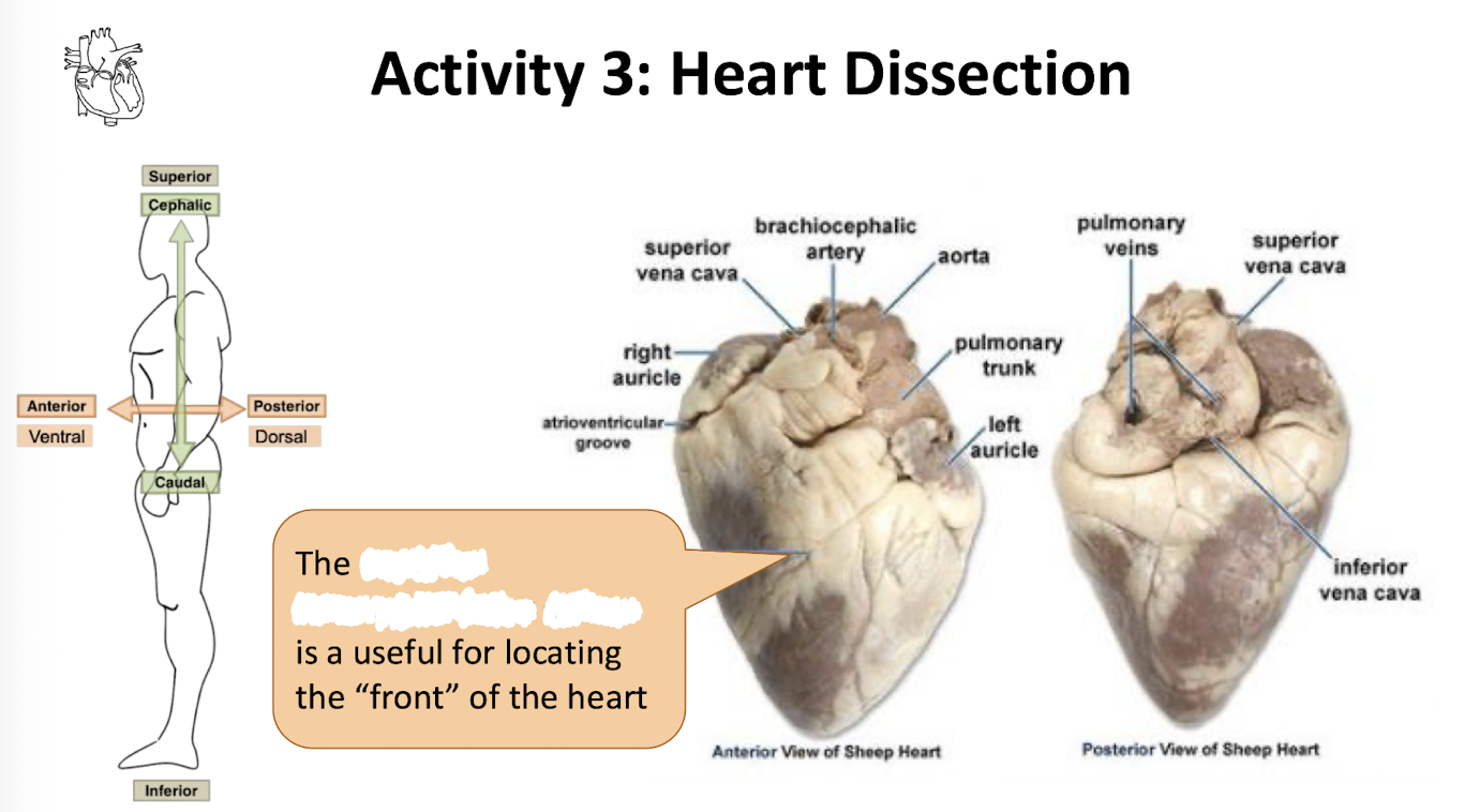
This is showcasing the
anterior interventricular sulcus

What is number 1?
superior vena cava

What is number 2?
right pulmonary artery

What is the star?
Pulmonary Trunk

What is number 3?
Right atrium

What is number 4?
right pulmonary veins

What is number 5?
Pectinate Muscles

What is number 6?
Right atrioventricular valve
Tricuspid Valve

What is number 7?
Right Ventricle

What is number 8?
Chordae Tendineae

What is number 9?
Trabeculae Carneae

What is number 10?
Inferior Vena Cava

What is number 11?
Aorta

What is number 12?
Left Pulmonary artery

What is number 13?
Left Atrium

What is number 14?
Left Pulmonary Veins

What is number 15?
Left atrioventricular valve
Bicuspid valve
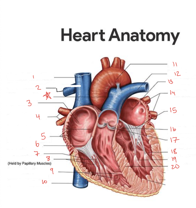
What is number 16?
Aortic Semilunar Valve

What is number 17?
Pulmonary Semilunar Valve

What is number 18?
Left Ventricle

What is number 19?
Papillary Muscles

What is number 20?
Interventricular Septum
What are the chordae tendineae ?
“ Heart Strings”
tendons connecting papillary muscles to tricuspid & bicuspid valves, helping prevent prolapse
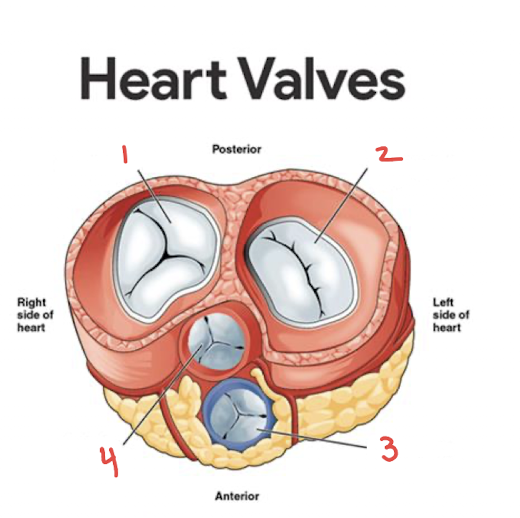
What is number 1?*
Right atrioventricular valve*
tricuspid valve

What is number 2?*
Left atrioventricular valve*
Bicuspid Valve*

What is number 3?*
Pulmonary semilunar valve*

What is number 4?*
Aortic semilunar valve*

What is number 1? ^
Left Atrium ^

What is number 2? ^
Left Ventricle ^

What is number 3? ^
interventricular septum ^

What is number 4? ^
Right Ventricle ^

What is number 5? ^^
Left Atrium ^^
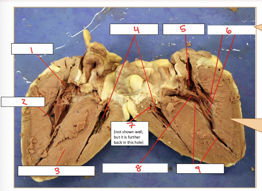
What is number 6?
Chordae Tendineae

What is number 7? ^
Right Atrium ^