CONOTRUNCAL DEFECT DORV
 Call Kai
 Learn
Learn Practice Test
 Spaced Repetition
 Match
Match Flashcards
 Knowt Play
1/83

There's no tags or description
Looks like no tags are added yet.
Name | Mastery | Learn | Test | Matching | Spaced | Call with Kai | Chat |
|---|
No analytics yet
Send a link to your students to track their progress
84 Terms
What is Double Outlet Right Ventricle (DORV)?
A ventriculoarterial connection in which both great arteries arise by more than 50% from the right ventricle.
List the 3 things used to categorize DORV.
Relationship of the great arteries
Location of the VSD (A VSD is usually present but the location varies)
Presence of conal tissue, or infundibulum
What is the relationship between the semilunar valves and inflow valves in DORV?
Neither semilunar valve is in fibrous continuity with an inflow valve.
How are the great arteries often related in DORV?
They are often normally related.
What congenital defect is DORV most often confused with? This is the #1 Differential
Tetralogy of Fallot. - #1 Differential b/c of PLAX

List the 4 possible positions of the aorta in DORV. & The spatial relationship of the great arteries is often what relationship?
How is it in D-TGA and L-TGA
Right anterior
Left anterior
Anterior-posterior
Side-by-side
(The spatial relationship of the great arteries is often normal - the Aorta is posterior and to the right of the PA)
D-TGA = Aorta is anterior and to the right of the PA
L-TGA = Aorta is anterior and to the left of the PA

List the 4 types of VSDs in DORV & which one is the most common? & Which one is the most rare?
Subaortic - Most common
Subpulmonic
Doubly committed
Remote - very rare

Are most VSDs in DORV restrictive or unrestrictive?
Unrestrictive
Why is the location of the VSD important in DORV?
The VSD location affects the pathophysiology of DORV.
VSD location → changes blood-flow pathway → changes hemodynamics → changes the clinical presentation of DORV.
What is the conus also called?
The infundibulum.(thick portion of the RVOT)
List the 4 conal morphologies in DORV.
Bilateral conus 2 muscular sleeves under both the pulmonary valve and aortic valve. It occurs in about 23%
Absent aortic conus
Absent subpulmonary conus
Bilateral absent conus
(if conal tissue is malformed it can cause obstruction - most common PS)
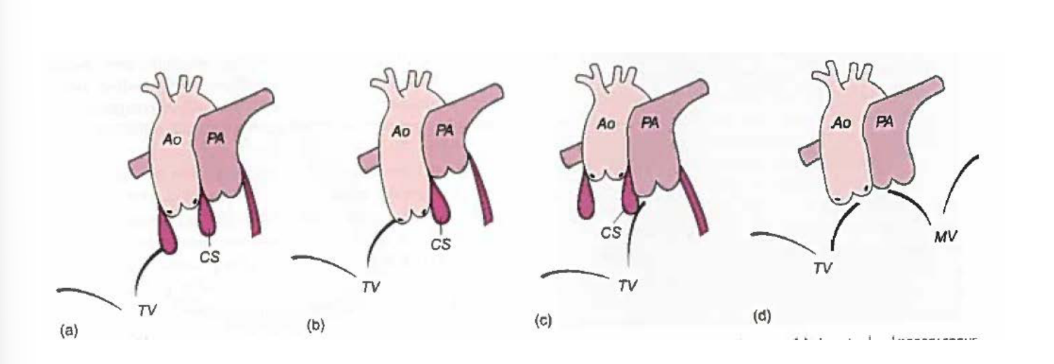
List the 3 main coronary artery patterns seen in DORV.
Normal
Abnormal as in TOF
Abnormal as in TGA
What is the most common coronary anomaly when the coronary pattern resembles TOF?
The LAD arises from the RCA.
What is the most common coronary anomaly when the coronary pattern resembles TGA?
The circumflex artery arises from the RCA.
What 2 types of stenosis may be present in DORV? & what does the presence of stenosis affects?
Subvalvar stenosis
Valvar stenosis
The presence of stenosis affects the patient's hemodynamics
Why may the cardiac conduction system be abnormal in DORV?
The conduction system is usually displaced because of the anatomic abnormalities of DORV.
In hearts with subpulmonic VSDs (such as the Taussig-Bing anomaly) or subaortic defects, what conduction abnormality is present?
The AV bundle is typically affected causing AV block
Embryology:
What outflow-tract defect contributes to DORV?
Impaired leftward migration of the conotruncus and/or abnormal persistence or absorption of the subarterial conus.
What normally happens during aortic wedging?
The aorta normally rotates and wedges between the mitral and tricuspid valves, connecting it to the LV.
What happens to aortic wedging in DORV?
Early in development, both vessels sit over the right ventricle. The normally rotation of the aorta in DORV, this rotation stops.
What are conal growth abnormalities in DORV?
The muscular subaortic and subpulmonary cones fail to absorb or position correctly, leaving both major vessels anchored to the RV.
Why does a VSD almost always form in DORV?
Because the great vessels do not align over the LV, the VSD forms as the only outlet for the LV.
Are most cases of DORV inherited or sporadic?
Most cases are sporadic.
It means most cases happen randomly, without a clear inherited or family pattern.
What percentage of DORV cases have chromosomal associations, and list the 3 associations given.
Approximately 41% have chromosomal associations:
Trisomy 13 - Patau syndrome
Trisomy 18 - Edwards syndrome
DiGeorge syndrome
List 4 Features of Patau Syndrome Trisomy 13
Slopping forehead / tiny ears
cleft Lip & palate
Polydactyly - extra fingers
Poor survival rate 95-100% chance have a heart defect (ASD, VSD, PDA)

List 3 Features of Edwards Syndrome Trisomy 18
Clenched fists
curved feet
Poor survival rate 95-100% chance have a heart defect (ASD, VSD, PDA)

List the 5 maternal factors associated with DORV.
Non-gestational diabetes
Reproductive assistance
Codeine use
Retinoic acid
Exposure to solvents
Non-gestational diabetes means diabetes that was already present before pregnancy or diabetes that is not caused by pregnancy.
Exposure to solvents means being around or coming into contact with certain chemical liquids or fumes, often from things like paint thinners, cleaners, degreasers, or industrial chemicals.
In pregnancy, some solvent exposures can be considered a risk factor because certain chemicals may affect fetal development.
What determines the hemodynamics of DORV?
The relationship of the VSD to the great vessels.
What does the position of the VSD determine?
It determines where LV outflow and blood exiting the LV will be directed.
What 2 factors are used to classify DORV based on its hemodynamics?
Location of the VSD
Presence or absence of obstruction
When is a VSD classified as subaortic?
When the VSD is more closely related to the aortic valve.
What is the blood-flow pathway in DORV with a subaortic VSD? for the LV and RV
LV blood travels through the VSD to the aorta.
RV blood travels mainly to the pulmonary artery.
Left to Right shunt, this is a good b/c oxygented blood is going to the Aorta
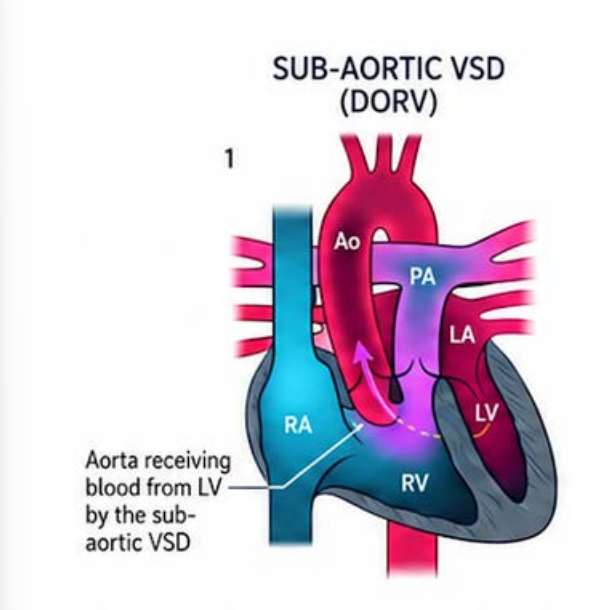
What is the most common type of DORV?
DORV with a subaortic VSD.
Which type of VSD for DORV has the potential to have an obstruction and what obstruction would it be?
Subaortic VSD - DORV, Pulmonary obstruction.
What are the hemodynamics of DORV with a subaortic VSD without pulmonary stenosis? List 3 - the Hemodynamics are similar to what defect?
Usually acyanotic
Hemodynamics similar to a large VSD
Increased pulmonary blood flow causes CHF, Tachypnea and poor growth
SKIP
How does pulmonary stenosis affect DORV with a subaortic VSD? & The hemodynamics are similar to what defect?
The degree of cyanosis depends on the severity of pulmonary stenosis, and the hemodynamics become similar to TOF.
When is a VSD classified as subpulmonary?
When it is more closely associated with the pulmonary valve.
What is another name for DORV with a subpulmonary VSD?
Taussig-Bing anomaly.
What is the blood-flow pathway in DORV with a subpulmonary VSD?
LV blood travels through the VSD to the pulmonary artery.
RV blood travels mainly to the aorta.

List the 3 structural findings described with the Taussig-Bing anomaly / DORV with subpulmonary VSD
Bilateral conus beneath both great vessels
Aorta and pulmonary artery are side-by-side
Aortic arch hypoplasia or coarctation
What are the hemodynamics of DORV with a subpulmonary VSD without pulmonary stenosis? & The hemodynamics are similar to what defect? - total list 3
Cyanotic
Hemodynamics similar to D-TGA
Increased pulmonary blood flow causes CHF
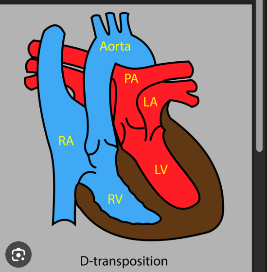
How does pulmonary stenosis affect cyanosis in DORV with a subpulmonary VSD?
The degree of cyanosis depends on the severity of pulmonary stenosis.
Where is LV outflow directed with a doubly committed VSD?
LV outflow is equally shared between the aorta and pulmonary artery.
Doubly committed VSD = LV outflow is directed toward both the aorta and pulmonary artery → both great arteries receive mixed oxygenated and deoxygenated blood.
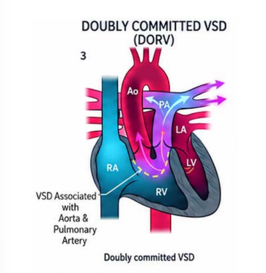
What happens to pulmonary blood flow with a doubly committed VSD?
Pulmonary overcirculation is present.
What causes a doubly committed VSD in DORV?
An absent infundibular septum.
How may cyanosis appear with a doubly committed VSD without pulmonary stenosis? & the Hemoydnamics are similar to what defect? t
Cyanosis may be mild or absent.
Hemoydnamics are similar to VSD -type
List the 4 signs of CHF associated with a doubly committed VSD. same for with or without PS
Tachypnea
Tachycardia
Hepatomegaly
Failure to thrive
How does pulmonary stenosis affect cyanosis with a doubly committed VSD?
The degree of cyanosis depends on the severity of the stenosis.
What is a remote VSD in DORV? & How does it affect the great vessel?
A VSD that is uncommitted to any specific great vessel.
There is no obstruction to any great vessel (located very bottom of the IVS muscle)

How does ventricular blood flow with a remote VSD without great-vessel obstruction?
Right- and left-ventricular blood mixes, with relatively equal amounts streaming to the aorta and pulmonary artery.

What is the typical postnatal physiology of DORV with a remote VSD?
Similar to a large VSD. It means that after birth, a remote VSD in DORV usually behaves hemodynamically like a large VSD
How common is DORV with a remote VSD?
It is a rare form of DORV.
What determines the clinical presentation of DORV with a remote VSD? (without and with PS) & The hemodynmaics are similar to what defect?
Location of the VSD
Direction of flow through the VSD
hemodynmaics are similar to single ventricle pathway
Why can the clinical presentation of DORV vary widely from severely low oxygen levels to signs of heart overload?
It depends on:
Position of the VSD
Whether an obstruction is present
List the 7 common symptoms of DORV.
Cyanosis
Tachypnea
Tachycardia
Feeding difficulties
Poor growth
Diaphoresis- sweating while feeding
Clubbing
List the 2 physical-exam findings of DORV.
Murmurs from VSD and/or pulmonary stenosis- cardiac cycle is systole
Decreased femoral pulses
List the 7 associated defects of DORV.
Pulmonary stenosis
ASD and PDA
Coarctation of the aorta
Mitral atresia
HLHS
AV canal defects
Heterotaxy
What should you look for when a large VSD is present?
Straddling or overriding valves.
List the 2 defects that should be ruled out from the suprasternal-notch view.
Aortic coarctation
PDA
List the 3 key measurements obtained during the DORV echo assessment.
Peak velocity and gradients across the pulmonary and aortic valves
Peak velocities across the ASD, VSD, and PDA
PAP by BP
What is the formula for calculating LV systolic pressure in DORV?
LVSP = RVSP + VSD pressure gradient
RVSP may be obtained from the TR jet.
If RVSP is 30 mmHg and the VSD gradient is 64 mmHg, what is the LVSP?
30 + 64 = 94 mmHg
What does an LVSP of 94 mmHg in the example indicate?
The LV is experiencing significant pathological afterload because the VSD is restrictive.
List the 3 differential diagnoses for DORV.
Tetralogy of Fallot
D-TGA
Truncus arteriosus
What determines the surgical approach for DORV?
The surgical approach varies based on the specific DORV findings.
What is the overall surgical goal when repairing DORV?
To connect:
LV → aorta
RV → pulmonary artery
How is DORV with a subaortic VSD repaired?
An intraventricular tunnel is created between the VSD and the aortic valve.
How is DORV with a subaortic VSD and pulmonary stenosis repaired? - the first is to do what?
An intraventricular tunnel using a VSD patch directs blood from the LV to the aorta.
How is the RVOT reconstructed in DORV with a subaortic VSD and pulmonary stenosis? - step 2
With a transannular patch.
List the 4 repair options for DORV with a subpulmonary VSD.
Kawashima procedure
Baffle VSD to PA combined with Jatene
Rastelli procedure
Nikaidoh procedure
DORV with subpulmonary VSD:
What is done to the VSD during the Kawashima procedure?
The VSD is baffled to the aortic valve with patch enlargement.
DORV with subpulmonary VSD:
How is the RVOT enlarged during the Kawashima procedure?
A second patch is placed to enlarge the RVOT.
Double patch
What repair is most commonly used for DORV with a subpulmonary VSD and TGA when the pulmonary valve is normal size?
Baffle the VSD to the pulmonary artery combined with an arterial switch operation, or Jatene.
List the 2 main parts of the Rastelli procedure. for DORV with a subpulmonary VSD and with PS
Close/baffle the VSD to the aorta
Place a valved RV-to-PA conduit for RVOT reconstruction
When is the Rastelli procedure preferred?
When the pulmonary valve is not sufficient to support systemic circulation because of stenosis.
What procedure may be used for DORV with a doubly committed VSD?
The Kawashima procedure:
VSD is baffled to the aortic valve with patch enlargement.
A second patch enlarges the RVOT.
(Double intraventricular baffle)
What type of procedure is initially needed for DORV with a remote VSD?
A palliative procedure.
List the 3 initial palliative options for DORV with a remote VSD.
Systemic BT shunt
PDA stent if RVOTO is present
Pulmonary artery band to prevent pulmonary overcirculation
How does a biventricular repair begin for a remote VSD without pulmonary stenosis?
By creating an intraventricular tunnel from the VSD to the aorta.
What valve apparatus may need modification during a biventricular repair for a remote VSD?
The tricuspid valve apparatus.
How is the remote VSD connected to the aorta when pulmonary stenosis is present?
Intraventricular Baffle:
Surgeons construct an extensive intracardiac patch or tunnel from the remote VSD to the aortic valve.
How may pulmonary stenosis be repaired in DORV with a remote VSD?
Resecting obstructing muscle bundles, or
Placing an RVOT patch,
Sometimes using a valved conduit depending on the anatomy.
List the 7 things evaluated during postoperative DORV echo interrogation.
LV and RV size and function
Residual obstruction
Residual ASD
Baffle or tunnel leak or stenosis
Jatene switch procedure
Inflow valves
PAEDP and RVSP