Skeletal Muscle
 Call Kai
 Learn
Learn Practice Test
 Spaced Repetition
 Match
Match Flashcards
 Knowt Play
1/41

There's no tags or description
Looks like no tags are added yet.
Name | Mastery | Learn | Test | Matching | Spaced | Call with Kai | Chat |
|---|
No analytics yet
Send a link to your students to track their progress
42 Terms
Structure of skeletal muscle
Striated
In what way are muscles excitable tissues
Membrane potential is linked to activation of that cell’s function (contraction)
What percentage of an adult’s body weight is skeletal muscle (& smooth muscle)
Skeletal muscle = 40%
Heart & Smooth muscle = 10%
Under what 3 headings are muscles classified
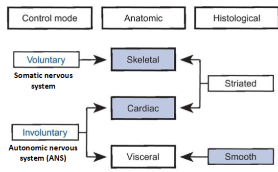
Location & function of skeletal muscle
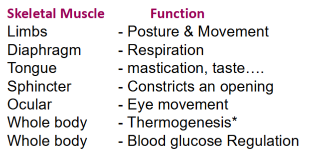
What controls shivering vs non shivering thermogenesis
Shivering - muscle
Non shivering - brown fat
Name a muscular dystropy
Duchenne Muscular dystrophy
Name a Neuromuscular disorder of skeletal muscle
Myasthenia gravis
name a muscular atrophy
Sarcopenia / Cachexia
what is defective in Type II Diabetes
Insulin stimulated glucose uptake into skeletal muscle is defective
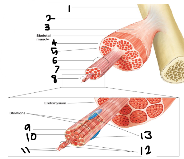
Label
Periosteum
Tendon
Fascia
Epimysium
Perimysium
Fasciculus
Endomysium
Muscle Fibre
Sarcolemma
Sarcoplasm
Filaments
Myofibrils
Nuclei
Sarcolemma
Plasma membranes
Unique feature of skeletal muscle
Multinucleated
Name of light & dark bands
Light = I bands
Dark = A bands
What are myofibrils
Thick (myosin) & thin (actin) myofilaments that run the length of the muscle fibre
3 types of muscle proteins
Contractile
Regulatory
Structural
Name 2 contractile muscle proteins
Myosin & actin
Name 2 regulatory muscle proteins
Troponin
Tropomyosin
Name 5 regulatory muscle proteins
Titin
Nebulin
Alpha-actin
Myom
Which muscle protein is the largest
Titin
Which muscle protein has the largest gene
Dystrophin
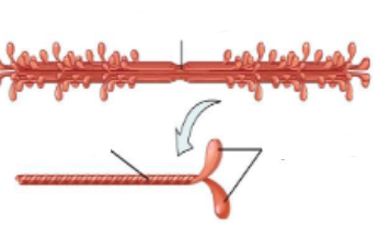
Identify
Thick filament with myosin molecule
Identify
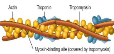
Portion of a thin filament
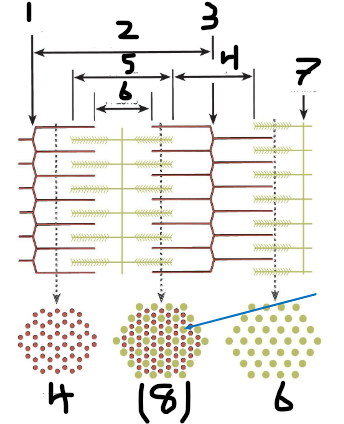
1 and 3 are the same
Z line
One sarcomere
Z line
I band
A band
H zone
M line
Thick & thin filaments overlap
One myosin filament is surrounded by how many actin filaments in 3D arrangement
6
What do sarcomeres do when a muscle contracts
Sarcomeres shorten
What do A and I bands do when a muscle contracts
A bands do not alter in size (they reflect the myosin filament length within a sarcomere).
I bands decrease in size (they reflect actin filament that has no overlapping myosin filaments on adjacent ends of two sarcomeres)
Do thin filaments slide towards to / away from the H zone
Towards
What are thick myofilaments composed of
Composed of the protein myosin
(Each protein has two globular heads with actin-binding sites and ATP-binding sites)
What are thin myofilaments composed of
Composed of the protein actin with proteins called tropomyosin and troponin
Role of tropomyosin and troponin
prevent myosin binding at rest
What are formed between myosin & actin during sliding
Cross bridges
Role of myosin head when muscle is stimulated
The myosin head serves as a myosin ATPase enzyme, splitting ATP into ADP + Pi.
This allows the head to bind to actin when the muscle is stimulated due to electrical & chemical events
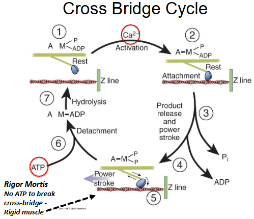
Cross bridge cycle
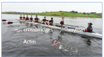
How do myosin heads on myosin filaments move
like oars knifing through water, but do not move synchronously
What percentage of crossbridges make contact with actin to form the protein complex actomyosin
50%
How is contraction regulated
Tropomyosin physically blocks cross bridges
How does troponin complex regulate contraction
Troponin I inhibits binding of myosin.
Troponin T binds to tropomyosin.
Troponin C binds to calcium.
When is Ca2+ released inside the muscle fibre
When muscle cells are stimulated, Ca2+ is released inside the muscle fibre
What happens when some Ca2+ attaches to troponin C
Conformational change in troponin and tropomyosin occurs
Myosin is allowed access to form cross bridges
What 2 things are required for muscle contraction
ATP & Calcium