MLS 411
 Call Kai
 Learn
Learn Practice Test
 Spaced Repetition
 Match
Match Flashcards
 Knowt Play
1/53

There's no tags or description
Looks like no tags are added yet.
Name | Mastery | Learn | Test | Matching | Spaced | Call with Kai | Chat |
|---|
No analytics yet
Send a link to your students to track their progress
54 Terms
Define antigen and antibody
Antigen: Foreign substance that can trigger an immune response
Antibody: Protein secreted by plasma that specifically bind to an antigen
What is complement?
A series of 25 serum proteins responsible for
Cell lysis
Opsonization
Mediation of inflammation
When complete: pore forms in RBC, causing intravascular hemolysis
What is zeta potential?
Electric charge difference between RBC surface and surrounding ionic cloud. Keeps RBCs separated and prevents agglutination. Lower zeta potential allows agglutination
What is dosage?
Some antibodies react more strongly when an antigen is present in a double dose (homozygous inheritance) than in a single dose (heterozygous inheritance)
Explain the process of agglutination, as it applies to immunohematology.
When corresponding antibodies bind to antigens on the surface of RBCs, the Fc regions become close in proximity, causing them to clump
Discuss the conditions that must exist to see hemolysis as a positive endpoint in immunohematology tests
Must be complement-binding antibodies (IgM or close-paired IgG)
Fresh serum
Presence of free Ca2+ and Mg2+ ions
Corresponding target antigen on RBC surface
Discuss the clinical significance of the Rh system as applied to transfusion medicine
Rh antigens are integral transmembrane proteins only found on the surface of RBCs. They play a role in transfusion medicine because they trigger a strong immune response if there is incompatibility between a recipient and donor.
List the major antigens of the Rh system using Fisher-Race terminology.
D, C, c, E, e
Describe the inheritance of Rh genes.
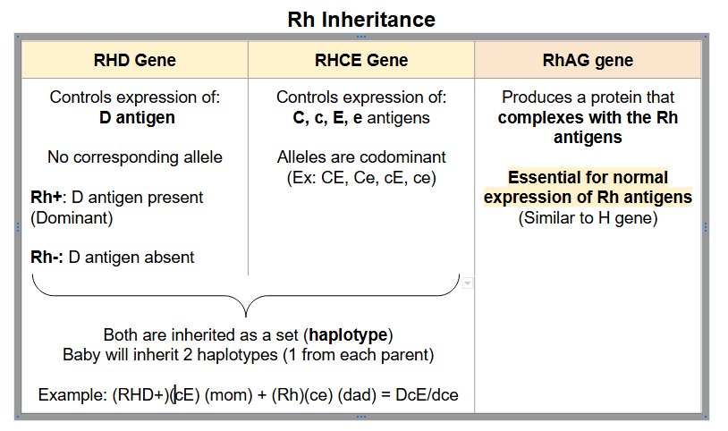
Discuss the structure and immunogenicity of Rh antigens.
Rh antigens are integral transmembrane proteins only found on the surface of RBCs
Highly immunogenic
Well developed at birth
Display antigen variability - can be weak/strong
Differentiate the three mechanisms that result in the presence of the weak D antigen.
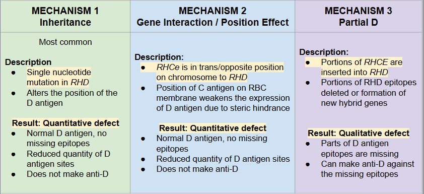
Discuss situations when weak D testing would be appropriate
If testing with anti-D at the IS phase of testing is negative, incubate at 37C and test with AHG reagent to test again
Blood, organ, tissue donors
Rh-negative infants born to Rh-negative mothers (to see if the baby is positive)
Individuals who historically typed as Rh-positive, but currently type as Rh-negative
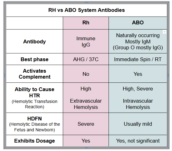
Compare Rh and ABO antibodies based on:
Type of antibody
Phase of reactivity
Ability to activate complement
Ability to cause HTR (hemolytic transfusion reaction)
Ability to cause HDFN (hemolytic disease of the fetus and newborn)
Exhibits dosage
List acceptable specimens for Rh type determinations.
EDTA or clot tube
Explain the principle of Rh testing, including the weak D test
Rh Testing
Performed at the IS phase, where patient RBCs have anti-D added to them
After centrifugation, the presence of agglutination = a positive test
An inert control (negative result) is always performed (if performing JUST Rh typing)
Can use: Rh-hr control, 6% bovine serum albumin (BSA), or DAT
Control not needed if ABO/Rh typing is performed together, unless all reactions are positive (AB+)
Weak D Testing
If the Rh test result is negative at the IS phase, then the sample is incubated at 37C
Sample is washed multiple times with saline to rid of any unbound antibodies
AHG reagent is added to the sample and spun
Agglutination = Weak D positive = Rh positive
List the order of immunogenicity of the Rh antigens from most to least immunogenic
D > c > E > C > e
Discuss the significance of the ABO system as applied to transfusion medicine
The ABO system is the most critical blood classification in transfusion medicine.
Matching donor and recipient ABO types prevents acute hemolytic transfusion reactions, where the recipient's immune system attacks and destroys incompatible donor red blood cells, which can be fatal.
List the most common to least common ABO types
O > A > B > AB
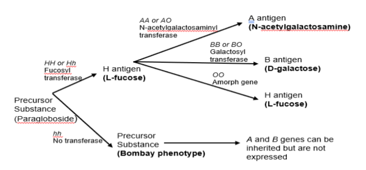
Explain the inheritance of the A, B and H genes
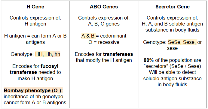
List the concentration of H substance in each ABO phenotype from most to least
O > B > A > AB
What genes are needed to express ABO antigens?
Hh, ABO, Sese (secretor)
Codes for transferases that add immunodominant sugars that form the antigen
Outline the development of A, B and H antigens including the specific transferases and terminal sugars
List acceptable specimens for ABO grouping determinations.
EDTA or clot tube
Explain the principle of ABO testing
ABO Testing involves forward and reverse typing
Reagents used for monoclonal, which are hybridoma-derived
Testing methods: tube, microtiter, gel method
Forward Typing
Patient RBCs are tested with antisera (Anti-A, Anti-B which contains antibodies) to detect antigens
Anti A,B is used only sometimes
Agglutination = positive test
Reverse Typing
Patient plasma is tested with known reagent red cells (A1 and B) to detect antibodies
Agglutination = positive test
Inert Control: If a patient’s results are AB, then an inert control must be used (6% BSA)
6% BSA is a protein base which mimics antisera but doesn’t have the antibody
MUST yield a negative result for the AB result to be valid
What else can serve as a positive endpoint in immunohematology tests instead of agglutination?
Hemolysis
Conditions:
Complement activation & complement-fixing antibodies (IgG)
Fresh serum
Antigen presence
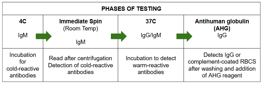
Describe the phases of testing
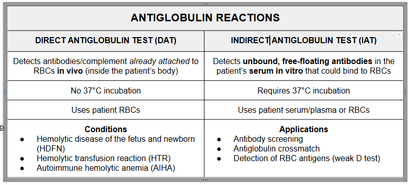
Describe the antiglobulin reaction/tests
The antiglobulin reaction (Coombs test) detects non-agglutinating antibodies or complement proteins bound to red blood cells (RBCs).
Compare polyspecific vs monospecific AHG reagent
Polyspecific AHG = contains both anti-IgG + anti-C3
Used for screening during IAT/DAT
Created by blending/mixing rabbit polyclonal or mouse monoclonal anti-IgG with mouse monoclonal anti-C3
Monospecific AHG contains either anti-IgG / anti-C3
Used for identification after a positive IAT/DAT
Created by isolating or preparing individual antibody specificities (ex: utilizing monoclonal hybridoma cells)
What is used as controls during the antiglobulin test?
Performed when AHG test is negative (no agglutination)
Check cells (IgG or C3-coated RBCs)
Must yield a positive reaction for a valid result
If negative, test must be fully repeated
Discuss how the following factors influence antigen-antibody reactions in immunohematology tests:
Antigen/ Antibody ratio
Temperature
Reaction time/Incubation
Ionic strength of test medium
Number and position of antigens
pH
Antigen/ Antibody ratio
Best when in equal amounts; if not, then lattice can’t be formed
Prozone: excess antibody
Postzone: excess antigen
Temperature
IgM: 4-25C (wide thermal amplitude)
IgG: 36-38C
Reaction time/Incubation
IgM: requires little to no incubation
IgG: requires incubation at 37C
Too long incubation = antibody dissociation
Too short incubation = insufficient sensitization
Ionic strength of test medium
Want low zeta potential, ionic strength, and shielding effect to allow antibodies to sensitize RBC and decrease distance between the RBCs
Add potentiators to the medium for this (Albumin, LISS, PEG)
Number and position of antigens
Surface-exposed antigens react more readily
Homozygous/double-dose antigen expression generally produce stronger reactions than a heterozygous/single-dose
pH
Physiological pH: 6.5 - 7.5
Adjusting pH can enhance or decrease some reactivities
Discuss the action of the following potentiators:
Albumin
Low Ionic Strength Solution
Polyethylene Glycol
Albumin
Reduces zeta potential
Incubation: 15-30 min
Low Ionic Strength Solution (LISS)
Lowers zeta potential and shielding effect, promoting antibody uptake onto RBCs
Incubation: 15-20 mins
Polyethylene Glycol (PEG):
Same as LISS, removes water from test system, concentrating antibodies
Incubation: 15-30 mins
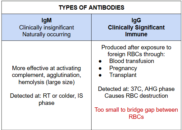
Compare IgM and IgG
Hemolysis can only be observed in what type of sample?
Serum (red or gold top)
EDTA tubes bind and remove Ca2+ and Mg2+, which are needed to activate complement
Antithetical antigens
Antithetical antigens are two different antigens encoded by different alleles of the same gene.
Immunogenicity
Ability of an antigen to stimulate antibody production
Factors include: non-human antigen sources, host tolerance
The more foreign an antigen, the more likely an antigen-___ individual will make an antibody if exposed
negative
Alloantibody
Antibody that attacks foreign RBC antigens
Autoantibody
Antibody that attacks self RBCs
Who needs an antibody screen?
Pregnant (prenatal patients): screens for maternal IgG which could cross the placenta and attach to fetal RBC antigens
Pre-Transfusion Patients: if the transfusion recipient has a clinically significant, then the donor unit must lack the corresponding antigen
Blood, Organ, and Tissue Donors: donors that have alloantibodies in their plasma could passively transfer that antibody to the recipient
The antibody screen is an application of what method?
Indirect Agglutination Test (IAT)
Patient plasma is combined with a set of RBCs (screen cells)
Positive: if the antibody is present in plasma and the screen cells have the antigen, agglutination/hemolysis occurs
Negative: Add check cells
Performed during AHG phase to bridge sensitized RBCs
What are screening cells (used for antibody screens) composed of?
2-4 vials of RBC suspensions in a preservative, which inhibits hemolysis and preserves Ag expression
Group O (always); Rh negative and Rh positive
Tube testing: 2-5% suspension; Gel 0.8%
Homozygous/double-dose of antigens in other major blood group systems
Each set of screen cells comes with an antigram: profile/matrix that lists Ag present on each reagent RBC
What specimens are used for antibody screens?
EDTA (specimen of choice)
Clot tubes
No gel tubes
Complement-dependent antibodies
Only detected in serum specimens
Describe the phases of testing during an antibody screening
IS (Optional)
Detects cold reacting IgM antibodies
Often omitted to limit detection of clinically insignificant antibodies
37C (Required)
37C required for RBC sensitization
Enhancement media can be used (Album, LISS, PEG)
Screen cells may or may not be read for agglutination or hemolysis at this phase
Strong IgG antibodies can be detected, some cold reacting antibodies (wide temp amplitude)
AHG (Required)
Detects clinically significant IgG antibodies
Can also detect insignificant IgM antibodies that can activate complement (if using serum/polyAHG)
CC (Check Cell Contro - if negative AHG)
Must see macroscopic agglutination (positive result)
Proves: AHG reagent was added and adequate, and the wash step was adequate to remove any unbound antibodies
Compare and contrast the following methods for antibody detection and identification:
Gel (microcolumn agglutination)
Solid phase adherence
Gel Technology (Column Agglutination): Uses microtubes pre-filled with a dextran-acrylamide gel matrix. Patient serum/plasma and reagent RBCs are added to the top of the column and centrifuged. If antibodies are present, they bind to the cells and form agglutinates that get trapped in the gel. The denser the agglutinate, the higher up the cell stays in the gel
Solid Phase (Capture-R) Reagent RBC antigens are bound to the bottom of microwells. Patient plasma and indicator RBCs (coated with anti-IgG) are added. If antibodies are present, they attach to the antigens on the well wall, creating a diffuse "carpet" of RBCs (positive). If no antibodies exist, the cells settle into a distinct, tight button at the bottom of the well (negative)
What are limitations of the antibody screen?
Will not detect ABO incompatibility
Will not detect antibodies to antigens that are not present on the screen cells
May not detect antibodies exhibiting dosage
May not detect antibodies that are too low in titer
Discuss quality control requirements for reagents used in the antibody screen.
For prenatal and potential transfusion recipients: Each vial must be tested separately
For donors: can use 2 screening cells that have been pooled into 1 vial
QC performed every day with plasma or commercial control material that contains antibodies
T/F: Autoantibodies can also be detected in an antibody screen because they can react equally well with all screen cells; can also mask the presence of alloantibodies
True
If an autocontrol is included with an antibody screen
Patient plasma tested with patient RBCs in all phases alongside the screen
If AC is reactive = indicative of autoantibodies present
Rosenfield Nomenclature
Assigns a number to each antigen in the system and uses letter combinations to represent the blood group system (Ex: Rh:1)
ISBT Nomenclature
Each antigen is assigned a unique 6-digit number (first 3 digits = blood group system, last 3 = antigen
High frequency antigen
Occurs in 99.9% or more of the population
Low frequency antigen
Occurs in 10% or less of the population
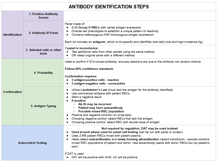
Outline the antibody identification steps
ANTIBODY IDENTIFICATION STEPS | ||
Identification | 1. Positive Antibody Screen |
|
2. Antibody ID Panel | Panel made of:
Each lot includes an antigram, which is lot-specific and identifies rare cells (low and high-incidence Ag) | |
3. Selected cells or other tools | If panel is inconclusive:
| |
Confirmation | 4. Probability | Used to confirm if ID’d correct antibody; ensures reactions are due to the antibody not random chance Follow 95% confidence standards Confirmation requires:
|
5. Antigen Typing |
| |
Autocontrol Testing | Not required by regulation; DAT may be used instead
If DAT is used
| |
Explain the importance of patient history when evaluating antibody screen and panel results.
History of Transfusion - important!
History of antibodies
Transplant
Pregnancy
Medications
Diagnosis
Ethnicity