3.2.10 CELLS DIVIDE BY BINARY FISSION AND MITOSIS
1/195
There's no tags or description
Looks like no tags are added yet.
Name | Mastery | Learn | Test | Matching | Spaced | Call with Kai |
|---|
No analytics yet
Send a link to your students to track their progress
196 Terms
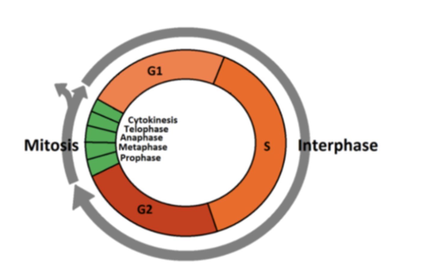
What are the steps of a cell cycle?
G1, S, G2, Mitosis, Cytokinesis.
What is mitosis?
Cell division that generates new cells for growth and repair.
- Parent cell divides = two genetically identical daughter cells, containing identical/exact copies of DNA of the parent cell.
- 1 division, 2 daughter cells
What are the stages of mitosis?
PMAT:
prophase, anaphase, metaphase, telophase, (cytokinesis)
TIP to remember PMAT: Paedophiles make adolescents terrified
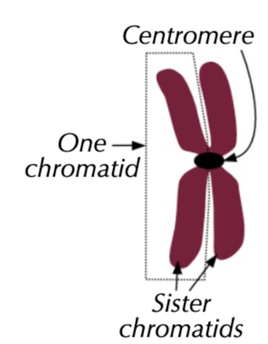
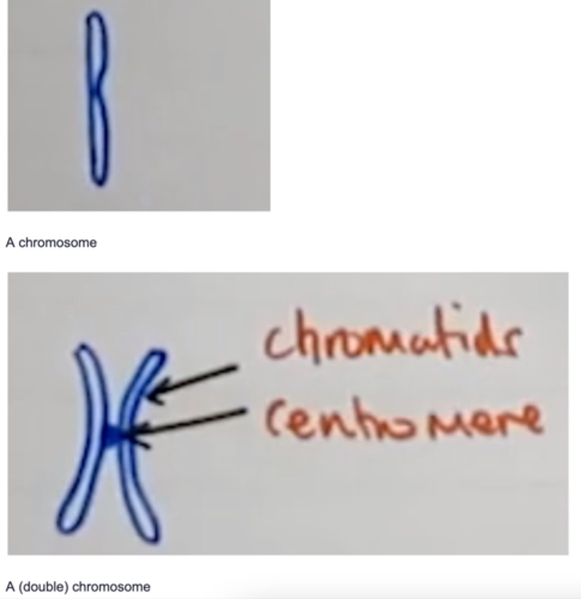
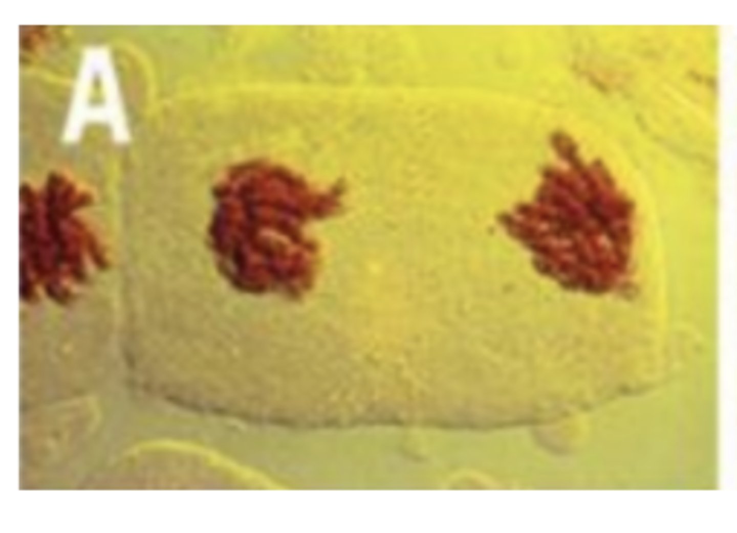
Draw the structure of a chromosome, labelling each part.
Chromosomes are made of two strands joined in the middle by a centromere. The separate strands are called chromatids. Two strands on the same chromosome are called sister chromatids. There are two strands because each chromosome has already made an identical copy of itself during interphase. When mitosis is over, the chromatids end up as one-strand chromosomes in the new daughter cells.
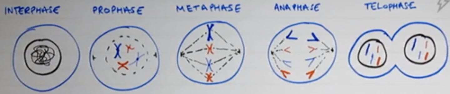
Draw out all the stages of mitosis.
Look at the image attached.
What is the S phase of the cell cycle?
Synthesis; DNA replicates semi-conservatively leading to two sister chromatids
What is the G1 phase of the cell cycle?
- Cell grows in size
- increases its protein content (protein synthesis)
- number of organelles such as mitochondria and ribosomes, and volume of cytoplasm increases.
What is the G2 phase of the cell cycle?
Cell keeps grows rapidly and proteins needed for cell division are synthesised, energy stores are increased (ATP)
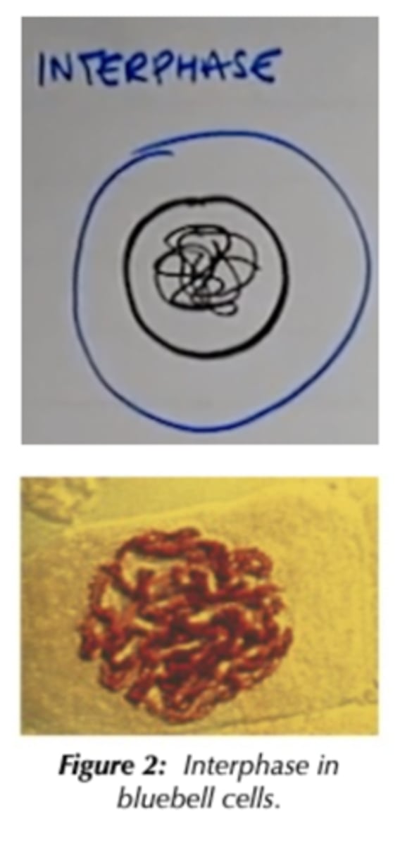
Describe the interphase stage of the cell cycle. Draw a diagram as well.
G1 = Growth; cell grows in size and increases its protein content and the number of organelles such as mitochondria and ribosomes
S = Synthesis; DNA replicates semi-conservatively leading to two sister chromatids
G2 = Growth; cell keeps grows rapidly and proteins needed for cell division are synthesised, energy stores are increased
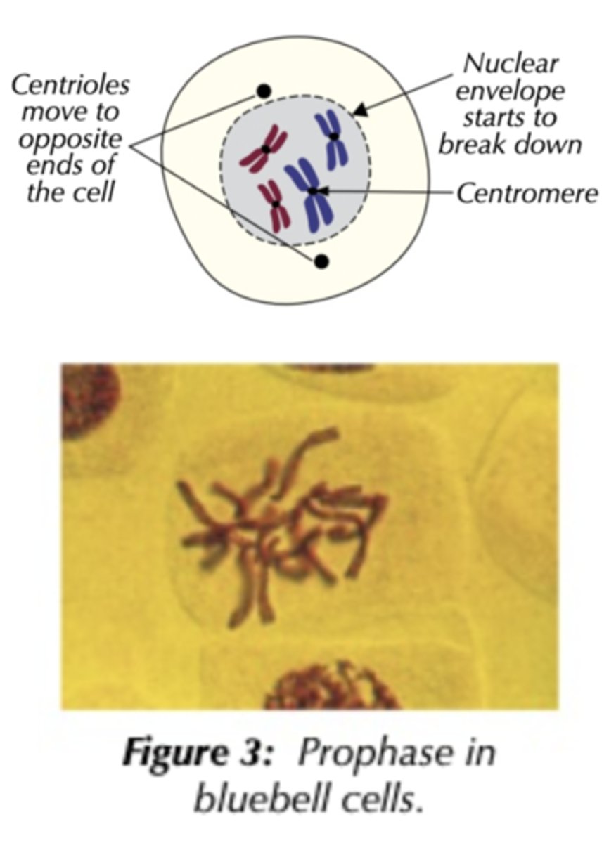
Describe the prophase stage of the cell cycle. Draw a diagram as well.
- Chromosomes condense & become visible, becoming shorter and thicker = appear as two sister chromatids joined by a centromere
- Nuclear envelope then breaks down (disappears)
- Nucleolus disappears
- Centrioles move to opposite poles forming spindle network
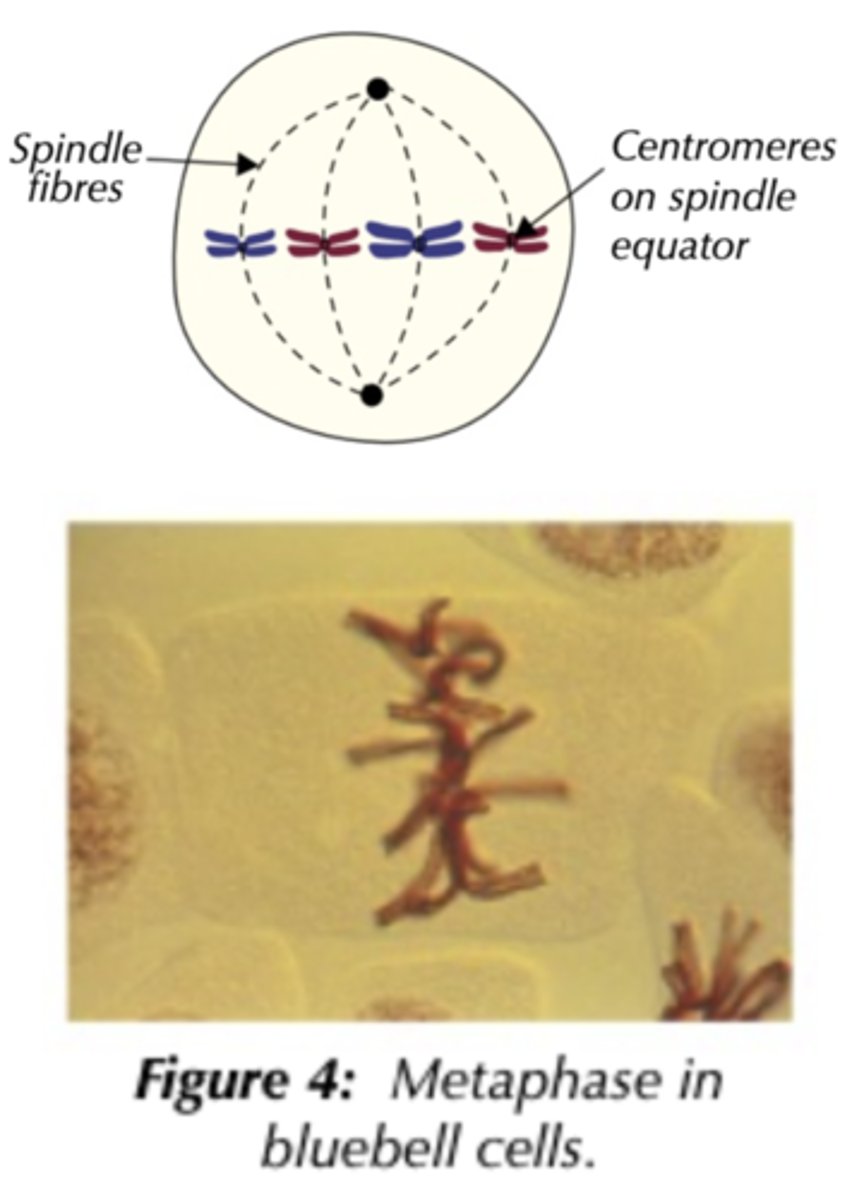
Describe the metaphase stage of the cell cycle. Draw a diagram as well.
- Spindle fibres attach to the centromeres and chromatid
- Chromosomes align along equator (centre of cell)
- Spindle forms
- No nuclear membrane
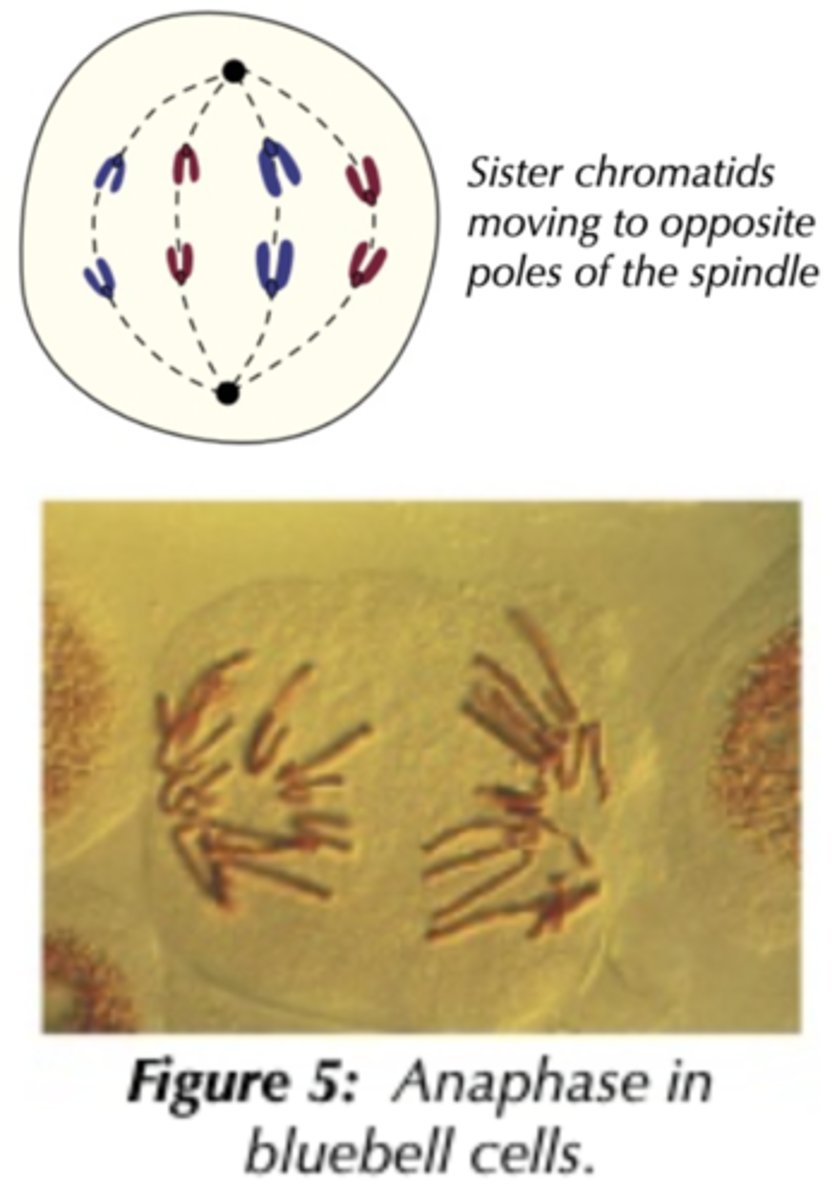
Describe the anaphase stage of the cell cycle. Draw a diagram as well.
- Spindle fibres contract, pulling the centromere and sister chromatids apart to opposite poles of the cell
- Centromere divides
- This stage requires ATP
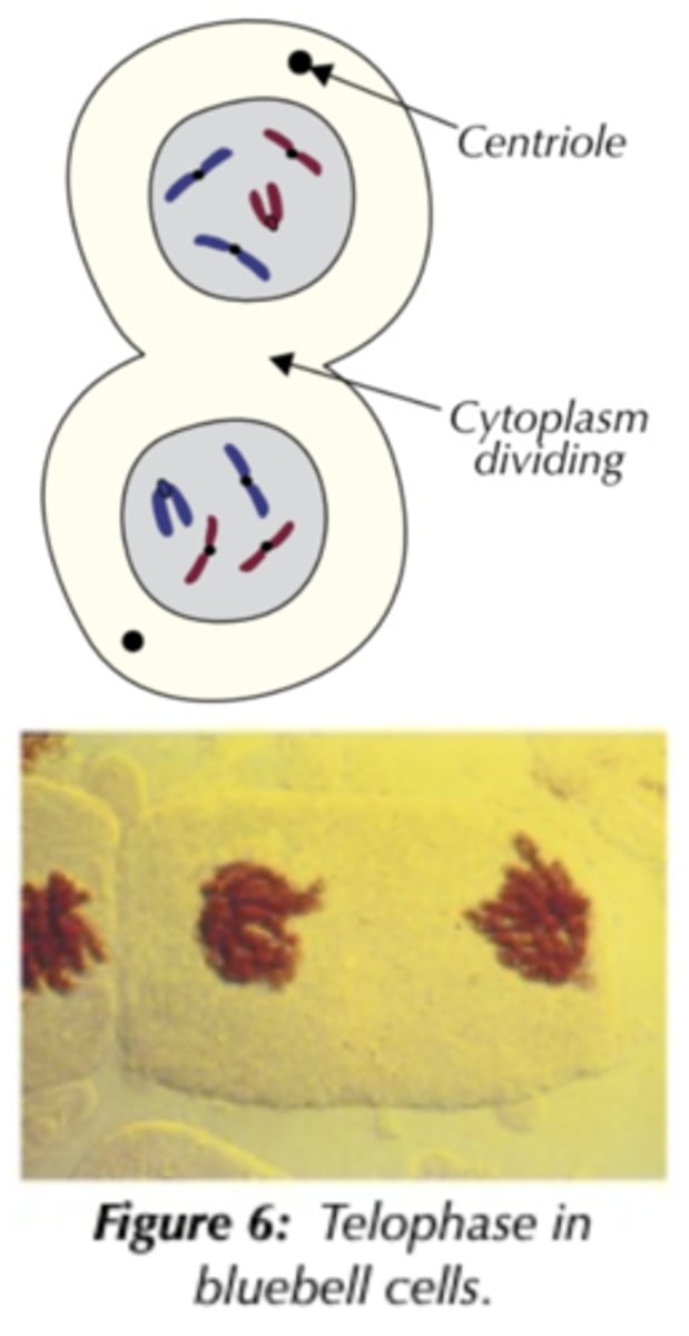
Describe the telophase stage of the cell cycle. Draw a diagram as well.
- Chromosomes uncoil, becoming longer and thinner
- Nuclear envelope reforms = two nuclei
- Nucleolus reforms
- Spindle fibres break down (disintegrate)
- Cytokinesis: The division of the cytoplasm, usually occurs, producing two new cells
What is cytokinesis?
The division of the cytoplasm, usually occurs, producing two new cells
TIP!
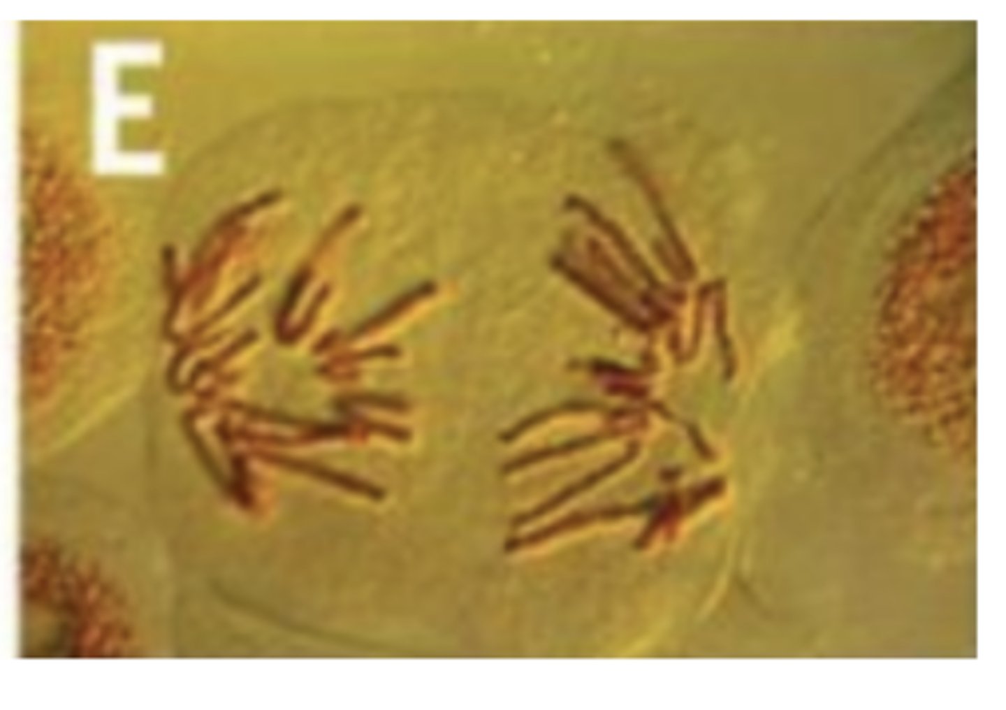
Look at image, both are called chromosomes, but the second (double) chromosome i only seen in mitosis before the sister chromatids are pulled apart.
Describe the importance of mitosis in the life of an organism.
Parent cell divides to produce 2 genetically identical daughter cells for...
- Growth of multicellular organisms by increasing cell number
- Repairing damaged tissues / replacing cells
- Asexual reproduction / cloning
- Genetically identical cells
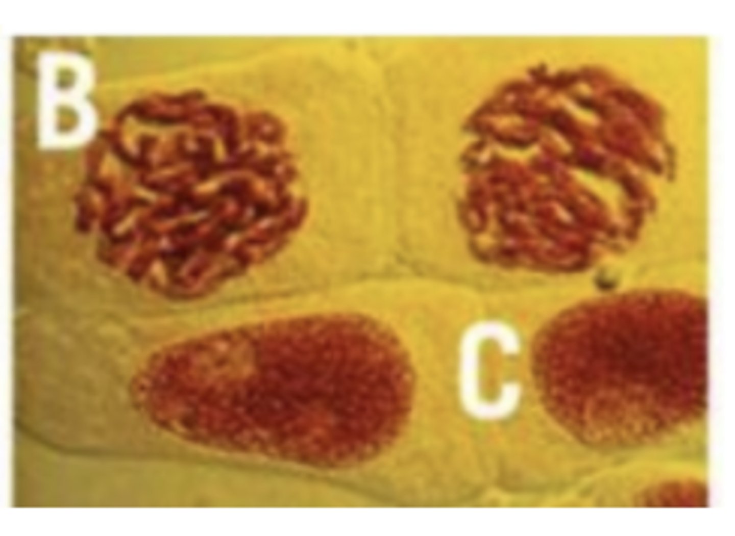
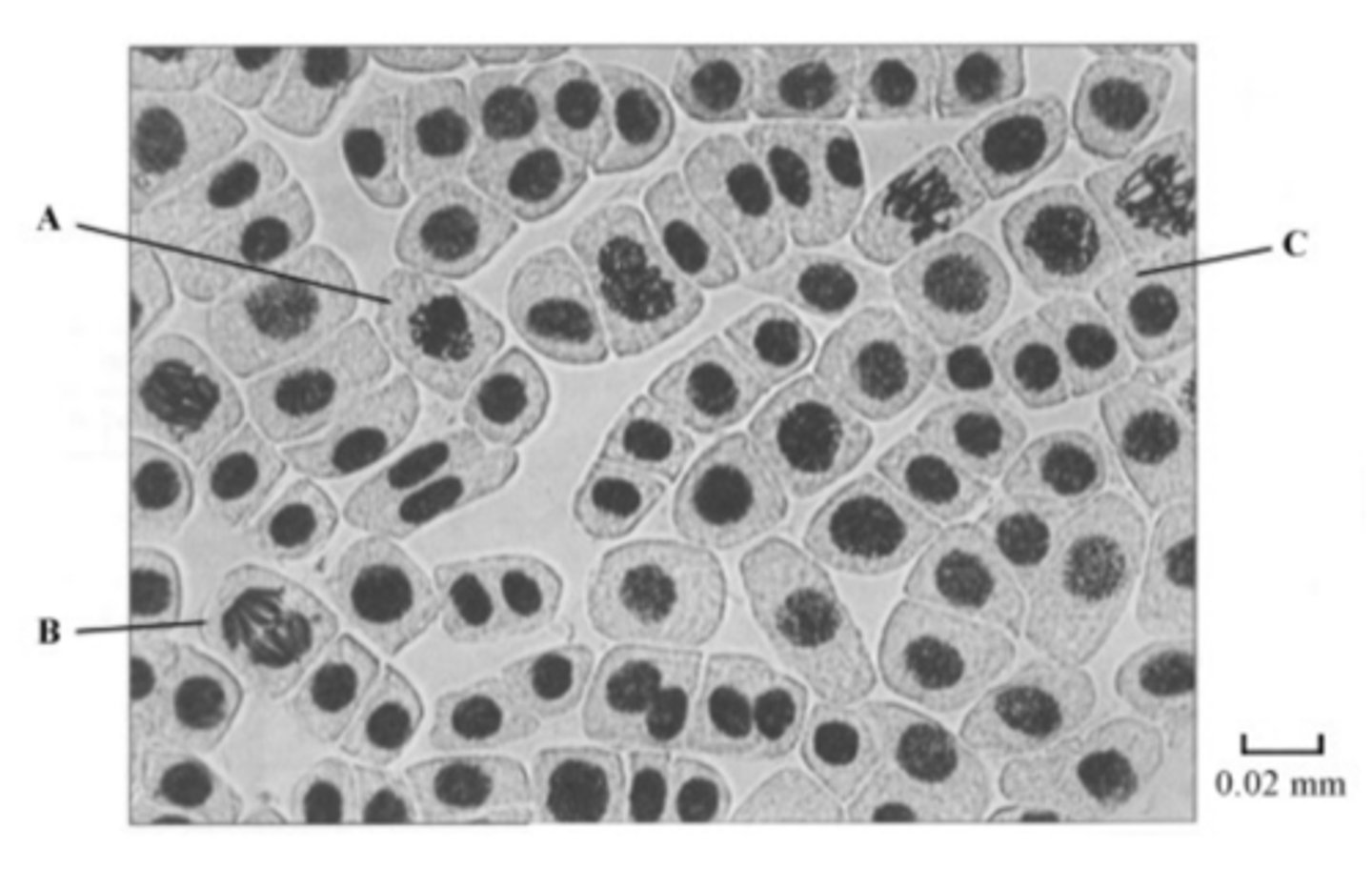
SKILL: What stage of mitosis is this cell in?
Telophase → chromosomes in two sets, one at each pole
SKILL: What stage of mitosis is this cell in?
- Interphase - C → no chromosomes visible (visible nucleus)
- Prophase - B → chromosomes visible but randomly arranged
SKILL: What stage of mitosis is this cell in?
Metaphase → chromosomes lined up on the equator
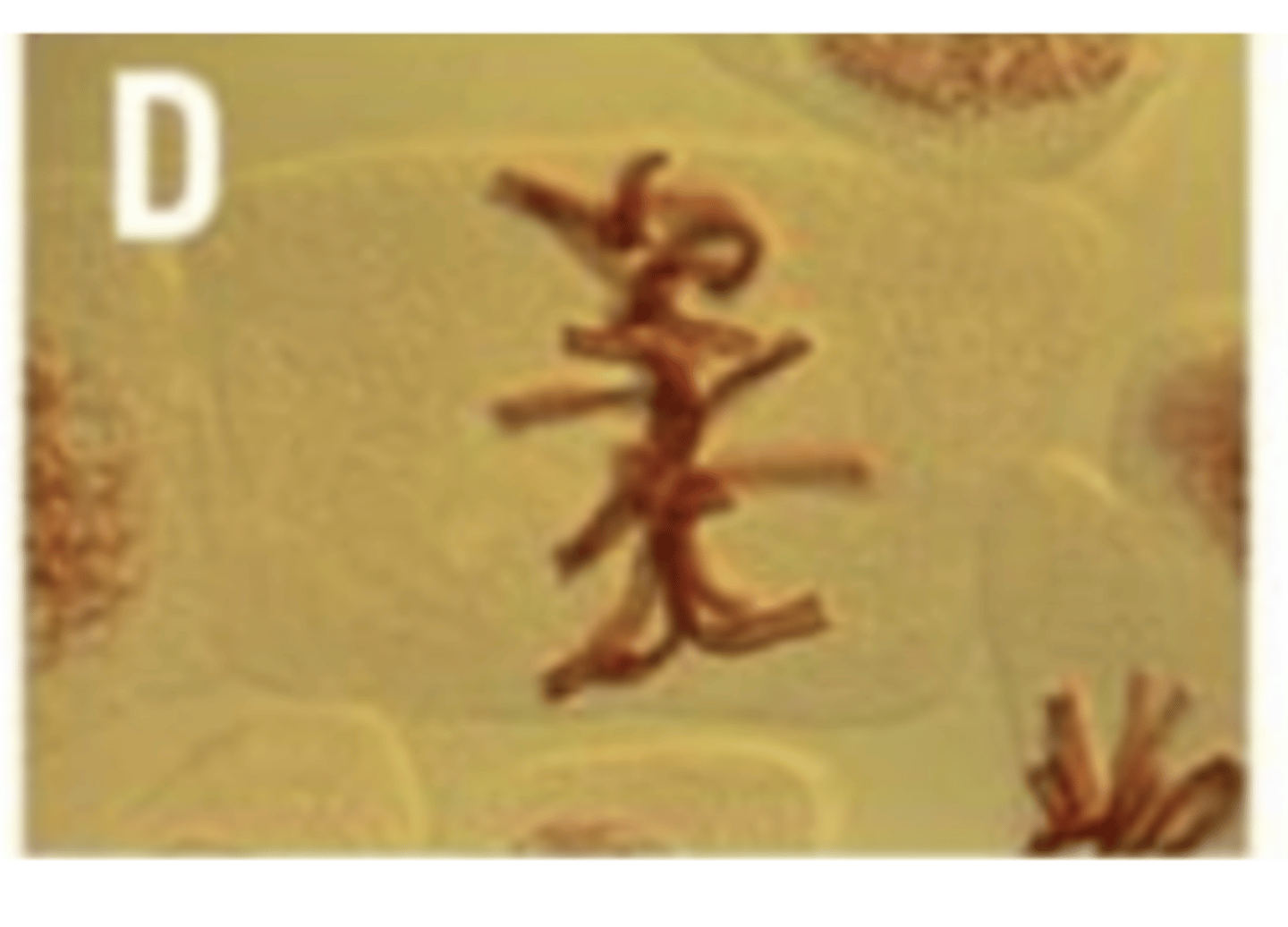
SKILL: What stage of mitosis is this cell in?
Anaphase → chromatids (in two sets) being separated to opposite poles by spindles, V shape shows sister chromatids have been pulled apart at their centromeres
By what method do prokaryotic cells replicate?
Binary fission.
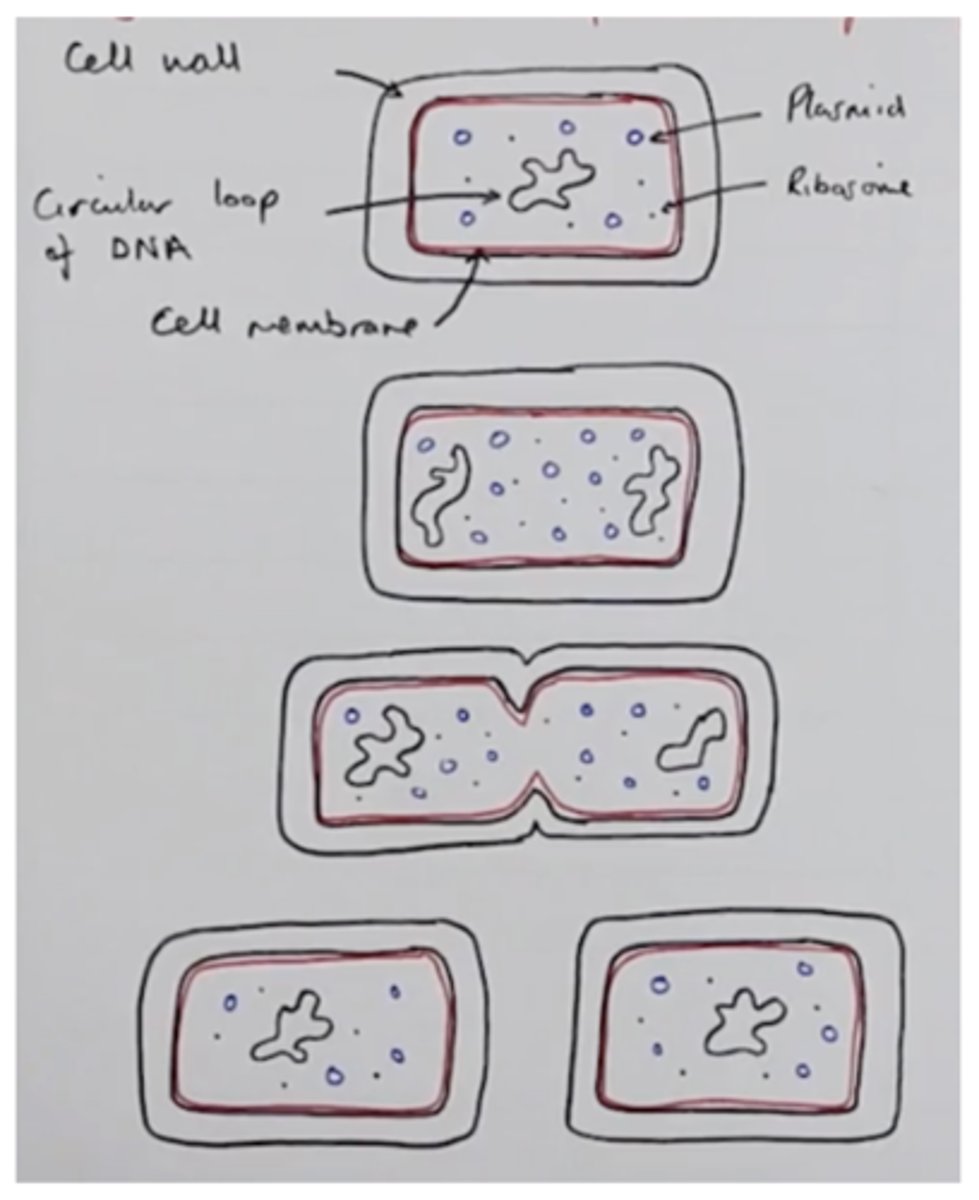
What is binary fission?
Asexual reproduction of single celled organisms eg bacteria
Describe and explain, with use of diagrams, the stages of binary fission.
- Bacteria divide by binary fission
- Circular loop of DNA replicates first → moves to opposite ends of cell (circular DNA replicates once)
- Plasmids replicate (plasmids can be replicated many times)
- Cytoplasm expands (cell gets bigger) as each DNA molecule moves to opposite poles of the cell
- Cytoplasm divides to form two daughter cells
- Each has a single copy of the circular DNA and a variable number of plasmids
What is conjugation?
DNA being passed from one species of bacterium to another. This is horizontal gene transmission.
What is horizontal gene transmission?
DNA in the form of genes can be passed from one species to another species in conjugation.
Describe the stages of conjugation in bacteria.
DNA can be transferred from one bacterial cell to another:
- One cell produces a thin projection that meets another cell and forms a thin conjugation between the two cells
- The donor cell replicates one of its small circular piece of DNA (plasmid)
- The circular DNA is broken to make it linear before it passes along the tube into the recipient cell
- Contact between the cells is brief, leaving only time for a portion of the donor's DNA to be transferred
- The recipient cell acquires new characteristics from the donor cell
What is vertical gene transmission?
When genes are passed down from one generation of a species to the next generation of the same species (during reproduction).
why don't viruses undergo cell division?
They are non-living.
Describe viral replication.
1) Attachment proteins bind to complementary receptor molecules on the cell-surface membrane of the host cells
2) Inject their RNA/DNA into the host cell
3) Genetic material and proteins are replicated by the host cells 'Enzymes'
4) Viral components are assembled and are released from the host cell
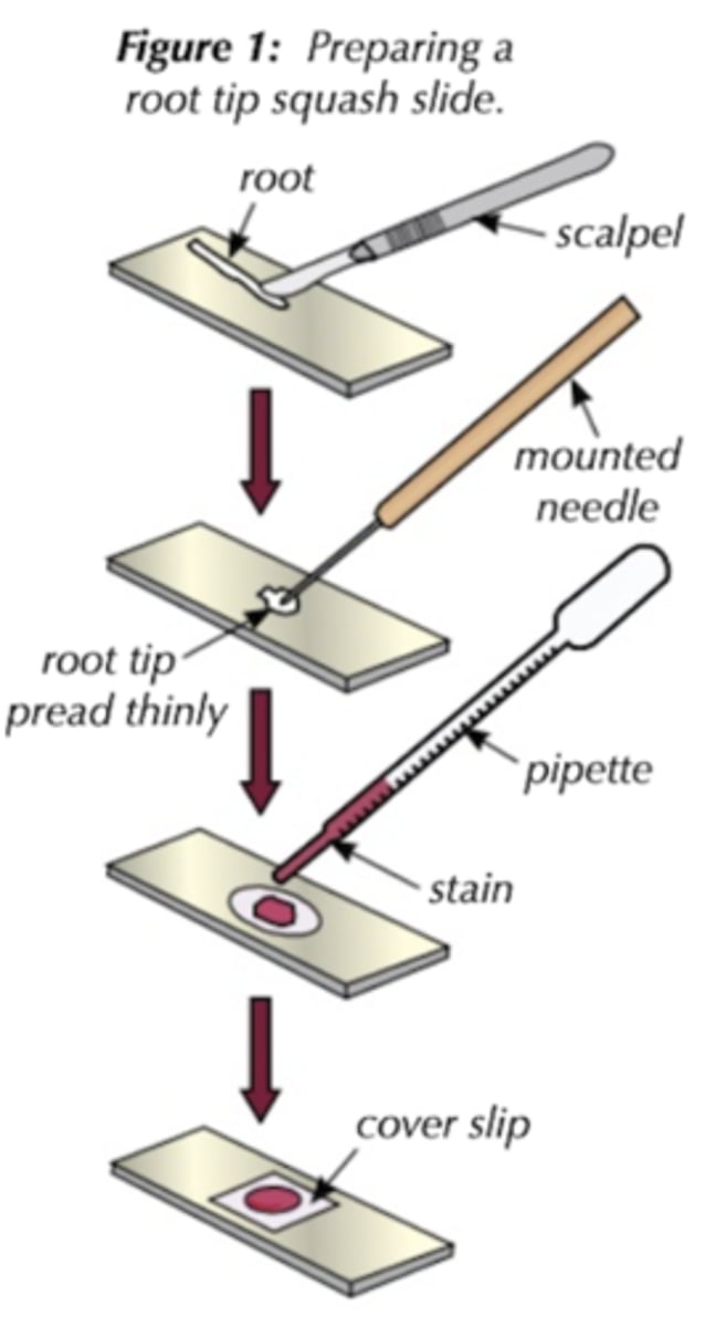
Required practical 4: Preparation of stained squashes of root tips and examination of these with a microscope. Observation of the stages of mitosis and calculation of a mitotic index. (Root tip squash): METHOD
Use a fixative eg acetic acid → prevent enzyme action to preserve cells
1. Add some 1M hydrochloric acid to the boiling tube. There should be just enough acid in the tube to cover the root tip. Put the tube in a water bath that has been allowed to reach 60 C.
2. Use a scalpel to cut 1cm from the tip of a growing root (e.g. an onion); needs to be the tip because that's where growth occurs and so that's where mitosis takes place
3. Carefully transfer the root tip into a boiling tube containing the acid. Incubate it for 5 minutes
4. Use tweezers to remove the root tip from the tube and use a pipette to rinse it with water. Leave the tip to dry on a paper towel
5. Place the root tip on a microscope slide and cut 2mm from the very tip of it. Get rid of the rest
6. Use a mounted needle to break the tip open and spread the cells out thinly
7. Add a few drop of stain to highlight the DNA (eg nile blue, aceto-orcein stain) and leave it for a few minutes. Stain will make chromosomes easier to see under a microscope.
8. Place a cover slip over the cells without air bubbles and put a piece of folded paper on top.
9. Push down firmly to squash the tissue + vertically → make it thinner and allow light to pass through
10. Don't smear coverslip sideways or you will damage the chromosomes
11. Now you can look at all the stages of mitosis under a optical microscope
Required practical 4: Preparation of stained squashes of root tips and examination of these with a microscope. Observation of the stages of mitosis and calculation of a mitotic index. (Root tip squash): COMMON PROBLEMS
- No fixative used → enzymes hydrolyse cells
- No stain used
- No cells dividing
- Multiple layers of cells → not squashed enough
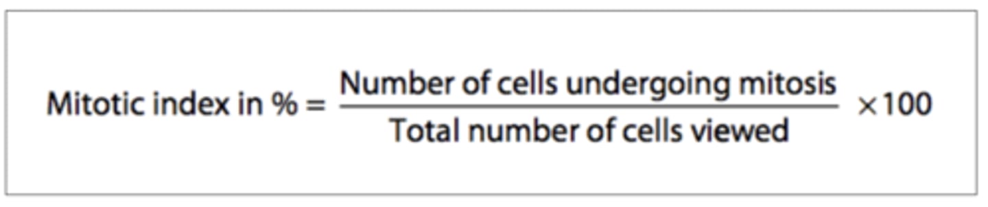
Define mitotic index.
The percentage of cell undergoing mitosis or the ratio between the number of cells undergoing mitosis to the number of cells in interphase.
How do you calculate mitotic index?
- Count the number of cells undergoing mitosis (Prophase, Metaphase, Anaphase & Telophase)
- Count the total number of cells in the field of view
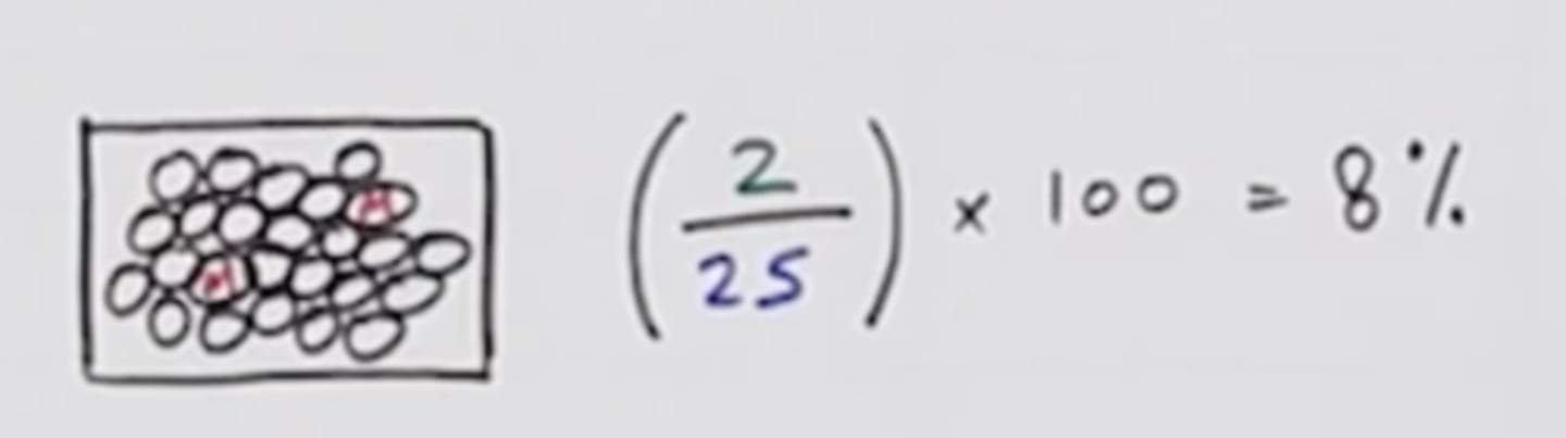
Calculate the mitotic index using the formula attached.
How does the mitotic index differ in different places in a plant?
- High in growing regions (meristem)
- E.g. root tip, shoot tip → growing
- Low away from growing region
- E.g. trunk
Mitotic Index: Example calculation 1.
Look at image attached.
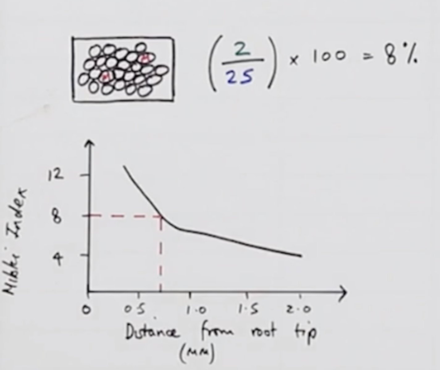
Mitotic Index: Example calculation 2.
- First calculate mitotic index
- Then use graph to calculate distance from root tip
How do you investigate the use of a herbicide with mitotic index?
- Use a range of at least 5 different herbicide concentrations
- Control all other variables; Eg species of plant same distance from root / shoot tip, Temperature
- Plot mitotic index on a graph
- Minimum concentration where the mitotic index = 0%
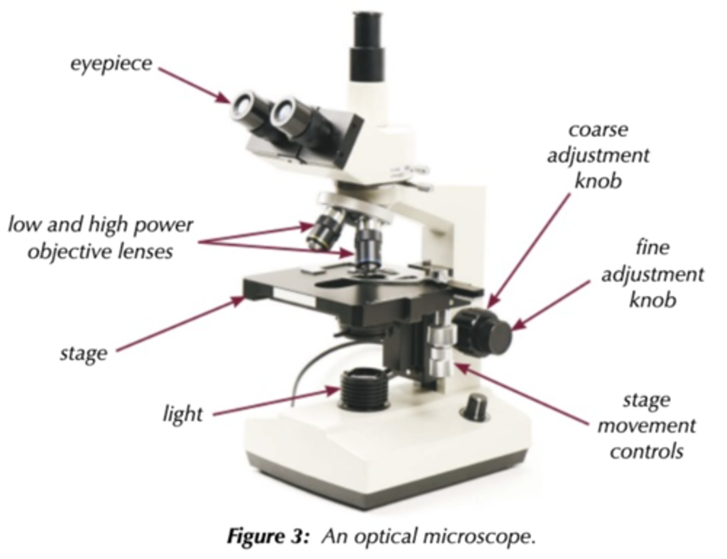
Describe how to use an optical microscope.
- Start with the lowest power (magnification) objective lens
- Put the slide onto the stage and hold it in place with stage clips
- Use the course focus to move the stage as close to the lens as possible (without touching)
- Look through the eyepiece and move focus so that the stage moves away from the lens and comes into focus
- Adjust fine focus (and light intensity) for clearest image
- Repeat with higher power if needed
Describe how to use an eye piece graticule & stage micrometer.
- Line up eyepiece graticule with stage micrometer
- Use stage micrometer to calculate the size of divisions on eyepiece graticule at a particular magnification
- Take the micrometer away and use the graticule to measure how many divisions make up the object
- Calculate the size of the object by multiplying the number of divisions by the size of division
- Recalibrate eyepiece graticule at different magnifications
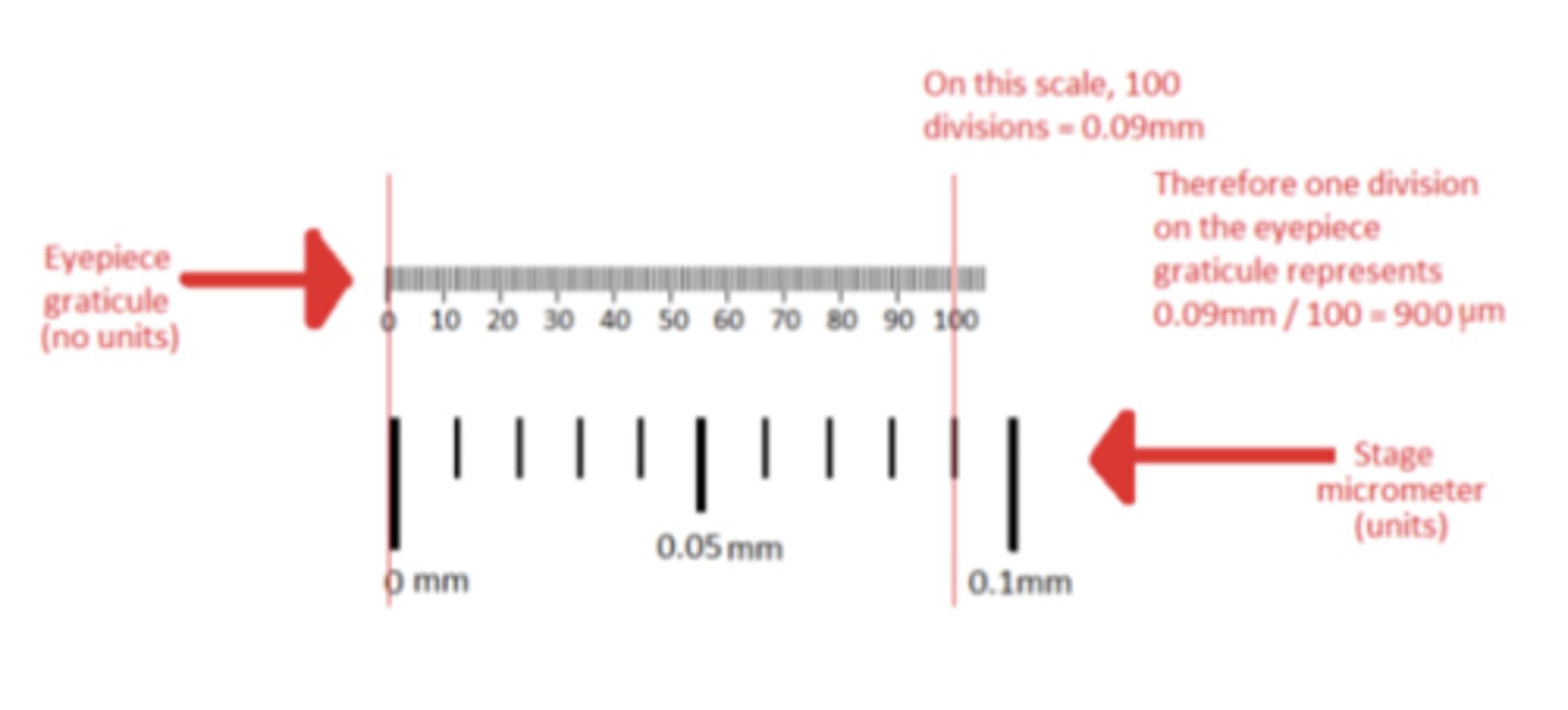
What is an eyepiece graticule?
-It is fitted onto the eyepiece.
-Like a transparent ruler with numbers, but no units. (varies with magnification)
What is a stage micrometer?
A microscope slide with a finely divided scale marked on the surface. (Tiny ruler embedded in the glass).
- Usually 1 micrometer markings on it.
Q: You could use an optical microscope and a slide of stained muscle tissue to find the diameter of one of the muscle fibres. Explain how. (2 MARKS)
- Measure with graticule/eyepiece scale
- Calibrate against something of known size: [ Last point could be a
calibrated slide/haemocytometer/red blood cell or reasonable
alternative]
Describe how you would prepare a 'temporary mount' of a specimen on a slide.
- Add few drops of water to blank microscope slide
- Use forceps / tweezers to place a thin section of specimen onto microscopic slide
- Must be thin so light can shine through
- Add a drop of a stain using a pipette e.g. iodine in potassium iodide solution used to stain starch grains in plant cells
- Add a cover slip by carefully tilting and lowering it using a mounted needle, trying not to get any air bubbles
- Now put slide on microscope stage and adjust magnification
Why is staining used when preparing a slide?
- Identify cellular components ge organelles
- Identify different cell types in a tissue
- We only see parts that absorb light / electrons
- Stains make parts of the cell absorb more light --> become more visible
- Use different stains for different cells / tissues
What stain is used for a nuclei?
Nile blue
What stain is used for the cytoplasm?
eosin
What stain is used for cell membranes?
Sudan Red
Q: In the production of a root tip squash to observe mitosis, suggest why the tip of the onion root is used. (1 MARK)
Site of cell division /mitosis /actively dividing cells /meristem
Q: Mitosis and meiosis are both forms of nuclear division. Mitosis can be observed in root tip squashes from a plant such as garlic. The stain used in a root tip squash can be intensified by…
Gently heating
Q: There are various risks associated with the production of a root tip squash. Suggest two risks and the precautions you would take to minimise each risk. (2 MARKS)
- cut and appropriate precaution e.g. replace blade covers when not in use/ use care when handling coverslips OR acid and appropriate precaution e.g. gloves/safety goggles OR heat and appropriate precaution OR stain and appropriate precaution e.g. gloves/safety goggles
- Award second mark for a second option from the answers above.
Q: A student prepared a root tip squash to observe the stages in mitosis. Describe how the student could distinguish between a cell in metaphase and a cell in anaphase. (3 MARKS)
- Metaphase - chromatids/chromosomes at equator /middle of cell. Anaphase - Not at equator/ separated/ pulled apart
- Metaphase - chromatids joined/attached to each other. Anaphase - chromatids separated /pulled apart
- Metaphase - centromere complete/intact. Anaphase -centromere splits OR Anaphase - fibres shorter / shortening / contracting
Q: Mitosis and meiosis are both forms of nuclear division. Mitosis can be observed in root tip squashes from a plant such as garlic. Mitosis occurs in…
Stem cells
Q: Describe the appearance of a cell in telophase of mitosis as seen in a root tip squash. (3 MARKS)
- idea that chromosomes will be in the process of decondensing /uncoiling/ becoming invisible
- idea that the nucleus / nuclear envelope(s) is visible OR idea that a nucleolus may be present
- idea that spindle has contracted / broken down / absent OR two separate nuclei/masses of chromatin now visible OR idea that there will be evidence of cell plate formation
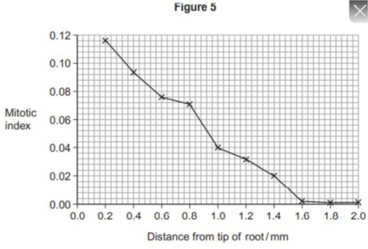
Q: A student cut thin sections of tissue at different distances from the tip of a root. She stained the sections and viewed them with an optical microscope. For each section, the student counted the number of cells in mitosis and the total number of cells in each field of view. She then calculated a mitotic index for each section using the equation:
Mitotic index = number of cells in mitosis/ total number of cells.
Figure 5 shows the student's results.
1. The student cut thin sections of tissue to view with an optical microscope. Explain why it was important that the sections were thin. (2 MARKS)
2. What does Figure 5 show about the growth of roots? Use the data to explain your answer. (3 MARKS)
1. - To allow (more) light through
- A single/few layer(s) of cells to be viewed
2. - More/faster mitosis/division near tip/at 0.2 mm
- (Almost) no mitosis/division at/ after 1.6 mm from tip
- (so) roots grow by mitosis/adding new cells to the tip
Q: Describe the role of the centromere in mitosis. (2 MARKS)
- Holds chromatids together
- Attaches (chromatids) to spindle/ (Allows) chromatids to be separated/move to (opposite) poles
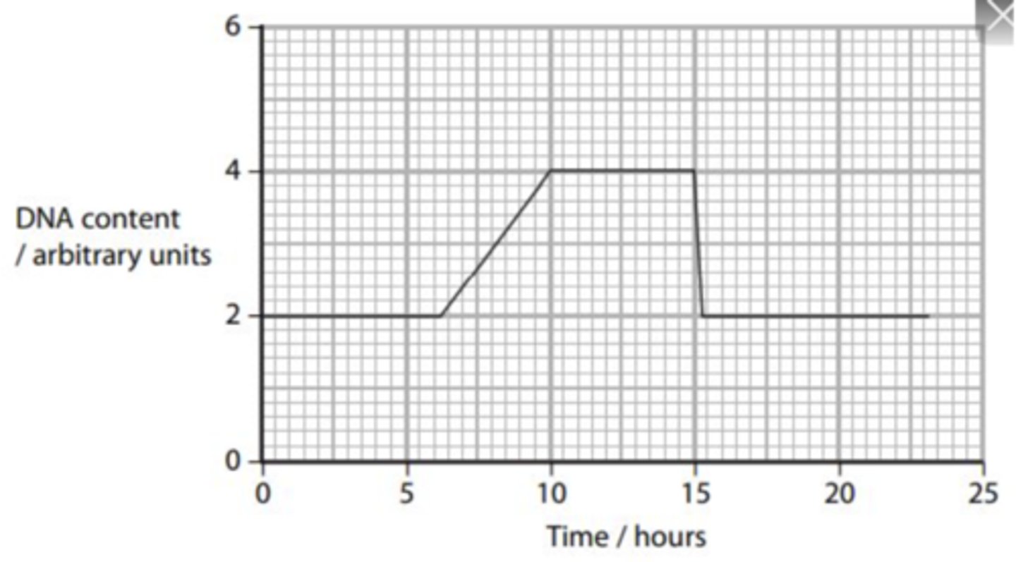
Q: The graph shows the changes in the DNA content of an onion cell, during one cell cycle.
1. Explain why the DNA content of the cell doubles. (2 MARKS)
2. Using the graph, state how long the S phase (DNA synthesis) takes. (1 MARK)
1. - DNA replication
- So that it can halve OR idea that new cells will have same amount as original /original (DNA) content restored
2. 3.5 to 3.75 hours
Q: Fig. 4.1 shows a cell during mitosis.
1. Name the stage of mitosis shown. (1 MARK)
2. From the start of mitosis, describe the events that have taken place in this cell to enable it to reach the stage shown in Fig. 4.1. (4 MARKS)
1. Anaphase
2. - Chromosomes coil/supercoil/condense OR nuclear envelope disintegrates OR nucleolus no longer visible/disappears
- Centrioles move to opposite ends of cell/poles OR chromosomes attached to spindle fibres at centromere
- Chromosomes align at equator
- Chromosomes move towards opposite poles /ends of cell OR spindle fibres change length/shorten
Q: How does the shape of DNA in bacteria differ to the double helix found in eukaryotes? (2 MARKS)
- Single stranded
- Circular/ loop
Q: In 1982, genetically engineered insulin, misleadingly named 'human' insulin, was the first genetically engineered drug to be marketed. Plasmids are used to introduce the gene into the bacterium. Plasmids have features that make them useful for this technique. One of these features is that the plasmid passes to both daughters in binary fission. Why is this important in genetic engineering? (2 MARKS)
- all offspring genetically modified
- all cells produce insulin / product
Q: A student investigated the stages of mitosis in a garlic root. The root tip was placed on a microscope slide with a stain. A cover slip was placed on top and the root tip was firmly squashed.
Explain why:
1. a root tip was used;
2. a stain was used;
3. the root tip was firmly squashed.
(3 MARKS)
1. where mitosis / division / growing / occurs (reject growing cells)
2. to distinguish chromosomes / chromosomes not visible without stain;
3. Create thin layer of cells to let light through sample and give clear image
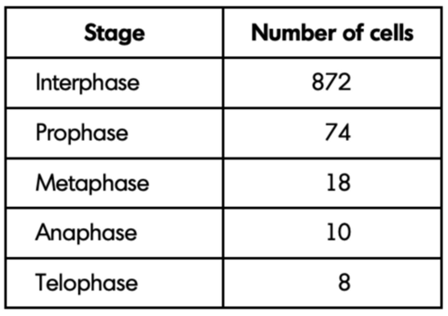
Q: The student examined the cells in the garlic root tip under the microscope, and obtained the following data.
Calculate the percentage of these cells in which the chromosomes are visible and would consist of a pair of chromatids joined together. Show your working. (2 MARKS)
- 74 + 18 / 982;
- = 9.4% / 9%;
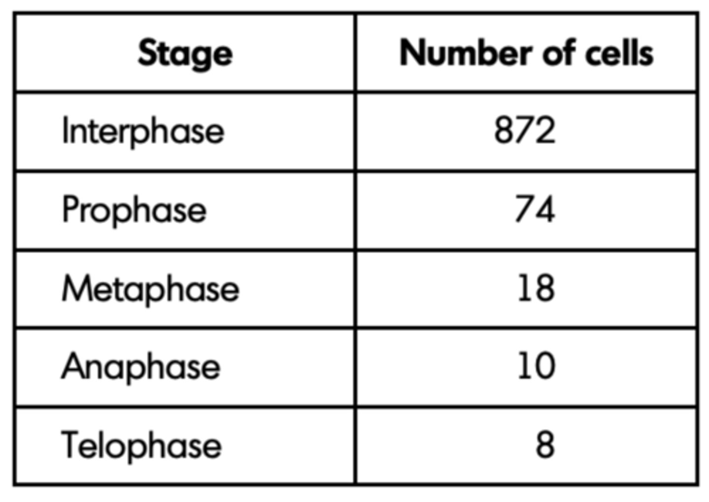
Q: A student examined the cells in the garlic root tip under the microscope, and obtained the following data.
A different set of results was obtained when the count was repeated on another occasion with a different garlic root tip. Give two reasons for the difference in results. (2 MARKS)
- genetic differences / different types of garlic;
- time of day;
- chance;
- age of root tip;
- water availability;
- temperature;
- nutrient availability;
Q: Cells lining the human intestine complete the cell cycle in a short time. Explain the advantage of these cells completing the cell cycle in a short time. (1 MARK)
Form / replace cells quickly / rapidly / divide / multiply / replicate rapidly;
Q: The time required for a cell to complete the cell cycle was 4 hours 18 minutes. Calculate the time required in minutes for this cell to multiply to produce eight cells. Show your working. (2 MARKS)
Correct answer = 774 minutes / 12 hours 54mins = 2 marks;;
Q: A biologist used a microscope to investigate plant tissue where some of the cells were dividing by mitosis. She examined 200 cells and counted the number of cells in interphase and in each stage of mitosis.
The table shows some of the cells she saw, and the percentage of cells in interphase and in two stages of mitosis, A and B.
Explain why the biologist chose to examine 200 cells. (1 MARK)
(Ensures) representative (sample);
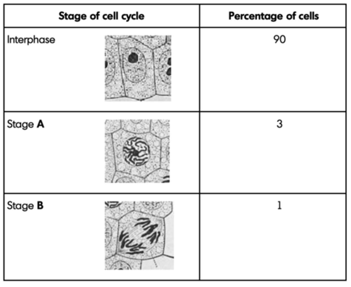
Q: A biologist used a microscope to investigate plant tissue where some of the cells were dividing by mitosis. She examined 200 cells and counted the number of cells in interphase and in each stage of mitosis.
The table shows some of the cells she saw, and the percentage of cells in interphase and in two stages of mitosis, A and B.
Name Stage A and Stage B. Give the evidence from the photograph that you used to identify the stage. (4 MARKS)
1. A = metaphase;
2. Chromosome / chromatids lie on equator; Reject homologous chromosomes Allow centre / middle
3. B = anaphase;
4. Chromatids / chromosomes separating / moving apart / moving to poles;
Q: A biologist used a microscope to investigate plant tissue where some of the cells were dividing by mitosis. She examined 200 cells and counted the number of cells in interphase and in each stage of mitosis.
The table shows some of the cells she saw, and the percentage of cells in interphase and in two stages of mitosis, A and B.
In this tissue one complete cell cycle took 20 hours. Using information from the table, calculate the mean time for these cells to complete mitosis. Show your working. (2 MARKS)
2 hours / 120 minutes;

Q: The photograph shows cells from an onion root tip. The root tip has been squashed and stained to show the stages of mitosis.
At what stage of mitosis is cell A? (1 MARK)
Prophase;
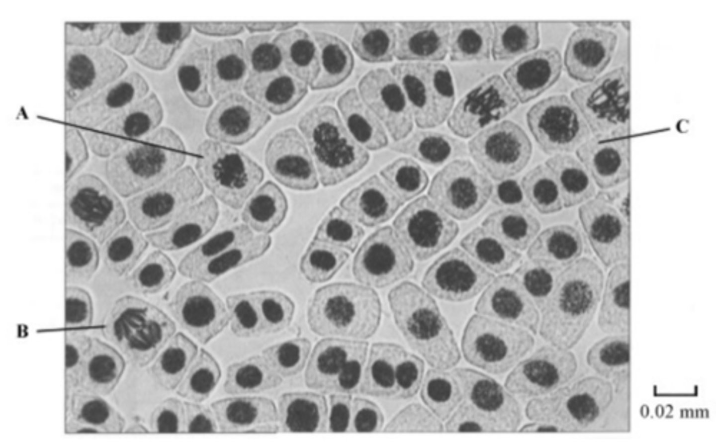
Q: The photograph shows cells from an onion root tip. The root tip has been squashed and stained to show the stages of mitosis.
What is the evidence that cell B is in anaphase? (1 MARK)
Chromosomes / chromatids moved apart;
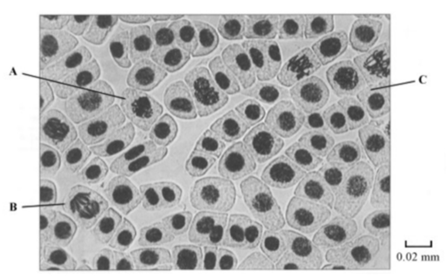
Q: The photograph shows cells from an onion root tip. The root tip has been squashed and stained to show the stages of mitosis.
Cell C is in interphase. Give two processes which occur during interphase that enable cell division to occur. (2 MARKS)
- A wide range of processes occurs during interphase. This list is by no means exhaustive, but we would expect to see answer such as:
- Increase in volume of cell / volume of cytoplasm / increase in mass / cell bigger;
- Increase in number of organelles;
- Synthesis of protein / named protein;
- DNA replication / increase / chromosomes copied;
- ATP synthesis / respiration;
Q: The photograph shows cells from an onion root tip. The root tip has been squashed and stained to show the stages of mitosis.
Explain how you would calculate the magnification of the photograph. (1 MARK)
Divide real length of bar (in mm) / 10 by 0.02
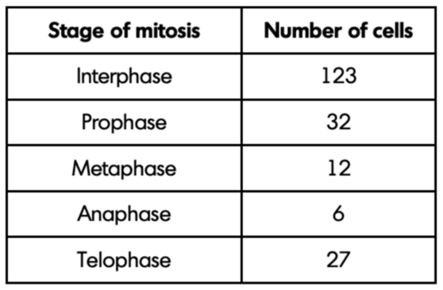
Q: The number of cells at each stage of mitosis was counted. The results are shown in the table.
One complete cell cycle takes 24 hours. The number of cells at each stage is proportional to the time spent at that stage. Calculate the length of time spent in metaphase. Show your working. (2 MARKS)
- 12 / 200 × 24 / single error in otherwise correct method;
- 1.44 hours (1 hour 26 min);
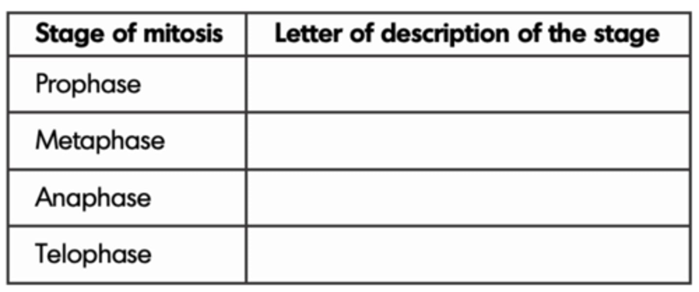
Q: The following statements describe stages of mitosis.
A chromosomes align at the centre of the cell attached to spindle fibres
B chromatids are in groups at the poles
C chromosomes become visible
D chromatids move towards the poles
Complete the table by entering the appropriate letter
(3 MARKS)
Sequence: C,A,D,B;
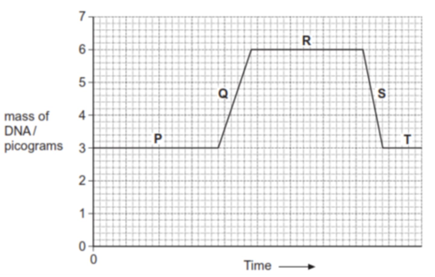
Q: The graph shows changes in the mass of DNA in a cell during one cell cycle. Five stages have been identified on the graph.
1. Which letter represents the stage when DNA is replicating? (1 MARK)
2. Explain the change in the DNA content during stage S. (1 MARK)
1. Q;
2. Cell/nucleus has divided / is dividing (into two);
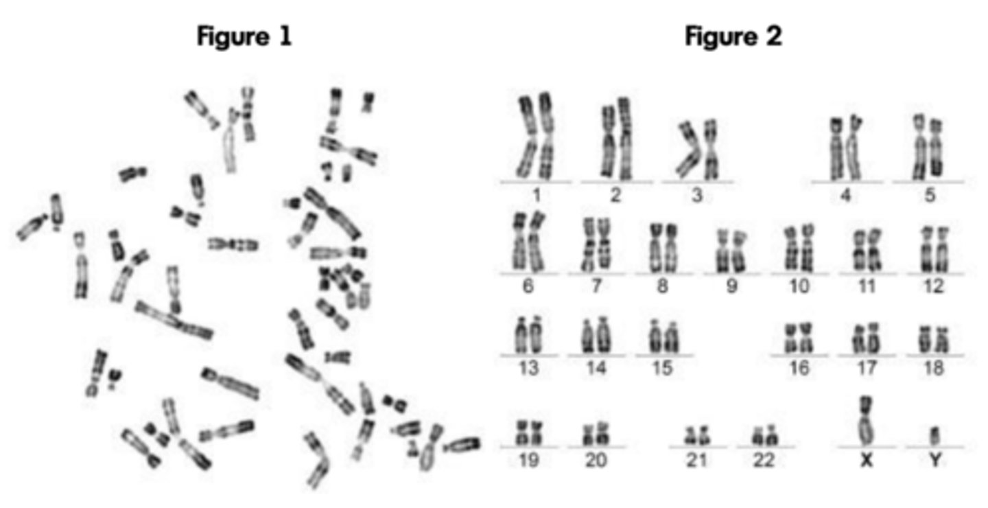
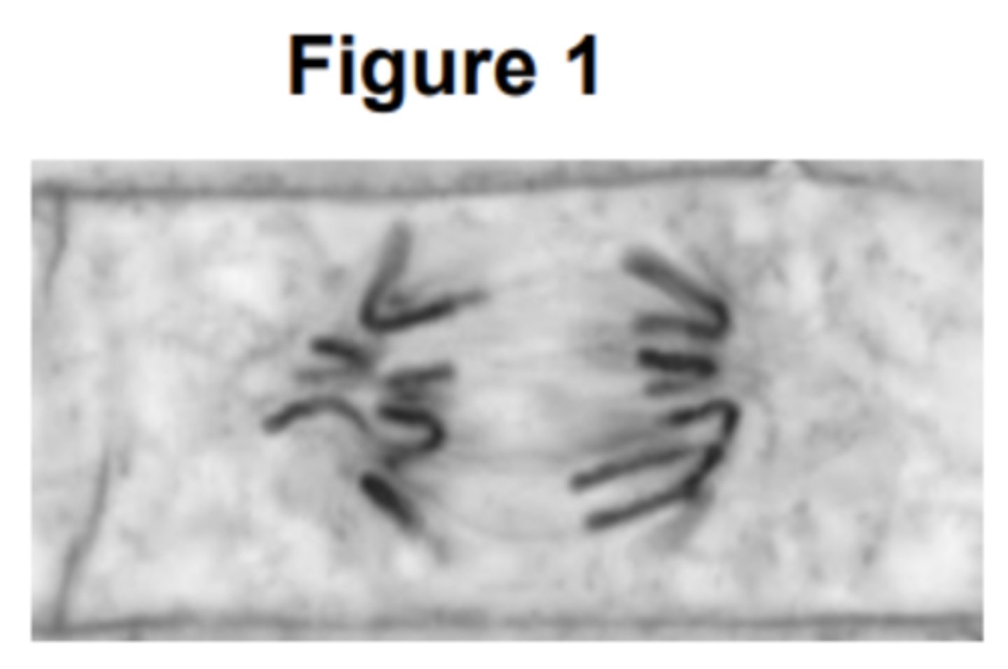
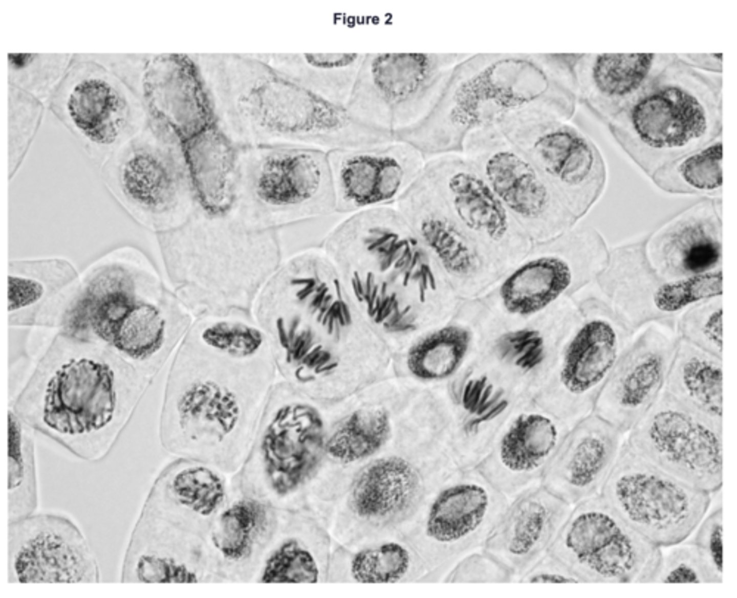
Q: Figure 1 shows all the chromosomes present in one human cell during mitosis. A scientist stained and photographed the chromosomes. In Figure 2, the scientist has arranged the images of these chromosomes in homologous pairs.
Give two pieces of evidence from Figure 1 that this cell was undergoing mitosis. Explain your answers. (2 MARKS)
1. The (individual) chromosomes are visible because they have condensed;
2. (Each) chromosome is made up of two chromatids because DNA has replicated;
3. The chromosomes are not arranged in homologous pairs, which they would be if it was meiosis;

Q: Figure 1 shows all the chromosomes present in one human cell during mitosis. A scientist stained and photographed the chromosomes. In Figure 2, the scientist has arranged the images of these chromosomes in homologous pairs.
Name the stage of mitosis shown in Figure 1. (1 MARK)
Prophase
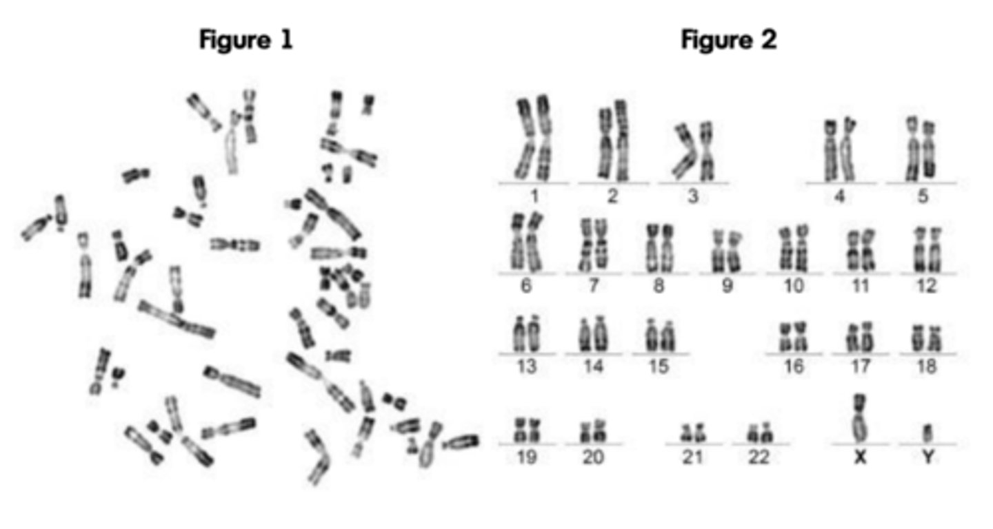
Q: When preparing the cells for observation the scientist placed them in a solution that had a slightly higher (less negative) water potential than the cytoplasm. This did not cause the cells to burst but moved the chromosomes further apart in order to reduce the overlapping of the chromosomes when observed with an optical microscope.
Suggest how this procedure moved the chromosomes apart. (2 MARKS)
1. Water moves into the cells/cytoplasm by osmosis;.
2. Cell/cytoplasm gets bigger;
Q: The dark stain used on the chromosomes binds more to some areas of the chromosomes than others, giving the chromosomes a striped appearance. Suggest one way the structure of the chromosome could differ along its length to result in the stain binding more in some areas. (1 MARK)
- Differences in base sequences
- OR Differences in histones/interaction with histones
- OR Differences in condensation/(super)coiling;
Q: Figure 1 shows all the chromosomes present in one human cell during mitosis. A scientist stained and photographed the chromosomes. In Figure 2, the scientist has arranged the images of these chromosomes in homologous pairs.
In Figure 2 the chromosomes are arranged in homologous pairs. What is a homologous pair of chromosomes? (1 MARK)
(Two chromosomes that) carry the same genes;
Q: Figure 1 shows all the chromosomes present in one human cell during mitosis. A scientist stained and photographed the chromosomes. In Figure 2, the scientist has arranged the images of these chromosomes in homologous pairs.
Give two ways in which the arrangement of prokaryotic DNA is different from the arrangement of the human DNA in Figure 1. (2 MARKS)
1. Circular (as opposed to linear);
2. Not associated with proteins/histones ;
3. Only one molecule/piece of DNA OR present as plasmids;
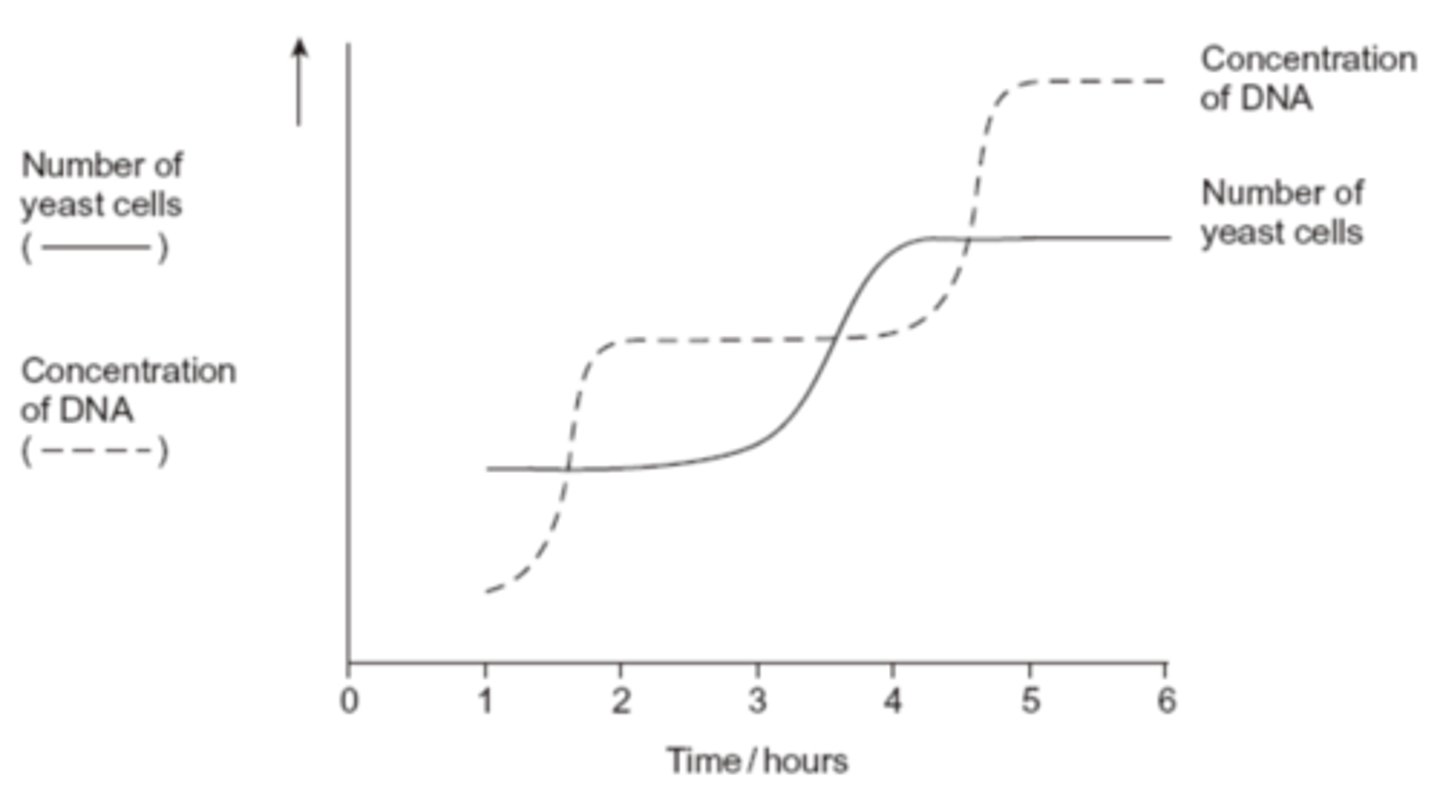
Q: Yeast is a single-celled eukaryotic organism. When yeast cells are grown, each cell forms a bud. This bud grows into a new cell. This allows yeast to multiply because the parent cell is still alive and the new cell has been formed.
Scientists grew yeast cells in a culture. They counted the number of cells present and measured the total concentration of DNA in the culture over a period of 6 hours. Their results are shown in the graph.
Use your knowledge of the cell cycle to explain the shape of the curve for the number of yeast cells
1. between 1 and 2 hours. (1 MARK)
2. between 3 and 4 hours. (1 MARK)
- Cells are in interphase;
- Cells undergoing mitosis / in telophase / cytokinesis;
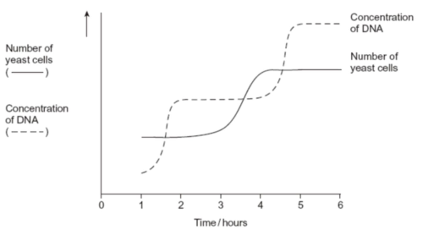
Q: Yeast is a single-celled eukaryotic organism. When yeast cells are grown, each cell forms a bud. This bud grows into a new cell. This allows yeast to multiply because the parent cell is still alive and the new cell has been formed.
Scientists grew yeast cells in a culture. They counted the number of cells present and measured the total concentration of DNA in the culture over a period of 6 hours. Their results are shown in the graph.
Use the curve for the concentration of DNA to find the length of a cell cycle in these yeast cells. Explain how you arrived at your answer. (3 MARKS)
1. 3 hours;
2. Time between beginnings / endings DNA replication / Increases / levelling outs of DNA concentration / for shape (of curve for replication) to be repeated;
3. (DNA) replication takes place once per cell cycle;
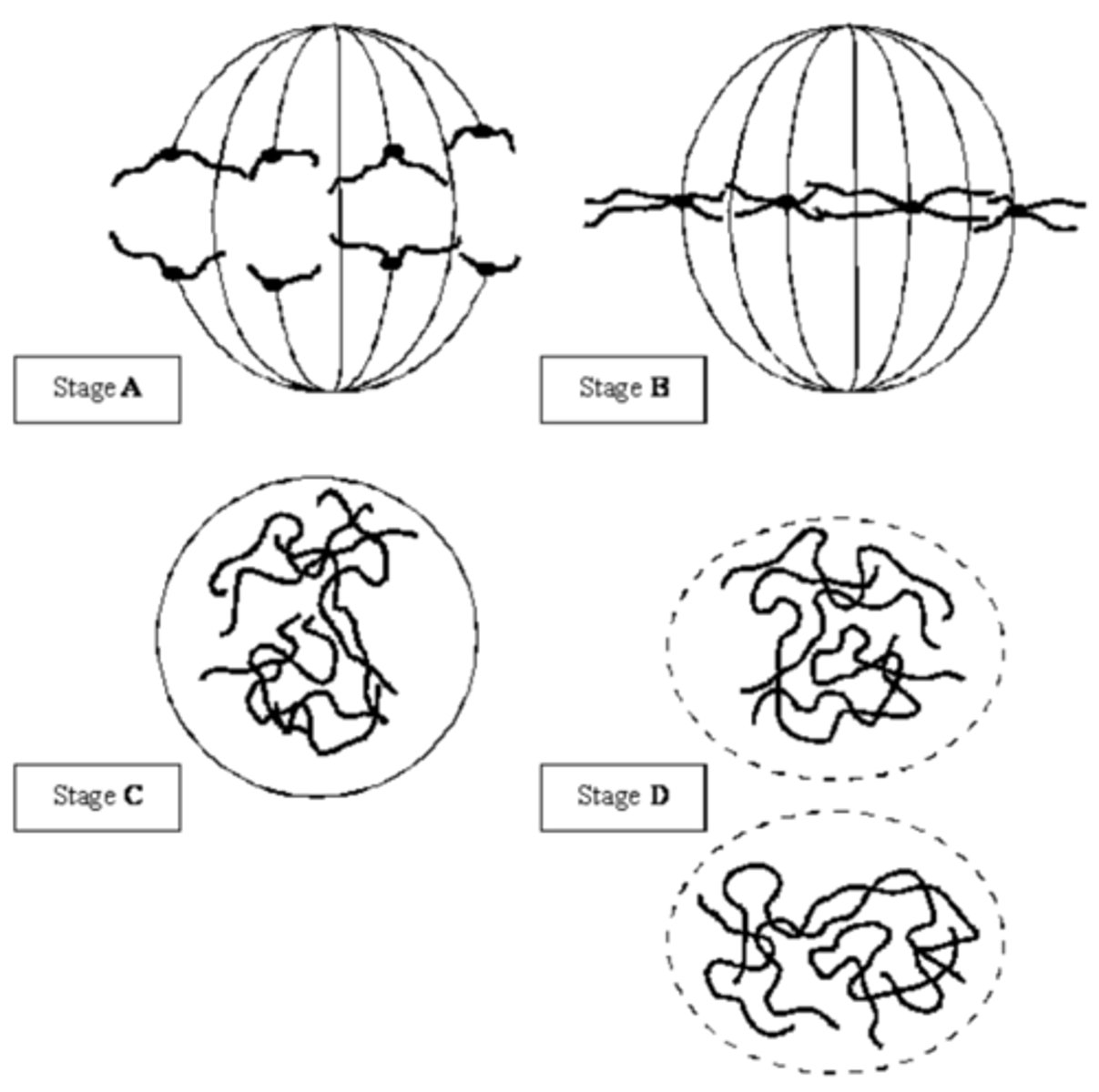
Q: The diagrams show four stages of mitosis.
Describe and explain the appearance of one of the chromosomes in stage B. (2 MARKS)
- (original) chromosome / DNA has been replicated;
- each chromosome consists of two chromatids / chromatids attached at centromere;
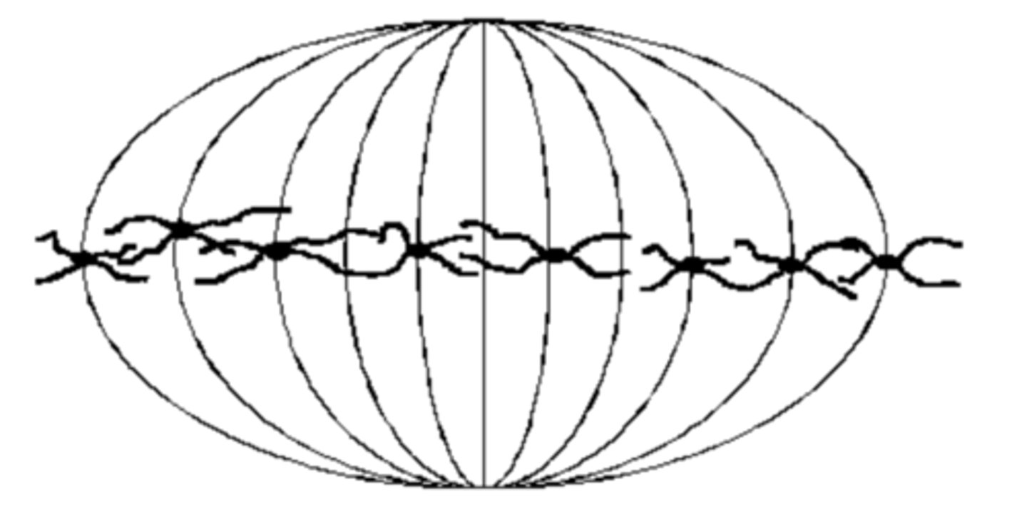
Q: Colchicine is a substance that prevents the formation of the spindle in mitosis. Dividing cells were treated with colchicine. This stopped them dividing. After a few hours, the colchicine was removed and the cells began to divide again. The diagram shows the chromosomes from one of the treated cells at stage B after the cell began dividing again.
1. What has happened to the chromosome number? (1 MARK)
2. Suggest an explanation for the change in the chromosome number. (1 MARK)
1. it has doubled / now 8;
2. chromosome / DNA replication but no separation / anaphase / cell division;
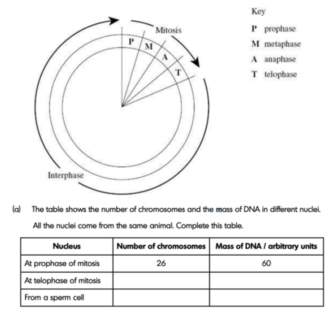
Q: The diagram shows a cell cycle.
The table shows the number of chromosomes and the mass of DNA in different nuclei. All the nuclei come from the same animal. Complete this table.
The table shows the number of chromosomes and the mass of DNA in different nuclei. All the nuclei come from the same animal. Complete this table.
(4 MARKS)
- Left to right;
- 26, 30
- 13, 15
Q: Describe the behaviour of chromosomes during mitosis in eukaryotes and explain how this results in the production of two genetically identical cells. (7 MARKS)
1. chromosomes shorten / thicken / supercoiling;
2. chromosomes (each) two identical chromatids / strands / copies (due to replication);
3. chromosomes / chromatids move to equator / middle of the spindle / cell;
4. attach to individual spindle fibres;
5. spindle fibres contract / centromeres divide / repel;
6. (sister) chromatids / chromosomes (separate) move to opposite poles / ends of the spindle;
7. each pole / end receives all genetic information / identical copies of each chromosome;
8. nuclear envelope forms around each group of chromosomes / chromatids / at each pole;
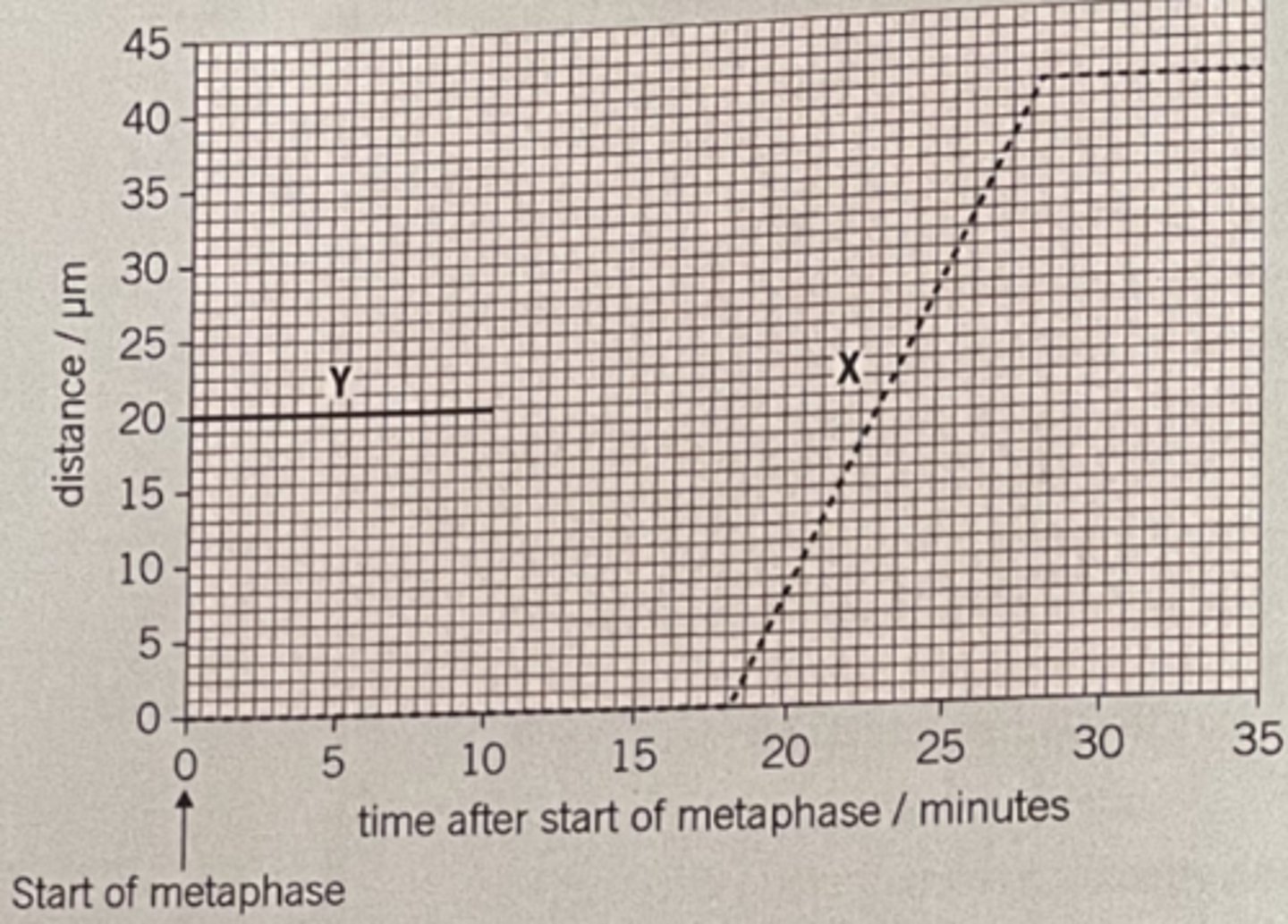
Q: The graph shows information about the movement of chromatids in a cell that has just started metaphase of mitosis
(i) What was the duration of metaphase in this cell in minutes (1 MARK)
(ii) Use line X to calculate the duration of anaphase in this cell in minutes (1 MARK)
(iii) Complete the line Y on the graph. (2 MARKS)
(i) 18
(ii) 10
(iii) 1. Horizontal until 18 minutes;
2. (Then) decreases as straight line to 0 μm at 28 minutes;
EQ: A student investigated mitosis in the tissue from an onion root tip. The student prepared a temporary mount of the onion tissue on a glass slide. She covered the tissue with a cover slip. She was then given the following instruction. “Push down hard on the cover slip, but do not push the cover slip sideways.” Explain why she was given this instruction. (2 MARKS)
1. Push hard - spread/squash tissue;
2. Not push sideways - avoid rolling cells together/breaking chromosomes;
EQ: Figure 1 shows one cell the student saw in the onion tissue.
The student concluded that the cell in Figure 1 was in the anaphase stage of mitosis. Was she correct? Give two reasons for your answer. (2 MARKS)
No (no mark)
Yes (no mark)
1. Chromosomes/chromatids are (in two groups) at poles of spindle/at ends of spindle;
2. V-shape shows that (sister) chromatids have been pulled apart at their centromeres/that centromeres of (sister) chromatids have been pulled apart;
EQ: The student counted the number of cells she observed in each stage of mitosis. Of the 200 cells she counted, only six were in anaphase. One cell cycle of onion root tissue takes 16 hours. Calculate how many minutes these cells spend in anaphase. Show your working. (2 MARKS)
(6 / 200) X (16 x 60) = 28.8 minutes / 29
EQ: A student prepared a stained squash of cells from the tip of an onion root and box observed it using an optical microscope.
During the preparation of the slide, he:
• cut the first 5 mm from the tip of an onion root and placed it on a glass slide
• covered this tip with a drop of stain solution and a cover slip
• warmed the glass slide
• pressed down firmly on the cover slip.
He identified and counted nuclei in different stages of the cell cycle. Explain why the student:
1. used only the first 5 mm from the tip of an onion root.
2. pressed down firmly on the cover slip.
(2 MARKS)
1. Where dividing cells are found / mitosis occurs;
OR No dividing cells / mitosis in tissue further away / more than 5mm from tip;
OR To get (soft) tissue that will squash;
OR Length that will fit under cover slip;
2. Single / thin layer of cells / spread out cells so light passes through (making cells / nuclei visible);
EQ: Figure 2 shows the cells the student saw in one field of view. He used this field of box view to calculate the length of time these onion cells spent in anaphase of mitosis.
Scientists have found the mean length of time spent by onion cells in anaphase of mitosis is 105 minutes. They also found the cell cycle of cells in the onion root shown in Figure 2 takes 1080 minutes.
32 whole cells are shown in Figure 2.
Use this information and Figure 2 to calculate the length of time the cells of this onion root are in anaphase and then calculate the percentage difference between your answer and the mean length of time found by the scientists. Show your working. (2 MARKS)
3.57 / 3.6 / 3.7 / 3.71 / 3.8 (%);;
EQ: Describe and explain what the student should have done when counting cells to make sure that the mitotic index he obtained for this root tip was accurate. (2 MARKS)
Description;
Explanation;
E.g,
1. Examine large number of fields of view / many cells;
2. To ensure representative sample;
OR
3. Repeat count;
4. To ensure figures are correct;
OR
5. Method to deal with part cells shown at edge /count only whole cells;
6. To standardise counting;
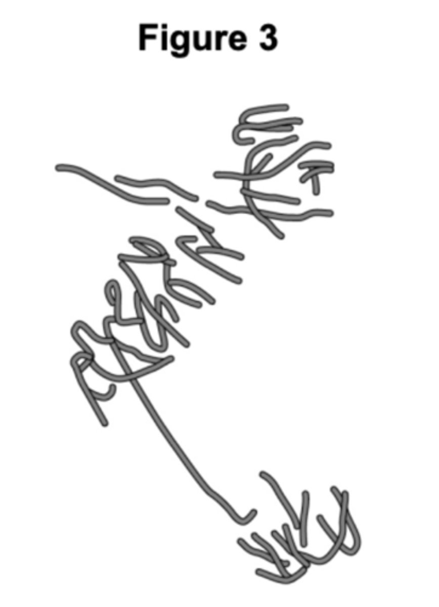
EQ: A scientist treated growing tips of onion roots with a chemical that stops roots box growing. After 24 hours, he prepared a stained squash of these root tips.
Figure 3 is a drawing showing the chromosomes in a single cell observed in the squash of one of these root tips in anaphase. This cell was typical of other cells in anaphase in these root tips.
Use all of this information to suggest how the chemical stops the growth of roots. (3 MARKS)
1. Stops anaphase / cell division / mitosis;
2. (By) stopping / disrupting / spindle fibres forming / attaching / pulling;
3. Preventing separation of (sister) chromatids;
4. (So) no new cells added (to root tip);
EQ: Some prokaryotic cells can divide every 30 minutes. A liquid culture contained a starting population of 1.35 × 10^4 cells. Assuming each cell divides every 30 minutes, calculate how many cells there will be after 3 hours. Assume no cells die during this time. (2 MARKS)
8.64 x 10⁵ ;
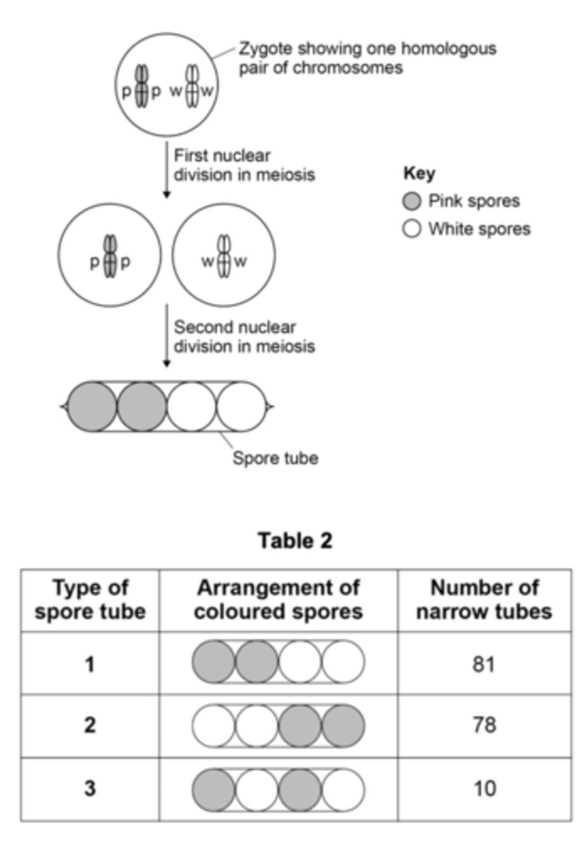
EQ: A scientist crossed a strain of the fungus Neurospora producing pink spores with a strain of Neurospora producing white spores.
To cross these strains, he used aseptic techniques. He moved a small agar cube containing one strain of the fungus onto a new agar plate. Then he placed a second agar cube containing the other strain of fungus next to the first agar cube.
Describe and explain three ways in which the scientist would ensure he used aseptic techniques to move each cube of agar onto a new agar plate. (3 MARKS)
1. Wash hands to remove/kill microbes
OR Wear gloves/apron to prevent contamination;
2. Burning Bunsen close by to create upward current of air;
3. Disinfect bench/work on disinfected cloth to kill microbes/prevent contamination;
4. Flame instrument/equipment to sterilise/kill microbes/prevent contamination;
5. Lift lid slightly to prevent entry of microbes;
EQ: Give two differences between mitosis and meiosis. (2 MARKS)
Mitosis given first
1. One division, two divisions in meiosis;
2. (Daughter) cells genetically identical, daughter cells genetically different in meiosis;
3. Two cells produced, (usually) four cells produced in meiosis;
4. Diploid to diploid/haploid to haploid, diploid to haploid in meiosis;
5. Separation of homologous chromosomes only in meiosis;
6. Crossing over only in meiosis;
7. Independent segregation only in meiosis;
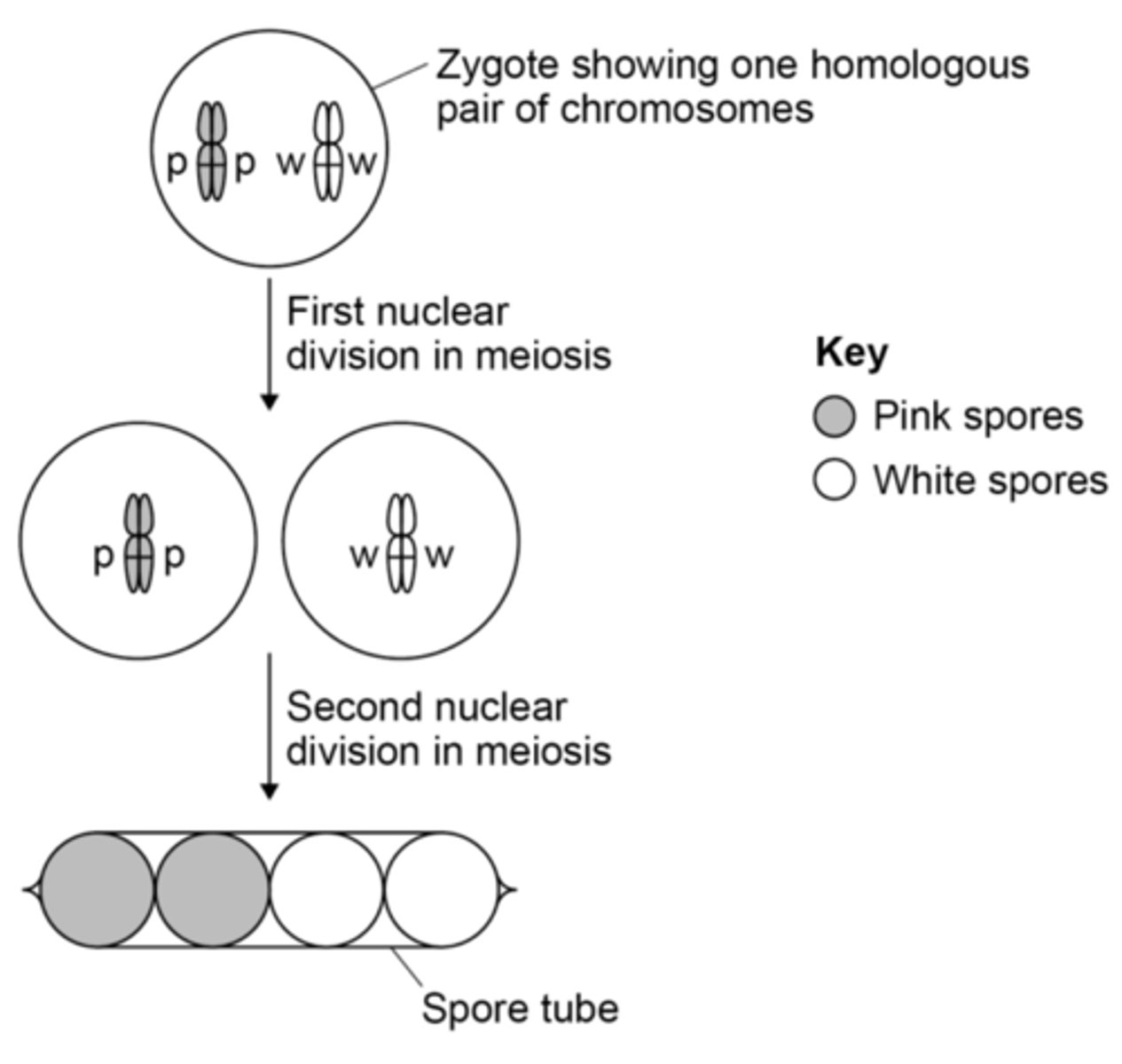
EQ: A scientist crossed a strain of the fungus Neurospora producing pink spores with a strain of Neurospora producing white spores.
To cross these strains, he used aseptic techniques. He moved a small agar cube containing one strain of the fungus onto a new agar plate. Then he placed a second agar cube containing the other strain of fungus next to the first agar cube.
In the life cycle of Neurospora most stages are haploid. Fusion of two haploid strains of this fungus produces diploid zygotes. Nuclear division in these zygotes occurs by meiosis.
At the end of meiosis, this fungus produces cells called spores. box The spores are produced in narrow tubes that restrict their movement. As a result, each tube contains a single line of spores. The spores are coloured either pink or white.
The spore colour gene is located on a pair of homologous chromosomes. Each zygote produced in this cross has one chromosome with a pink allele (p) and one chromosome with a white allele (w).
This is shown in Figure 7.
There are seven chromosomes in a spore nucleus. Place a tick (X) in the box next to the number that represents the number of chromatids present in the zygote shown in Figure 7.
- 7
- 14
- 21
- 28
28;
EQ: A scientist crossed a strain of the fungus Neurospora producing pink spores with a strain of Neurospora producing white spores.
To cross these strains, he used aseptic techniques. He moved a small agar cube containing one strain of the fungus onto a new agar plate. Then he placed a second agar cube containing the other strain of fungus next to the first agar cube.
In the life cycle of Neurospora most stages are haploid. Fusion of two haploid strains of this fungus produces diploid zygotes. Nuclear division in these zygotes occurs by meiosis.
At the end of meiosis, this fungus produces cells called spores. box The spores are produced in narrow tubes that restrict their movement. As a result, each tube contains a single line of spores. The spores are coloured either pink or white.
The spore colour gene is located on a pair of homologous chromosomes. Each zygote produced in this cross has one chromosome with a pink allele (p) and one chromosome with a white allele (w).
This is shown in Figure 7.
The scientist recorded the arrangement of coloured spores inside many narrow tubes. box His results are shown in Table 2.
Using all the information in this question, what can you conclude from the scientist’s results about the movement of chromosomes in meiosis in this fungus? (3 MARKS)
1. Separation of homologous chromosomes (occurred)
OR (Independent) segregation (occurred);
2. (Arrangement/separation/segregation of chromosomes is) random/ (almost) equally frequent (in tubes 1 and 2);
3. Crossing over occurred in tube 3/10 tubes;
4. (Crossing over) is rare/infrequent/in only 10 tubes;
EQ: Bacteria are often used in industry as a source of enzymes. One reason is because bacteria divide rapidly, producing a large number of them in a short time. Describe how bacteria divide. (2 MARKS)
1. Binary fission;
2. Replication of (circular) DNA;
3. Division of cytoplasm to produce 2 daughter cells;
4. Each with single copy of (circular) DNA;