Immunology - Lecture 17 - Function of Secondary Lymphoid Organs

0.0(0)
studied byStudied by 0 people
learnLearn
examPractice Test
spaced repetitionSpaced Repetition
heart puzzleMatch
flashcardsFlashcards
Card Sorting

1/21

encourage image

There's no tags or description

Looks like no tags are added yet.

Study Analytics
Name
Mastery
Learn
Test
Matching
Spaced

No study sessions yet.

22 Terms

1
New cards

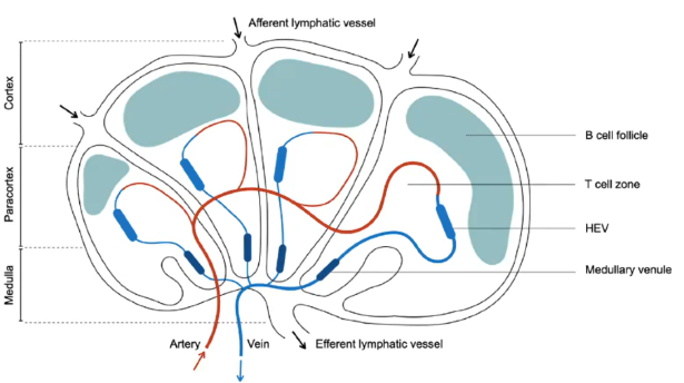
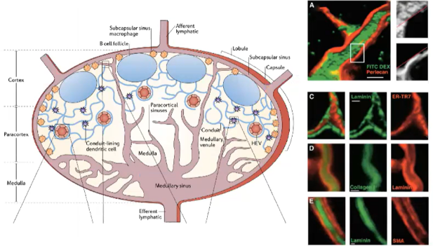
How do T cells and B cells enter secondary lymphoid organs, and what are their distinct zones within a lymph node?

  • B Cells outnumber T Cells for any given antigen:

    • B cells (100-1000) recognize antigens directly.

    • T cells (5-10) require antigen processing.

  • Zones:

    • B cells reside in outer B cell follicles.

    • T cells are in inner T cell zones.

    • Zones are adjacent but physically separated.

  • Entry:

    • Lymphocytes (T and B cells) enter via high endothelial venules (HEV).

    • HEVs express molecules to facilitate lymphocyte entry.

  • Lymphatic Vessels:

    • Afferent lymphatic vessels carry fluid, antigens, dendritic cells, and innate immune cells (not lymphocytes).

    • Efferent lymphatic vessels drain adjacent tissues.

  • This organization supports efficient immune cell interactions with antigens and each other.

<ul><li><p><strong>B Cells</strong> outnumber <strong>T Cells</strong> for any given antigen:</p><ul><li><p><strong>B cells</strong> (100-1000) recognize antigens directly.</p></li><li><p><strong>T cells</strong> (5-10) require <strong>antigen processing</strong>.</p></li></ul></li><li><p><strong>Zones</strong>:</p><ul><li><p><strong>B cells</strong> reside in <strong>outer B cell follicles</strong>.</p></li><li><p><strong>T cells</strong> are in <strong>inner T cell zones</strong>.</p></li><li><p>Zones are <strong>adjacent</strong> but <strong>physically separated</strong>.</p></li></ul></li><li><p><strong>Entry</strong>:</p><ul><li><p><strong>Lymphocytes</strong> (T and B cells) enter via <strong>high endothelial venules (HEV)</strong>.</p></li><li><p><strong>HEVs</strong> express molecules to facilitate lymphocyte entry.</p></li></ul></li><li><p><strong>Lymphatic Vessels</strong>:</p><ul><li><p><strong>Afferent lymphatic vessels</strong> carry fluid, antigens, dendritic cells, and innate immune cells (not lymphocytes).</p></li><li><p><strong>Efferent lymphatic vessels</strong> drain adjacent tissues.</p></li></ul></li><li><p>This organization supports efficient <strong>immune cell interactions</strong> with <strong>antigens</strong> and each other.</p></li></ul><p></p>
2
New cards

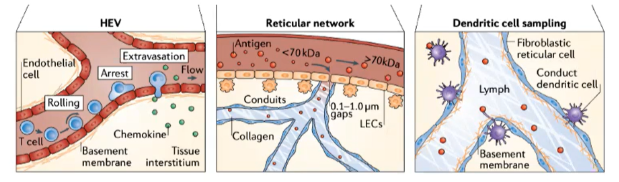
What makes HEVs unique in allowing lymphocyte entry into lymph nodes?

  • HEVs express addressins such as GlyCAM-1 and PNAd.

  • These addressins interact with L-selectin on lymphocytes.

  • Creates a sticky interaction with the endothelial wall, facilitating rolling and migration into lymph nodes.

<ul><li><p><strong>HEVs</strong> express <strong>addressins</strong> such as <strong>GlyCAM-1</strong> and <strong>PNAd</strong>.</p></li><li><p>These addressins interact with <strong>L-selectin</strong> on <strong>lymphocytes</strong>.</p></li><li><p>Creates a <strong>sticky interaction</strong> with the endothelial wall, facilitating <strong>rolling and migration</strong> into lymph nodes.</p></li></ul><p></p>
3
New cards

What initiates the tethering and fast rolling of lymphocytes on HEV endothelium?

  • L-selectin on B and T cells binds to GlyCAM-1 and PNAd on HEVs.

  • This binding enables lymphocytes to tether and roll along the endothelial wall.

<ul><li><p><strong>L-selectin</strong> on <strong>B and T cells</strong> binds to <strong>GlyCAM-1</strong> and <strong>PNAd</strong> on <strong>HEVs</strong>.</p></li><li><p>This binding enables <strong>lymphocytes</strong> to <strong>tether</strong> and <strong>roll</strong> along the endothelial wall.</p></li></ul><p></p>
4
New cards

How do chemokines on HEVs influence lymphocyte migration?

  • CCL21 binds to the CCR7 receptor on lymphocytes.

  • CXCL12 binds to the CXCR4 receptor on lymphocytes.

These interactions activate integrins, enhancing adhesion and stopping lymphocyte rolling.

<ul><li><p><strong>CCL21</strong> binds to the <strong>CCR7 receptor</strong> on <strong>lymphocytes</strong>.</p></li><li><p><strong>CXCL12</strong> binds to the <strong>CXCR4 receptor</strong> on <strong>lymphocytes</strong>.</p></li></ul><p>These interactions activate integrins, enhancing adhesion and stopping lymphocyte rolling.</p>
5
New cards

Which integrins are activated on lymphocytes to stop rolling and promote HEV entry?

  • VLA4 and LFA1 integrins are activated on lymphocytes.

  • VLA4 binds to VCAM1 and LFA1 binds to ICAM1 on HEVs.

  • This binding creates strong adhesion, stopping rolling and enabling transmigration into lymph nodes.

<ul><li><p><strong>VLA4</strong> and <strong>LFA1 integrins</strong> are activated on <strong>lymphocytes</strong>.</p></li><li><p><strong>VLA4</strong> binds to <strong>VCAM1</strong> and <strong>LFA1</strong> binds to <strong>ICAM1</strong> on <strong>HEVs</strong>.</p></li><li><p>This binding creates <strong>strong adhesion</strong>, stopping <strong>rolling</strong> and enabling <strong>transmigration</strong> into lymph nodes.</p></li></ul><p></p>
6
New cards

Describe the sequence of interactions that allow lymphocytes to enter lymph nodes through HEVs.

  • Rolling: L-selectin binds to GlyCAM-1 and PNAd.

  • Activation: CCR7 and CXCR4 receptors engage chemokines (CCL21 and CXCL12).

  • Arrest: Integrins VLA4 and LFA1 bind to VCAM1 and ICAM1.

  • Transmigration: Chemokine gradients guide lymphocytes into lymph nodes.

<ul><li><p><strong>Rolling</strong>: <strong>L-selectin</strong> binds to <strong>GlyCAM-1</strong> and <strong>PNAd</strong>.</p></li><li><p><strong>Activation</strong>: <strong>CCR7</strong> and <strong>CXCR4</strong> receptors engage <strong>chemokines</strong> (CCL21 and CXCL12).</p></li><li><p><strong>Arrest</strong>: <strong>Integrins VLA4</strong> and <strong>LFA1</strong> bind to <strong>VCAM1</strong> and <strong>ICAM1</strong>.</p></li><li><p><strong>Transmigration</strong>: <strong>Chemokine gradients</strong> guide lymphocytes into <strong>lymph nodes</strong>.</p></li></ul><p></p>
7
New cards

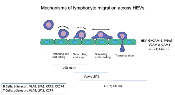
Which receptors do B and T cells share, and which are unique for lymph node migration?

  • Both B and T cells use:

    • L-selectin for initial rolling.

    • VLA4 and LFA1 integrins for adhesion.

    • CCR7 for CCL21 chemokine signaling.

  • B cells additionally use:

    • CXCR4 for CXCL12 chemokine signaling in lymph node migration.

<ul><li><p><strong>Both B and T cells</strong> use:</p><ul><li><p><strong>L-selectin</strong> for initial rolling.</p></li><li><p><strong>VLA4</strong> and <strong>LFA1</strong> integrins for adhesion.</p></li><li><p><strong>CCR7</strong> for <strong>CCL21</strong> chemokine signaling.</p></li></ul></li><li><p><strong>B cells</strong> additionally use:</p><ul><li><p><strong>CXCR4</strong> for <strong>CXCL12</strong> chemokine signaling in <strong>lymph node migration</strong>.</p></li></ul></li></ul><p></p>
8
New cards

What role do addressins play in lymphocyte migration across HEVs?

  • Addressins (e.g., GlyCAM-1 and PNAd) are sticky proteoglycans expressed on HEVs.

  • Recognized by the L-selectin receptor on B and T cells.

  • As lymphocytes pass through HEVs, L-selectin binds to GlyCAM-1 and PNAd, creating a sticky interaction.

  • This interaction initiates tethering and fast rolling along the endothelial wall.

<ul><li><p><strong>Addressins</strong> (e.g., <strong>GlyCAM-1</strong> and <strong>PNAd</strong>) are <strong>sticky proteoglycans</strong> expressed on <strong>HEVs</strong>.</p></li><li><p>Recognized by the <strong>L-selectin receptor</strong> on <strong>B and T cells</strong>.</p></li><li><p>As lymphocytes pass through HEVs, <strong>L-selectin</strong> binds to <strong>GlyCAM-1</strong> and <strong>PNAd</strong>, creating a <strong>sticky interaction</strong>.</p></li><li><p>This interaction initiates <strong>tethering</strong> and <strong>fast rolling</strong> along the endothelial wall.</p></li></ul><p></p>
9
New cards

How do chemokines CCL21 and CXCL12 influence lymphocyte migration across HEVs?

  • HEV endothelial cells express chemokines CCL21 and CXCL12.

  • As lymphocytes roll through HEVs, they encounter these chemokines:

    • CCL21 engages with CCR7 receptor.

    • CXCL12 engages with CXCR4 receptor.

  • This receptor engagement activates integrins on the lymphocyte.

  • Integrins provide strong adhesion to secure the lymphocyte to the endothelial wall.

  • Prepares the lymphocyte for migration into the lymph node.

<ul><li><p><strong>HEV endothelial cells</strong> express chemokines <strong>CCL21</strong> and <strong>CXCL12</strong>.</p></li><li><p>As <strong>lymphocytes</strong> roll through HEVs, they encounter these chemokines:</p><ul><li><p><strong>CCL21</strong> engages with <strong>CCR7</strong> receptor.</p></li><li><p><strong>CXCL12</strong> engages with <strong>CXCR4</strong> receptor.</p></li></ul></li><li><p>This <strong>receptor engagement</strong> activates <strong>integrins</strong> on the lymphocyte.</p></li><li><p><strong>Integrins</strong> provide <strong>strong adhesion</strong> to secure the lymphocyte to the endothelial wall.</p></li><li><p>Prepares the lymphocyte for <strong>migration into the lymph node</strong>.</p></li></ul><p></p>
10
New cards

How do integrins and ligands regulate lymphocyte migration across HEVs?

  • Integrins VLA4 and LFA1 on B and T cells facilitate the shift from fast to slow rolling and eventual stopping on HEV endothelium.

  • GPCR signaling activates these integrins, shifting them from a dense to a tight conformation.

  • Activated integrins bind to their ligands:

    • VLA4 binds VCAM1.

    • LFA1 binds ICAM1.

  • This firm adhesion halts lymphocyte rolling, enabling response to chemokine signals.

  • Lymphocytes then migrate from the blood into the lymph node.

  • Chemokines within the lymph node guide B and T cells to their specific zones.

<ul><li><p><strong>Integrins VLA4</strong> and <strong>LFA1</strong> on <strong>B and T cells</strong> facilitate the shift from <strong>fast to slow rolling</strong> and eventual stopping on <strong>HEV endothelium</strong>.</p></li><li><p><strong>GPCR signaling</strong> activates these integrins, shifting them from a <strong>dense</strong> to a <strong>tight conformation</strong>.</p></li><li><p>Activated integrins bind to their ligands:</p><ul><li><p><strong>VLA4</strong> binds <strong>VCAM1</strong>.</p></li><li><p><strong>LFA1</strong> binds <strong>ICAM1</strong>.</p></li></ul></li><li><p>This <strong>firm adhesion</strong> halts lymphocyte rolling, enabling response to <strong>chemokine signals</strong>.</p></li><li><p>Lymphocytes then <strong>migrate from the blood into the lymph node</strong>.</p></li><li><p><strong>Chemokines within the lymph node</strong> guide B and T cells to their specific <strong>zones</strong>.</p></li></ul><p></p>
11
New cards

How do lymphocytes migrate through High Endothelial Venules (HEVs) to enter lymph nodes?

  • HEVs are unique blood vessels that express addressins (GlyCAM-1 and PNAd), making the endothelium "sticky".

  • B and T cells use L-selectin to bind to these addressins, initiating tethering and fast rolling on the endothelial wall.

  • HEVs also express chemokines CCL21 and CXCL12:

    • CCL21 binds to CCR7 on lymphocytes.

    • CXCL12 binds to CXCR4 on lymphocytes.

  • Chemokine binding activates integrins (VLA4 and LFA1) through GPCR signaling, shifting integrins to an active conformation.

  • Active integrins tightly bind to VCAM1 (for VLA4) and ICAM1 (for LFA1), stopping lymphocyte rolling.

  • This strong adhesion enables lymphocyte migration from the blood into the lymph node.

  • Within the lymph node, specific chemokines direct B and T cells to their respective zones.

<ul><li><p><strong>HEVs</strong> are unique blood vessels that express <strong>addressins</strong> (<strong>GlyCAM-1</strong> and <strong>PNAd</strong>), making the endothelium <strong>"sticky"</strong>.</p></li><li><p><strong>B and T cells</strong> use <strong>L-selectin</strong> to bind to these addressins, initiating <strong>tethering and fast rolling</strong> on the endothelial wall.</p></li><li><p><strong>HEVs</strong> also express chemokines <strong>CCL21</strong> and <strong>CXCL12</strong>:</p><ul><li><p><strong>CCL21</strong> binds to <strong>CCR7</strong> on lymphocytes.</p></li><li><p><strong>CXCL12</strong> binds to <strong>CXCR4</strong> on lymphocytes.</p></li></ul></li><li><p>Chemokine binding activates <strong>integrins</strong> (<strong>VLA4</strong> and <strong>LFA1</strong>) through <strong>GPCR signaling</strong>, shifting integrins to an <strong>active conformation</strong>.</p></li><li><p>Active integrins tightly bind to <strong>VCAM1</strong> (for VLA4) and <strong>ICAM1</strong> (for LFA1), stopping lymphocyte rolling.</p></li><li><p>This <strong>strong adhesion</strong> enables lymphocyte <strong>migration from the blood into the lymph node</strong>.</p></li><li><p>Within the lymph node, <strong>specific chemokines</strong> direct <strong>B and T cells</strong> to their respective <strong>zones</strong>.</p></li></ul><p></p>
12
New cards

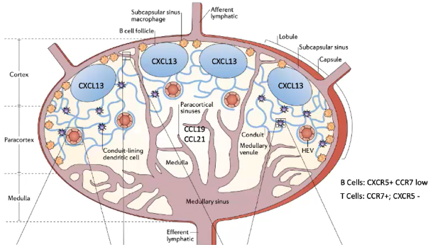
How do T cells remain in the T cell zone of the lymph node?

  • After entering the lymph node, T cells remain in the T cell zone due to CCR7 expression.

  • CCR7 detects chemokines CCL21 and CCL19 in the T cell zone.

  • This zone has a fibroblast network specialized in expressing these chemokines.

  • CCL19 is soluble, while CCL21 is more membrane-bound.

  • The combination helps retain T cells within the T cell zone.

<ul><li><p>After entering the <strong>lymph node</strong>, <strong>T cells</strong> remain in the <strong>T cell zone</strong> due to <strong>CCR7</strong> expression.</p></li><li><p><strong>CCR7</strong> detects chemokines <strong>CCL21</strong> and <strong>CCL19</strong> in the T cell zone.</p></li><li><p>This zone has a <strong>fibroblast network</strong> specialized in expressing these chemokines.</p></li><li><p><strong>CCL19</strong> is <strong>soluble</strong>, while <strong>CCL21</strong> is more <strong>membrane-bound</strong>.</p></li><li><p>The combination helps <strong>retain T cells</strong> within the <strong>T cell zone</strong>.</p></li></ul><p></p>
13
New cards

How do B cells localize to the B cell follicle in the lymph node?

  • B cells express low CCR7 but high CXCR5, which binds the chemokine CXCL13.

  • CXCL13 is produced by follicular dendritic cells (FDCs) in the B cell follicle.

  • Unlike T cell zones, the B cell follicle contains FDCs (fibroblasts, not hematopoietic dendritic cells).

  • FDCs display CXCL13 and preserve antigens on their surface.

  • This preservation allows B cells to recognize antigens without antigen destruction.

<ul><li><p><strong>B cells</strong> express low <strong>CCR7</strong> but high <strong>CXCR5</strong>, which binds the chemokine <strong>CXCL13</strong>.</p></li><li><p><strong>CXCL13</strong> is produced by <strong>follicular dendritic cells (FDCs)</strong> in the <strong>B cell follicle</strong>.</p></li><li><p>Unlike <strong>T cell zones</strong>, the <strong>B cell follicle</strong> contains <strong>FDCs</strong> (fibroblasts, not hematopoietic dendritic cells).</p></li><li><p><strong>FDCs</strong> display <strong>CXCL13</strong> and preserve <strong>antigens</strong> on their surface.</p></li><li><p>This preservation allows <strong>B cells</strong> to recognize antigens without <strong>antigen destruction</strong>.</p></li></ul><p></p>
14
New cards

How do dendritic cells and lymphocyte receptors influence cell movement and antigen processing in the lymph node?

  • In the T cell zone, blood-derived dendritic cells process antigens and express CCR7.

  • These dendritic cells are exclusive to T cell zones and are not found in B cell follicles.

  • Each dendritic cell can interact with about 5,000 T cells.

  • B and T cells migrate between lymph nodes:

    • T cells express CCR7, with a half-life of ~12 hours in a lymph node.

    • B cells express CXCR5, with a longer half-life of ~24 hours in a lymph node.

<ul><li><p>In the <strong>T cell zone</strong>, <strong>blood-derived dendritic cells</strong> process antigens and express <strong>CCR7</strong>.</p></li><li><p>These dendritic cells are exclusive to <strong>T cell zones</strong> and are <strong>not found in B cell follicles</strong>.</p></li><li><p>Each dendritic cell can interact with about <strong>5,000 T cells</strong>.</p></li><li><p><strong>B and T cells</strong> migrate between <strong>lymph nodes</strong>:</p><ul><li><p><strong>T cells</strong> express <strong>CCR7</strong>, with a half-life of ~<strong>12 hours</strong> in a lymph node.</p></li><li><p><strong>B cells</strong> express <strong>CXCR5</strong>, with a longer half-life of ~<strong>24 hours</strong> in a lymph node.</p></li></ul></li></ul><p></p>
15
New cards

How do T cells and B cells remain in their respective zones within the lymph node, and how are antigens presented to them?

  • T Cells:

    • Remain in the T cell zone due to CCR7, which binds to CCL21 (membrane-bound) and CCL19 (soluble) from specialized fibroblasts.

    • Blood-derived dendritic cells in the T cell zone also express CCR7 and present processed antigens.

    • Each dendritic cell can interact with up to 5,000 T cells.

    • CCR7 allows T cells to stay in a lymph node for about 12 hours before migrating to another.

  • B Cells:

    • Have low CCR7 but high CXCR5, which binds CXCL13 from follicular dendritic cells (FDCs) in the B cell follicle.

    • Unlike T-cell-interacting dendritic cells, FDCs are fibroblast-derived and not hematopoietic.

    • FDCs preserve antigens on their surface, enabling B cell recognition without antigen destruction.

    • CXCR5 allows B cells to stay in a lymph node for around 24 hours before moving to another.

<ul><li><p><strong>T Cells</strong>:</p><ul><li><p>Remain in the <strong>T cell zone</strong> due to <strong>CCR7</strong>, which binds to <strong>CCL21</strong> (membrane-bound) and <strong>CCL19</strong> (soluble) from specialized fibroblasts.</p></li><li><p><strong>Blood-derived dendritic cells</strong> in the T cell zone also express <strong>CCR7</strong> and present processed antigens.</p></li><li><p>Each dendritic cell can interact with up to <strong>5,000 T cells</strong>.</p></li><li><p><strong>CCR7</strong> allows T cells to stay in a <strong>lymph node</strong> for about <strong>12 hours</strong> before migrating to another.</p></li></ul></li><li><p><strong>B Cells</strong>:</p><ul><li><p>Have low <strong>CCR7</strong> but high <strong>CXCR5</strong>, which binds <strong>CXCL13</strong> from <strong>follicular dendritic cells (FDCs)</strong> in the <strong>B cell follicle</strong>.</p></li><li><p>Unlike T-cell-interacting dendritic cells, <strong>FDCs</strong> are <strong>fibroblast-derived</strong> and <strong>not hematopoietic</strong>.</p></li><li><p><strong>FDCs</strong> preserve antigens on their surface, enabling <strong>B cell recognition</strong> without antigen destruction.</p></li><li><p><strong>CXCR5</strong> allows B cells to stay in a <strong>lymph node</strong> for around <strong>24 hours</strong> before moving to another.</p></li></ul></li></ul><p></p>
16
New cards

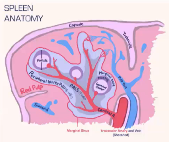
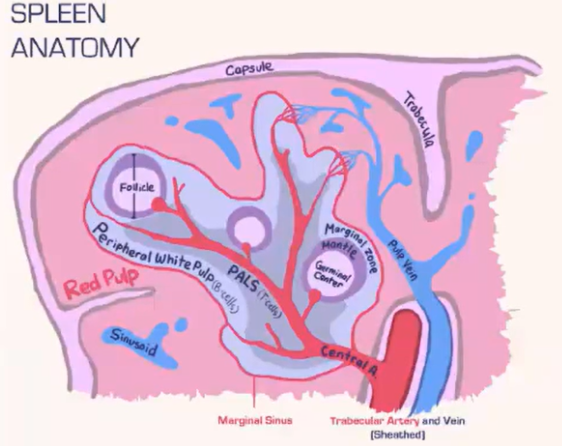
How does the spleen structure and function differ from lymph nodes, and how are T cells and B cells organized within it?

  • Spleen Structure:

    • Lacks a lymphatic system, relying solely on blood flow.

    • Consists of:

      • Red pulp: Contains red blood cells.

      • White pulp: Houses lymphocytes.

    • Blood is deposited at the marginal sinus (between red and white pulp), serving as a filter.

  • Lymphocyte Organization:

    • T Cells:

      • Move toward the T cell zone near the central arteriole.

      • Guided by CCR7 with support from fibroblastic reticular cells (FRCs).

    • B Cells:

      • Migrate to the B cell follicle in response to CXCR5.

      • Follicles contain follicular dendritic cells (FDCs), which preserve antigens for B cell recognition, similar to lymph nodes.

  • Lymphocyte Entry:

    • Lymphocytes passively enter the spleen, without selectins or rolling.

    • Integrins assist in adhesion at the marginal sinus area.

<ul><li><p><strong>Spleen Structure</strong>:</p><ul><li><p><strong>Lacks a lymphatic system</strong>, relying solely on <strong>blood flow</strong>.</p></li><li><p>Consists of:</p><ul><li><p><strong>Red pulp</strong>: Contains <strong>red blood cells</strong>.</p></li><li><p><strong>White pulp</strong>: Houses <strong>lymphocytes</strong>.</p></li></ul></li><li><p><strong>Blood</strong> is deposited at the <strong>marginal sinus</strong> (between red and white pulp), serving as a <strong>filter</strong>.</p></li></ul></li><li><p><strong>Lymphocyte Organization</strong>:</p><ul><li><p><strong>T Cells</strong>:</p><ul><li><p>Move toward the <strong>T cell zone</strong> near the <strong>central arteriole</strong>.</p></li><li><p>Guided by <strong>CCR7</strong> with support from <strong>fibroblastic reticular cells (FRCs)</strong>.</p></li></ul></li><li><p><strong>B Cells</strong>:</p><ul><li><p>Migrate to the <strong>B cell follicle</strong> in response to <strong>CXCR5</strong>.</p></li><li><p>Follicles contain <strong>follicular dendritic cells (FDCs)</strong>, which preserve antigens for <strong>B cell recognition</strong>, similar to lymph nodes.</p></li></ul></li></ul></li><li><p><strong>Lymphocyte Entry</strong>:</p><ul><li><p>Lymphocytes <strong>passively enter</strong> the spleen, without <strong>selectins</strong> or <strong>rolling</strong>.</p></li><li><p><strong>Integrins</strong> assist in <strong>adhesion</strong> at the <strong>marginal sinus</strong> area.</p></li></ul></li></ul><p></p>
17
New cards

How are antigens channeled to different locations in the lymph node?

  • Small Antigens:

    • <60-70 kilodaltons (e.g., soluble proteins) are transported through a conduit system in the lymph node.

    • Conduits extend from the capsule into the T cell zone, allowing direct access for T cells.

    • Dendritic cells in the T cell zone pick up, process, and present these antigens on MHC molecules to T cells.

  • Large Antigens:

    • Captured by dendritic cells at the infection site.

    • Dendritic cells, with processed antigens, enter the lymph node via the afferent lymphatic system.

    • Present antigens to T cells within the T cell zone.

  • This system allows effective delivery of both small and large antigens to initiate an immune response in the lymph node.

<ul><li><p><strong>Small Antigens</strong>:</p><ul><li><p><strong>&lt;60-70 kilodaltons</strong> (e.g., soluble proteins) are transported through a <strong>conduit system</strong> in the lymph node.</p></li><li><p><strong>Conduits</strong> extend from the <strong>capsule</strong> into the <strong>T cell zone</strong>, allowing direct access for T cells.</p></li><li><p><strong>Dendritic cells</strong> in the T cell zone pick up, process, and present these antigens on <strong>MHC molecules</strong> to T cells.</p></li></ul></li><li><p><strong>Large Antigens</strong>:</p><ul><li><p>Captured by <strong>dendritic cells</strong> at the infection site.</p></li><li><p>Dendritic cells, with <strong>processed antigens</strong>, enter the lymph node via the <strong>afferent lymphatic system</strong>.</p></li><li><p>Present antigens to T cells within the <strong>T cell zone</strong>.</p></li></ul></li><li><p>This system allows <strong>effective delivery</strong> of both small and large antigens to initiate an <strong>immune response</strong> in the lymph node.</p></li></ul><p></p>
18
New cards

How do conduits and dendritic cells contribute to antigen sampling in the lymph node T cell zone?

  • Conduits in the lymph node are narrow channels that exclude larger molecules.

  • Allow only small antigens (<70 kDa) to pass through.

  • Dendritic cells in the T cell zone continuously sample antigens from these conduits.

  • Enables constant surveillance of potential pathogens by dendritic cells.

<ul><li><p><strong>Conduits in the lymph node</strong> are <strong>narrow channels</strong> that exclude larger molecules.</p></li><li><p>Allow only <strong>small antigens (&lt;70 kDa)</strong> to pass through.</p></li><li><p><strong>Dendritic cells</strong> in the <strong>T cell zone</strong> continuously sample antigens from these conduits.</p></li><li><p>Enables <strong>constant surveillance</strong> of potential pathogens by dendritic cells.</p></li></ul><p></p>
19
New cards

What role do subcapsular sinus macrophages (SCS MΦ) play in antigen presentation to B cells within the lymph node?

  • Subcapsular sinus macrophages (SCS MΦ):

    • Located beneath the lymph node capsule near afferent lymphatic vessels.

    • Express complement receptors (e.g., CR1), allowing them to capture and retain opsonized antigens without internalizing or destroying them.

    • Displayed antigens provide B cells an opportunity for antigen encounter near the capsule.

  • Antigen Acquisition by B Cells:

    • B cells use CD21 (CR2) to "steal" opsonized antigens from SCS MΦ.

    • Interaction is independent of the B cell receptor (BCR) and relies solely on complement receptor engagement.

  • Antigen Transfer to Follicular Dendritic Cells (FDCs):

    • After acquiring the antigen, B cells carry it into the lymph node follicle.

    • B cells transfer the antigen to FDCs, which express CR1 (CD35) with high avidity, enabling strong antigen retention.

    • FDCs display antigens long-term, providing continuous exposure for B cells and supporting ongoing immune responses.

  • This pathway allows B cells to access and retain antigens without direct processing by traditional antigen-presenting cells.

20
New cards
<p>How does the spleen filter blood-borne pathogens, and what role do macrophages play?</p>

How does the spleen filter blood-borne pathogens, and what role do macrophages play?

  • The spleen has two main areas:

    • Red pulp: Contains red blood cells.

    • White pulp: Houses lymphocytes.

  • Marginal sinus separates red and white pulp, populated with phagocytic macrophages.

  • These macrophages filter blood-borne pathogens, acting as a primary defense.

  • Unlike lymph nodes, the spleen lacks non-phagocytic subcapsular sinus macrophages.

  • This structure enables the spleen to function as a primary blood pathogen filter.

<ul><li><p>The <strong>spleen</strong> has two main areas:</p><ul><li><p><strong>Red pulp</strong>: Contains <strong>red blood cells</strong>.</p></li><li><p><strong>White pulp</strong>: Houses <strong>lymphocytes</strong>.</p></li></ul></li><li><p><strong>Marginal sinus</strong> separates red and white pulp, populated with <strong>phagocytic macrophages</strong>.</p></li><li><p>These macrophages <strong>filter blood-borne pathogens</strong>, acting as a primary defense.</p></li><li><p>Unlike lymph nodes, the spleen <strong>lacks non-phagocytic subcapsular sinus macrophages</strong>.</p></li><li><p>This structure enables the spleen to function as a <strong>primary blood pathogen filter</strong>.</p></li></ul><p></p>
21
New cards
<p>How do marginal zone B cells in the spleen aid in antigen presentation?</p>

How do marginal zone B cells in the spleen aid in antigen presentation?

  • Marginal zone B cells are located beneath the marginal sinus in the B cell follicle.

  • They migrate between the red pulp and follicle approximately every 20 minutes.

  • Express high levels of CD21, efficiently capturing opsonized antigens missed by macrophages.

  • Movement is guided by S1P and CXCL13 gradients:

    • In the follicle, they sense S1P via S1PR1 and migrate to the S1P-rich red pulp.

    • In the red pulp, S1PR1 receptors internalize due to S1P saturation, guiding them back to the follicle via CXCR5 sensing CXCL13.

  • This cycle enables antigen deposition onto follicular dendritic cells (FDCs), supporting B cell immune responses against blood-borne pathogens.

<ul><li><p><strong>Marginal zone B cells</strong> are located beneath the <strong>marginal sinus</strong> in the <strong>B cell follicle</strong>.</p></li><li><p>They migrate between the <strong>red pulp</strong> and <strong>follicle</strong> approximately every <strong>20 minutes</strong>.</p></li><li><p>Express high levels of <strong>CD21</strong>, efficiently capturing <strong>opsonized antigens</strong> missed by macrophages.</p></li><li><p>Movement is guided by <strong>S1P</strong> and <strong>CXCL13</strong> gradients:</p><ul><li><p>In the <strong>follicle</strong>, they sense <strong>S1P</strong> via <strong>S1PR1</strong> and migrate to the <strong>S1P-rich red pulp</strong>.</p></li><li><p>In the <strong>red pulp</strong>, <strong>S1PR1</strong> receptors internalize due to <strong>S1P saturation</strong>, guiding them back to the <strong>follicle</strong> via <strong>CXCR5</strong> sensing <strong>CXCL13</strong>.</p></li></ul></li><li><p>This cycle enables <strong>antigen deposition onto follicular dendritic cells (FDCs)</strong>, supporting <strong>B cell immune responses</strong> against <strong>blood-borne pathogens</strong>.</p></li></ul><p></p>
22
New cards
<p>How do marginal zone B cells in the spleen contribute to antigen presentation?</p>

How do marginal zone B cells in the spleen contribute to antigen presentation?

  • Marginal zone B cells in the spleen's B cell follicle specialize in capturing opsonized antigens missed by macrophages.

  • Express high levels of CD21, efficiently picking up complement-bound antigens.

  • Migrate between the red pulp (S1P-rich) and the follicle (CXCL13-rich) approximately every 20 minutes, following S1P and CXCL13 gradients.

    • In the red pulp, high S1P levels cause S1PR1 internalization, directing cells back to the follicle using CXCR5 and CXCL13 signaling.

    • In the follicle, S1PR1 is re-expressed due to low S1P, guiding cells back to the red pulp.

  • During each cycle, they deposit captured antigens onto follicular dendritic cells (FDCs) in the follicle.

  • This process maintains a reservoir of antigens for presentation to B cells, supporting ongoing immune responses.

<ul><li><p><strong>Marginal zone B cells</strong> in the spleen's <strong>B cell follicle</strong> specialize in capturing <strong>opsonized antigens</strong> missed by macrophages.</p></li><li><p>Express high levels of <strong>CD21</strong>, efficiently picking up <strong>complement-bound antigens</strong>.</p></li><li><p>Migrate between the <strong>red pulp</strong> (S1P-rich) and the <strong>follicle</strong> (CXCL13-rich) approximately every <strong>20 minutes</strong>, following <strong>S1P</strong> and <strong>CXCL13</strong> gradients.</p><ul><li><p>In the <strong>red pulp</strong>, high <strong>S1P</strong> levels cause <strong>S1PR1 internalization</strong>, directing cells back to the <strong>follicle</strong> using <strong>CXCR5</strong> and <strong>CXCL13</strong> signaling.</p></li><li><p>In the <strong>follicle</strong>, <strong>S1PR1</strong> is re-expressed due to low S1P, guiding cells back to the <strong>red pulp</strong>.</p></li></ul></li><li><p>During each cycle, they deposit captured antigens onto <strong>follicular dendritic cells (FDCs)</strong> in the follicle.</p></li><li><p>This process maintains a <strong>reservoir of antigens</strong> for presentation to <strong>B cells</strong>, supporting ongoing immune responses.</p></li></ul><p></p>