04: Forelimb 1
1/82
There's no tags or description
Looks like no tags are added yet.
Name | Mastery | Learn | Test | Matching | Spaced |
|---|
No study sessions yet.
83 Terms
Modifications in cows and horses that makes weight bearing less energetically expensive
CT has replaced muscle in the distal limb
There are less bones and less bone mass at the distal limb
Where is most of the weight carried in a horse
Forelimbs
What happens if a rider is not aligned with the horse’s center of gravity
They eat dirt :)
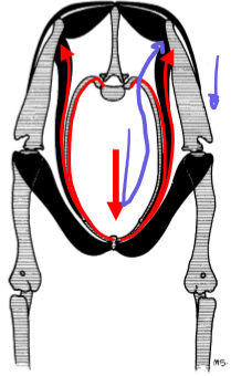
When a horse lands from a jump, how is the force from the torso transferred to the forelimb
Wt goes down → travels up the serratus ventralis and other ventral muscles to the medial scapula → down the leg
New/significant scapular structures in the horse
Spinal tuber
Scapular cartilage
New/significant scapular structures in the ox
Acromion
Scapular cartilage
Scapular cartilage function
Increases surface area for attachment of serratus ventralis and rhomboideus muscles
New/significant structures of the humerus in large animals
Cranial part of the greater tubercle
Caudal part of the greater tubercle
Extra structure of the horse humerus and function
Intermediate tuberosity; stabilizes the biceps tendon
Structure that reduces friction of the proximal biceps tendon in the bicipital/intertubercular groove
Intertubercular/bicipital bursa
Additional significant shoulder muscle in the horse
Subclavius muscle
Where is the subclavius
Just cranial to the supraspinatus
Subclavius function at rest and in movement
Rest: brings trunk forward
Movement: brings leg forward; adducts limb
How is the deltoideus different between the horse and ox
In the horse, the two heads of deltoideus are fused. In the ox, they are split by the acromion process of the scapular spine
Non-muscular structure in the horse that works with the serratus ventralis for shock absorption
Dorsoscapular ligament
Muscle that is key for keeping the scapula in place
Infraspinatus
Structure that reduces friction at the distal aspect of the infraspinatus muscle
Infraspinous bursa
Muscle that is key for standing up from recumbency
Triceps muscle
Muscle that lies caudomedial to the triceps
Tensor fascia antebrachii
T/F: the triceps muscle is kept engaged while the horse/ox is standing still
False, that’s too much energy
Triceps innervation
Radial nerve
Clinical signs of proximal radial nerve damage
Loss of extensor function
No weight bearing
Dropped elbows
Knuckling
Abn movement
Biceps brachii attachments
Supraglenoid tubercle (scapula)
Radial tuberosity (radioulna)
Metacarpal tuberosity (cannon)
What part of the biceps brachii attaches to the metacarpal tuberosity
Lacertus fibrosus
What muscle does the lacertus fibrosus join as it runs distally
Extensor carpi radialis
Where does the lacertus fibrosus come from
The CT core in the biceps
Lacertus fibrosus function in the stay apparatus
Limits flexion of the shoulder
Maintains extension of elbow and carpus
Why does the elbow joint favor extension in large animals
The collateral ligaments are located caudal to the axis of rotation, pulling the joint towards extension
Key muscles of the antebrachium
SDF and DDF
Extra muscle belly in the cow antebrachium that horses have reduced to CT
Pronator teres
What part of the digital flexors is most important
Tendons! We can’t really see the muscle bellies
SDF distal attachment
Distal P1 and proximal P2
SDF distal attachment in the ox
P2
DDF distal attachment
P3
DDF distal attachment in the ox
P3
Which forelimb joint commonly gets hyperextended
Fetlock/metacarpophalangeal joint
Which tendons stretch when the fetlock joint overextends
SDF and DDF
Components that keep the SDF and DDF from tearing under stress
Elasticity
Check ligaments
Check ligament function
Transfers some of the force from the muscle belly to the bone structure
Where does the SDF check ligament attach
Distal radius
Where does the DDF check ligament attach
Palmar carpal ligament
Which species has a full complement of carpal bones
Pigs
Carpal bones in the pig
Top row: radial, intermediate, ulnar, accessory
Bottom row: I, II, III, IV
Carpal bones in the horse
Top row: radial, intermediate, ulnar, accessory
Bottom row: maybe I, II, III, IV
Carpal bones in the cow
Top row: radial, intermediate, ulnar, accessory
Bottom row: II + III (fused), IV
How can you tell which side of the carpus is which
Accessory carpal bone is always lateral
List the carpal joints
Radiocarpal/antebrachiocarpal
Intercarpal
Carpometacarpal
Which carpal joint has the highest range of motion
Radiocarpal
Which carpal joint has the lowest range of motion
Carpometacarpal
Which carpal joints communicate with each other
Intercarpal and carpometacarpal joints
Structure that prevents hyperextension of the carpus
Palmar carpal ligament
Reduced carpal pad analog
Chestnut
Reduced metacarpal pad analog
Ergot
What bones make up the cannon bone in the cow
Met III and IV
Where are the other metacarpal bones in the cow
I and II are gone, V is rudimentary but visible
What bones make up the cannon bone in the horse
Met III
Where are the other metacarpal bones in the horse
I and V are gone, II and IV make up the splint bones
Landmarks at the end of the equine splint bones
Buttons (bulges at the distal splint bones)
Anatomic and layman’s terms for the digital bones
Proximal phalanx/P1 or long pastern bone
Middle phalanx/P2 or short pastern bone
Distal phalanx/P3 or coffin/pedal bone
Anatomic and layman’s terms for the digital joints
Metacarpophalangeal or fetlock joint
Proximal interphalangeal or pastern joint
Distal interphalangeal or coffin joint
Where are the hoof/ungual cartilages
Medial and lateral to the coffin bone, above the hoof capsule
Age related problems that can arise with the hoof cartilages
Ossification and overgrowth, AKA “sidebone”
Which digital joint has the highest range of motion (and rate of injury)
Fetlock/metacarpophalangeal joint
Why can’t we visualize the coffin joint
It is withing the hoof capsule
Where do we find sesamoid bones in the horse and cow
2 proximal sesamoid bones at the distal palmar aspect of the cannon bone
1 distal sesamoid bone between palmar P2 and the coffin bone
Sesamoid bone function
Helps redistribute force for shock absorption and joint stability
Equine term for the distal sesamoid bone
Navicular bone
Dorsal pouch of fetlock joint function
Reduces friction between the common digital extensor and the fetlock joint (P1 and P2)
Dorsal pouch of pastern joint function
Reduces friction between the common digital extensor and the pastern joint (P2 and p3)
Navicular bursa function
Reduces friction between the DDF and navicular bone
What other space is the navicular bursa connected to
None, it is isolated
Ligaments attached to the navicular bone
Collateral ligaments (medial and lateral)
Impar/distal navicular ligament
Collateral navicular ligament attachment
P1 → navicular bone
Distal navicular ligament attachment
Navicular bone → P3
Extra shock absorbing structure that supports the navicular and coffin bones
Digital cushion
Extra pedal structures in the cow
Dewclaws: rudimentary digits II and V attached by ligaments
Interdigital ligaments
Functional group associated with the suprascapular nerve
Lateral stabilizers of the shoulder
Functional group associated with the subscapular nerve
Medial stabilizers of the shoulder
Functional group associated with the musculocutaneous nerve
Flexors of the elbow
Functional group associated with the axillary nerve
Flexors of the shoulder
Functional group associated with the radial nerve
Extensors of the elbow, carpus, and digits
Functional group associated with the median nerve
Flexors of the carpus and digits
Functional group associated with the ulnar nerve
Flexors of the carpus and digits