Anatomy and Histology ID
 Call Kai
 Learn
Learn Practice Test
 Spaced Repetition
 Match
Match Flashcards
 Knowt Play
1/85

There's no tags or description
Looks like no tags are added yet.
Name | Mastery | Learn | Test | Matching | Spaced | Call with Kai | Chat |
|---|
No analytics yet
Send a link to your students to track their progress
86 Terms
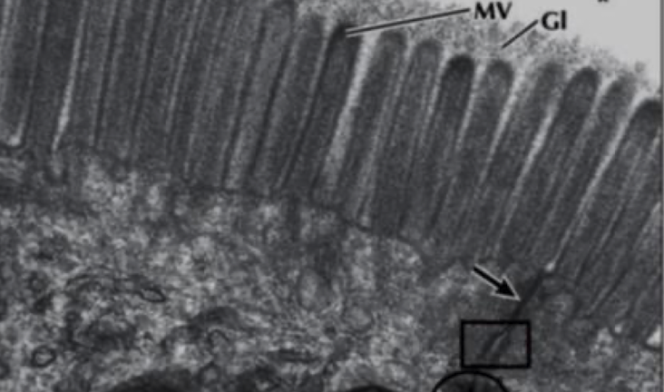
Brush Border
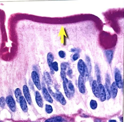
Microvilli
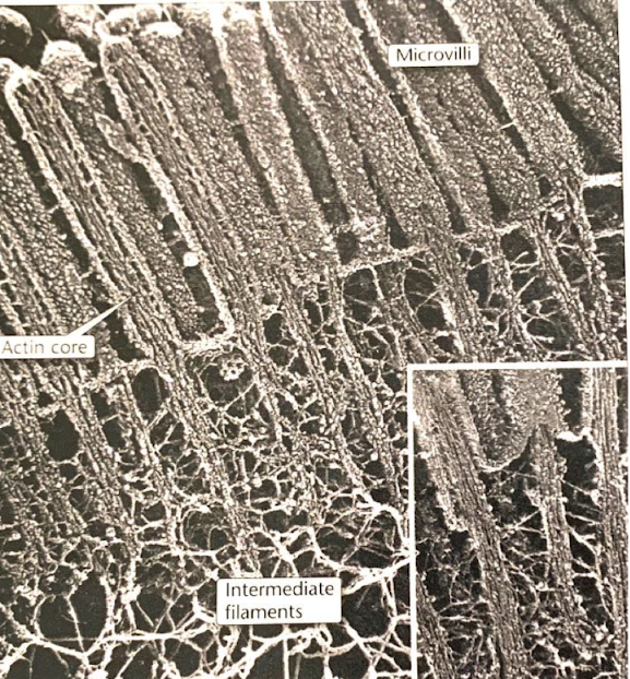
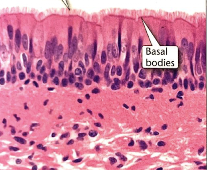
Cilia
cross section
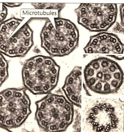
Cilia
top arrow
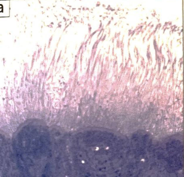
stereocilia (top of epididymis)
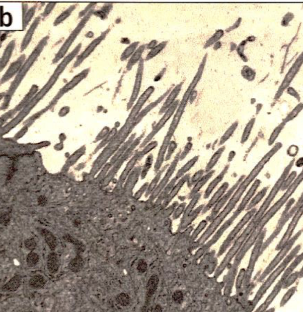
stereocilia (inner ear for balance)
endothelial cell | simple squamous epithelium
function: allows materials to pass by diffusion and filtration in sites where protection is not important
location: epithelium of blood vessels and lymphatic vessls; kidney glomeruli; air sac of lungs
longitudinal
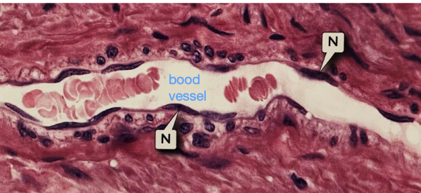
endothelial cell | simple squamous epithelium
function: allows materials to pass by diffusion and filtration in sites where protection is not important
location: epithelium of blood vessels and lymphatic vessls; kidney glomeruli; air sac of lungs
longitudinal
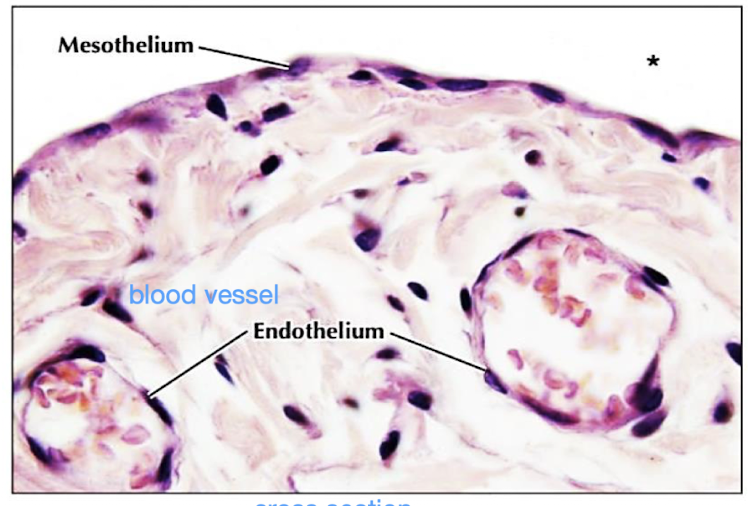
simple cuboidal epithelium
renal tubules
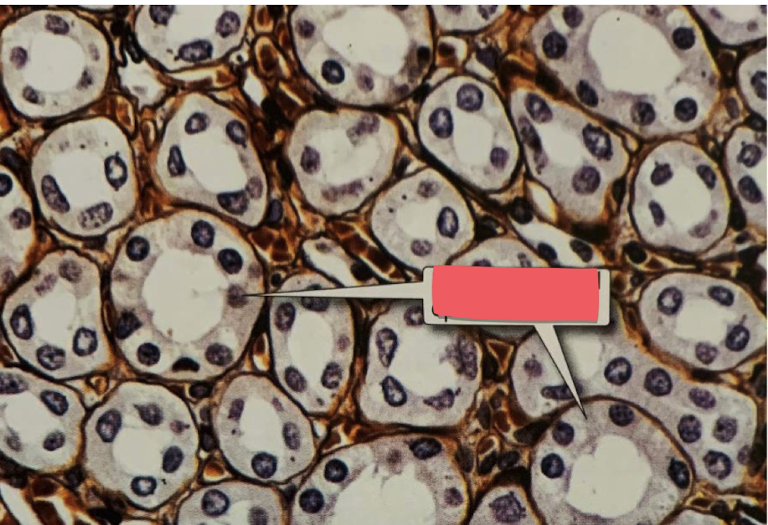
simple cuboidal epithelium
tubules in kidney
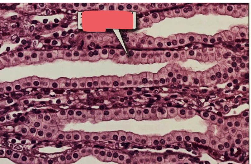
simple columnar epithelium
gallbladder
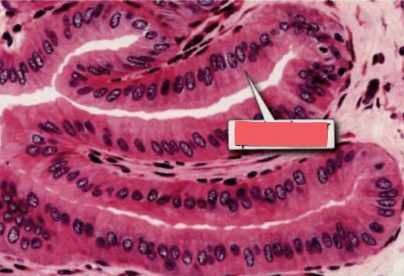
simple columnar epithelium with microvilli
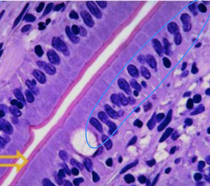
microvilli
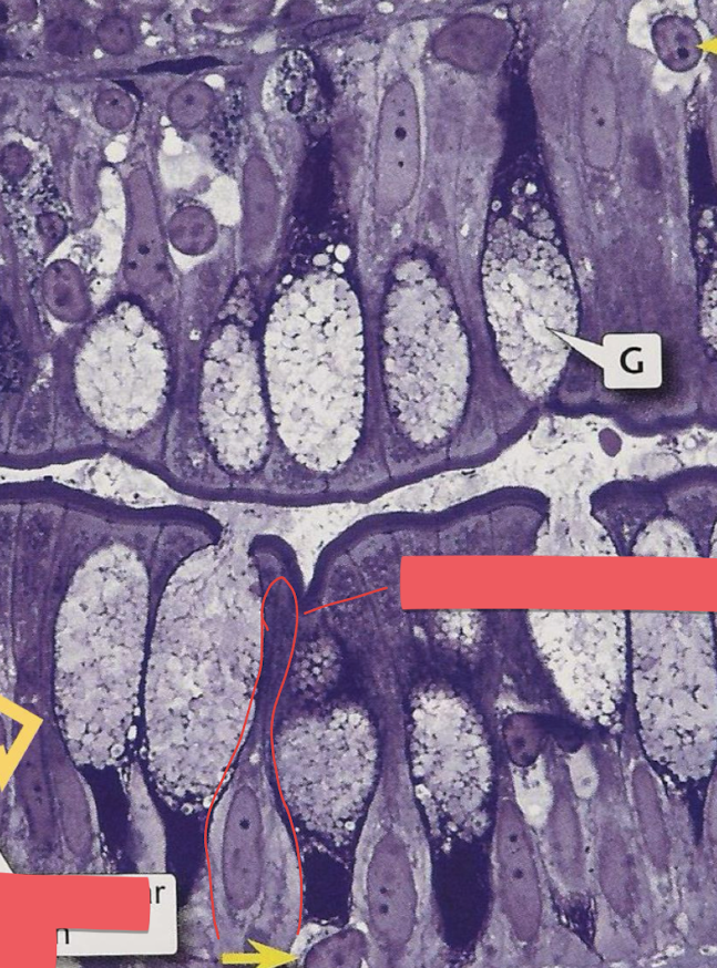
simple columnar cells with goblet cells and microvilli
yellow arrow = goblet cell
simple columnar cell in red border
brush border = thick purple line
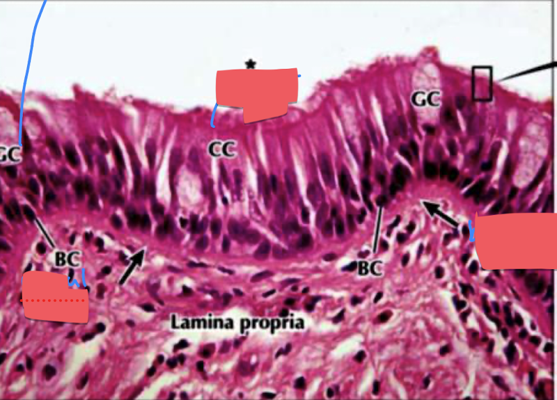
pseudostratified ciliated columnar epithelia with goblet cells
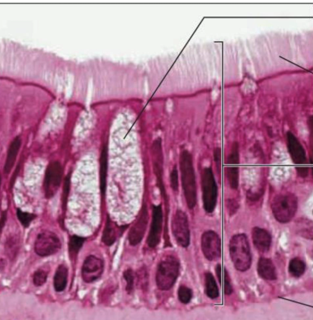
pseudostratified ciliated columnar epithelia with goblet cells
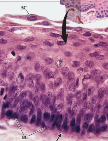
stratified squamous epithelium nonkeratinized
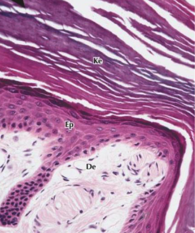
stratified squamous epithelium keratinized
stratified squamous epithelium keratinized
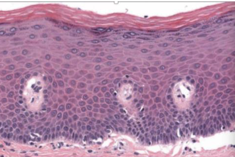
stratified squamous epithelium nonkeratinized
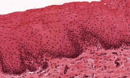
straitifed cuboidal epithelium
duct of salivary glands
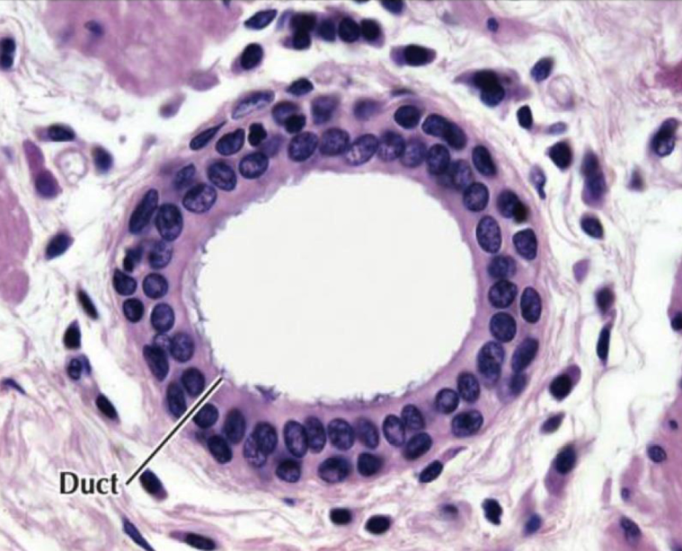
stratified columnar epithelium
male urethra, pharnyx
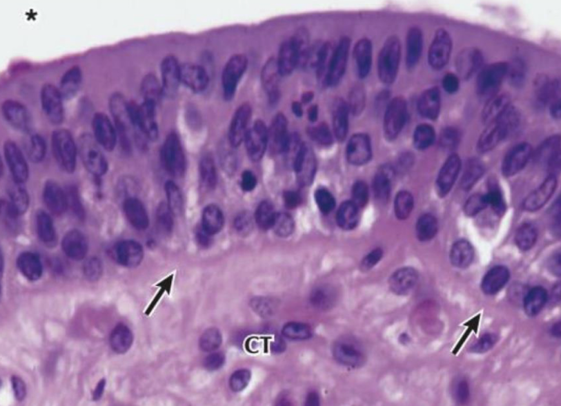
transitional epithelium relaxed
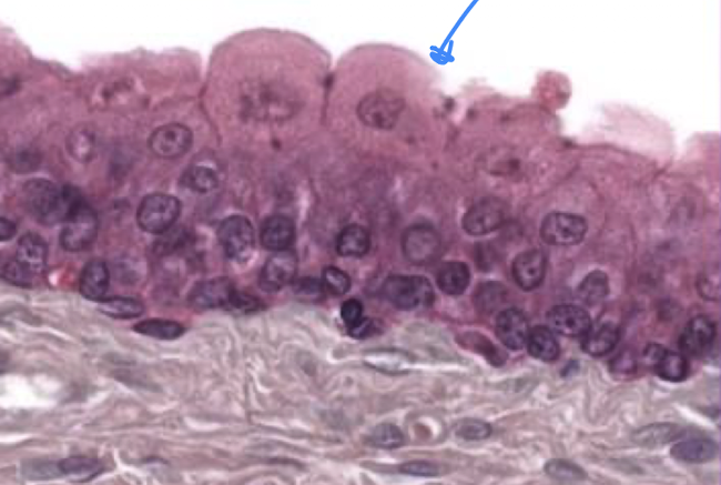
transitional epithelium extended state
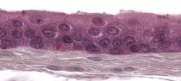
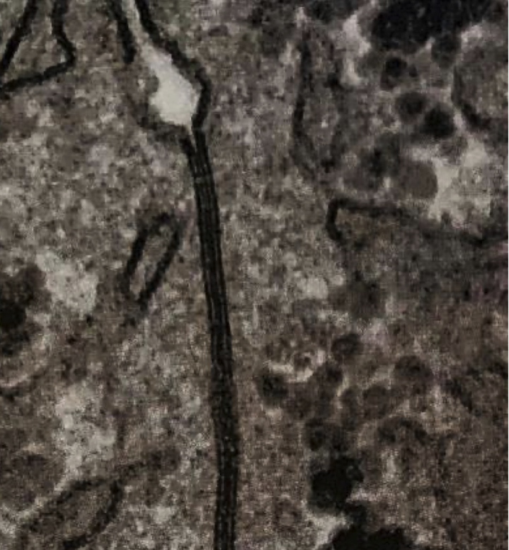
zonula adherens (bottom), zonula occludens (top)
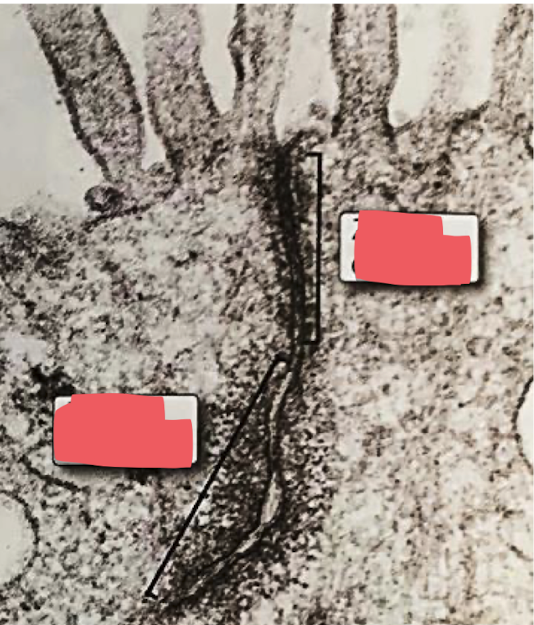
adherens junction
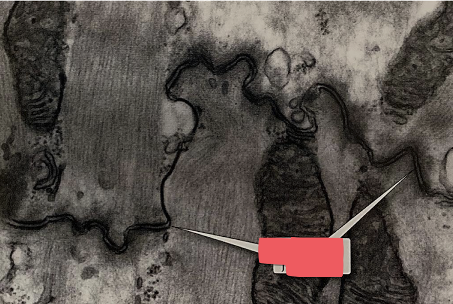
desmosomes
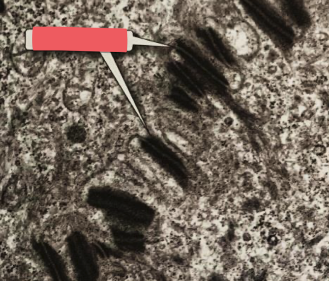
gap junction
lines across very hard to see
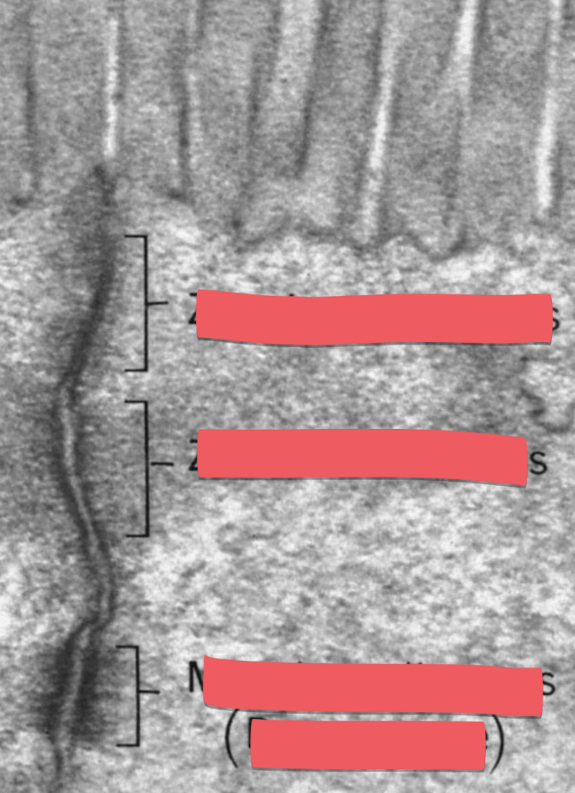
zonula occludes (top), zonula adherens (middle), macula adherens (desmosome bottom)
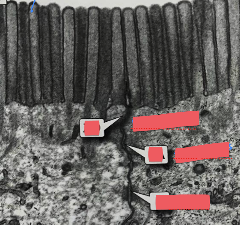
blue arrow (microvilli)
zonula occludens
zona adherens
desmosome
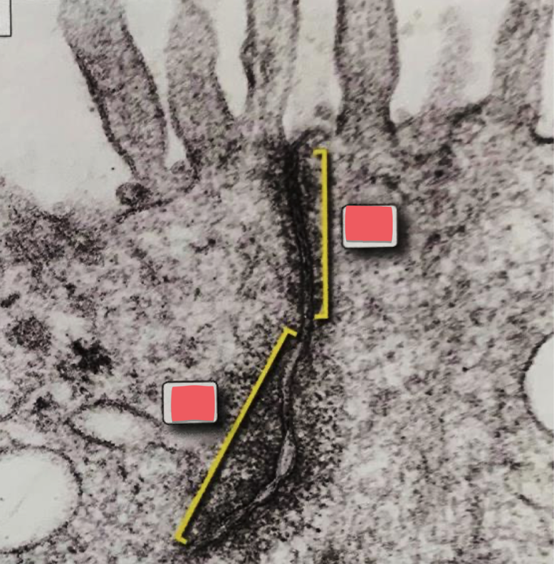
top: zonula occludens (tight junction)
bottom: zonula adherens
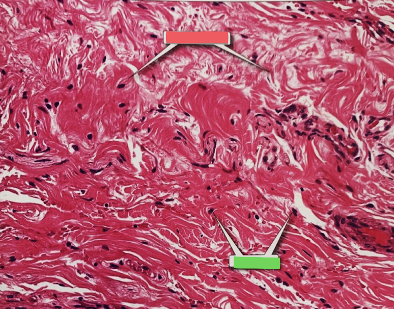
collagen fibers (red box), fibroblast (green box)
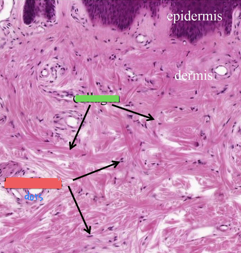
collagen (green box), fibroblasts (red box)
mature fibroblast
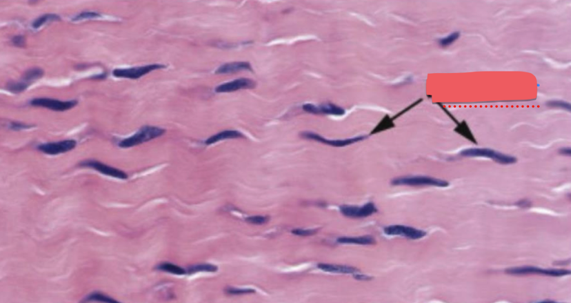
collagen fiber (with mature fibroblast)
pseudopodium (red box)
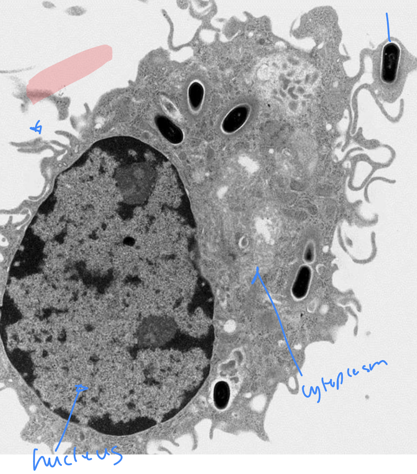
Macrophages
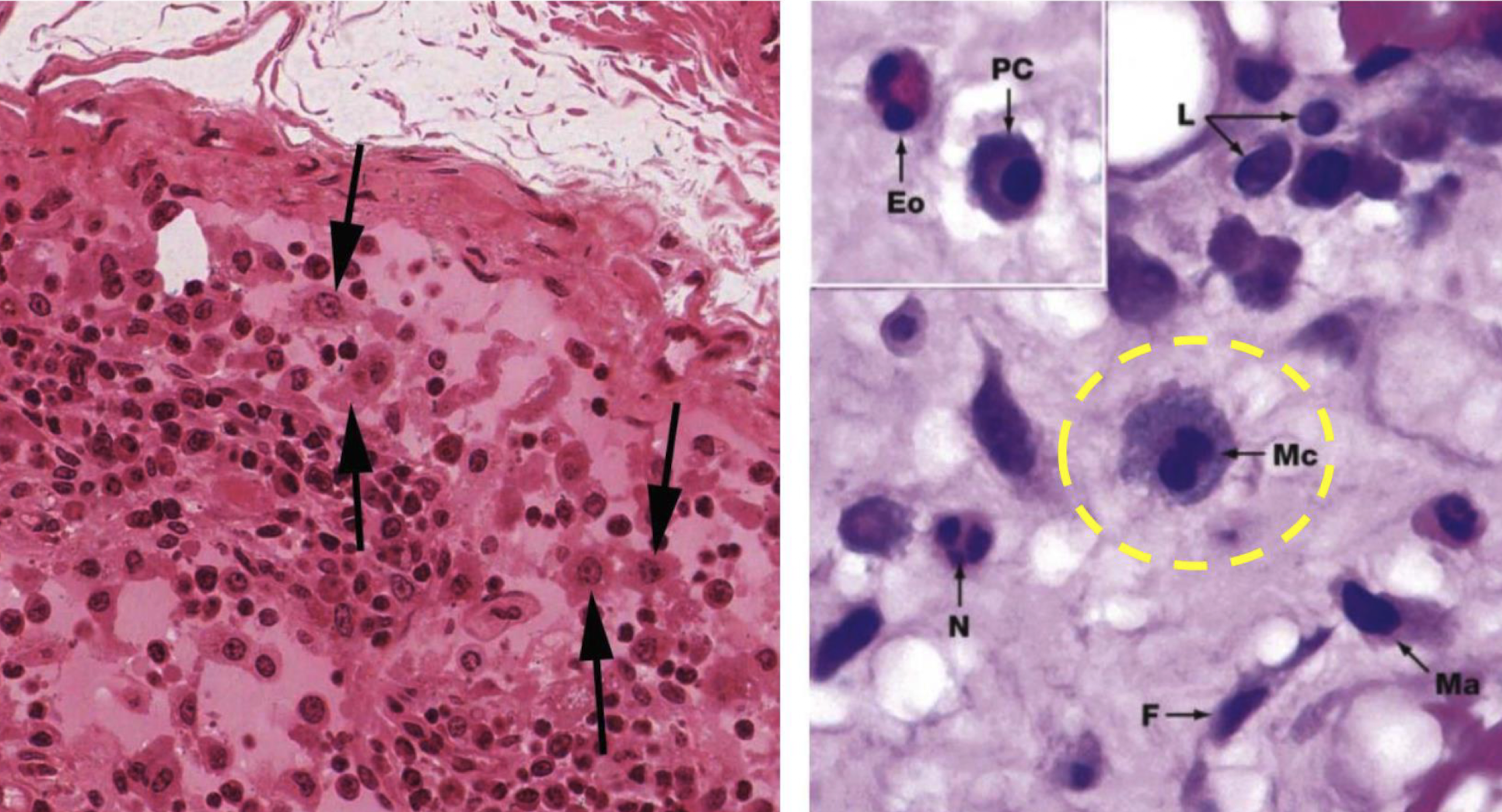
mast cell
look at the dark pink circles for the right image
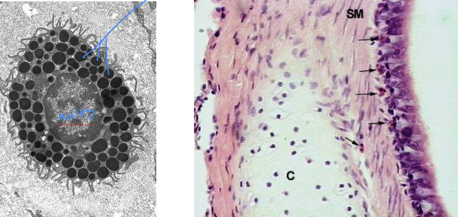
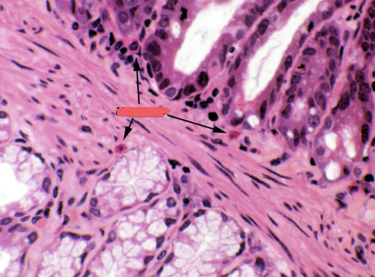
Mast cells
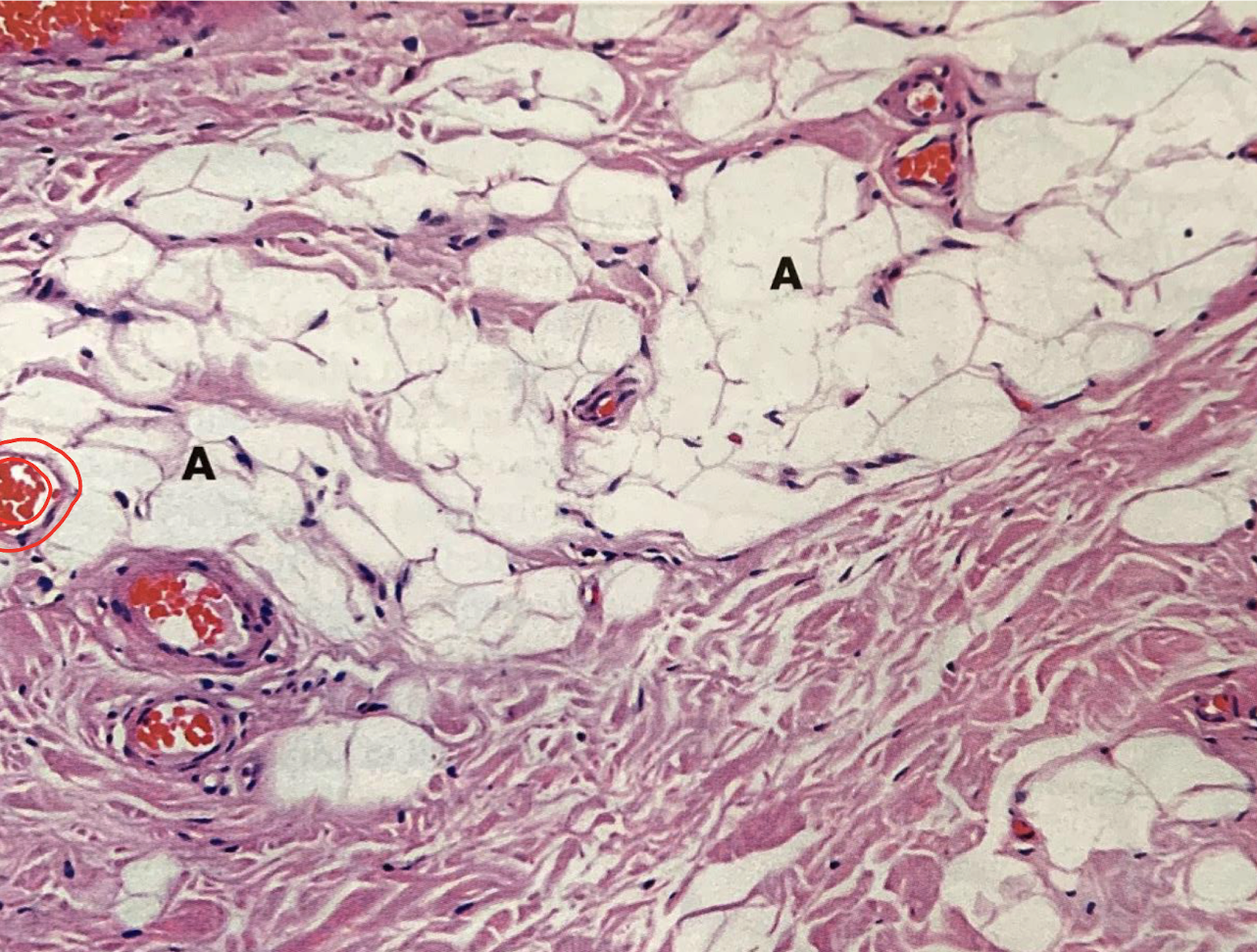
adipocytes
simple squamous epithelium (red border), blood vessels with erythryoctes (blue arrow), aidpocytes (A)
identify red, blue, and A site
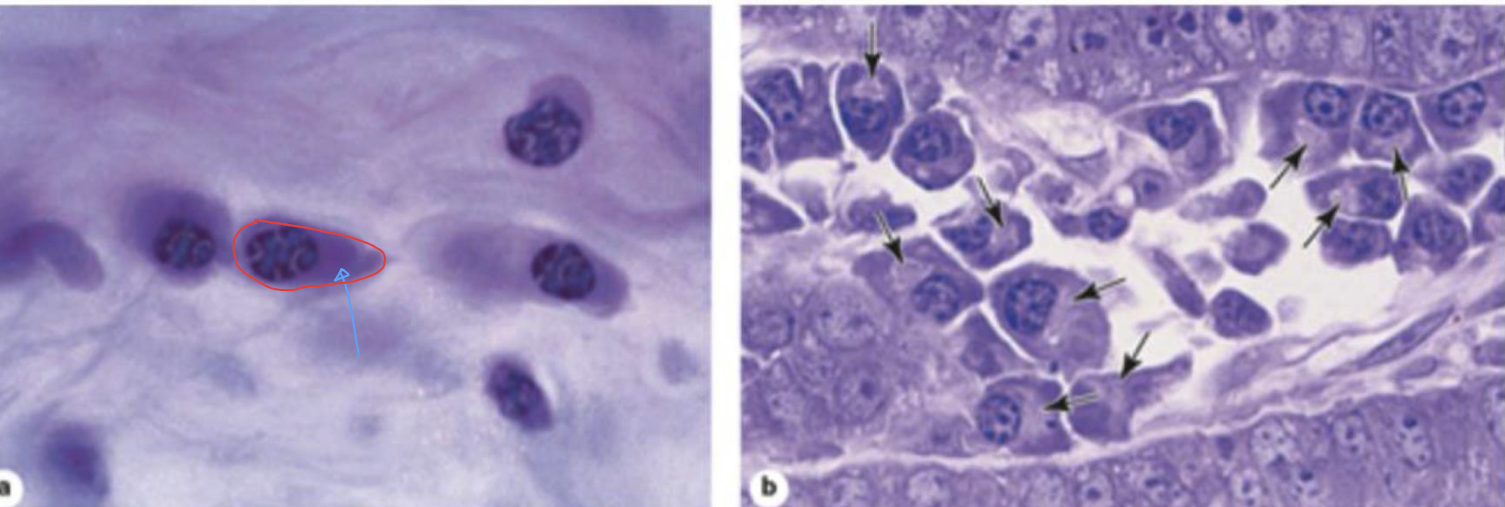
plasma cells, blue arrow (basophilic cytoplasm)
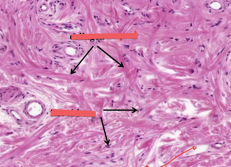
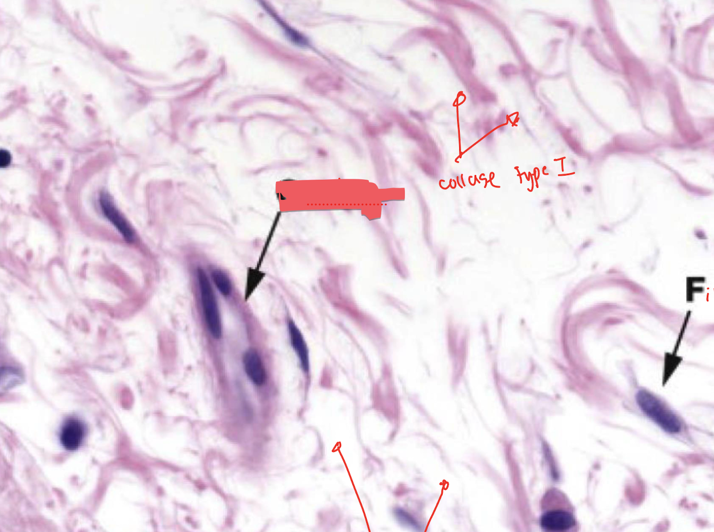
collagen type 1(top), fibroblast, ground substance (bottom)
capillary
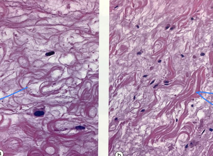
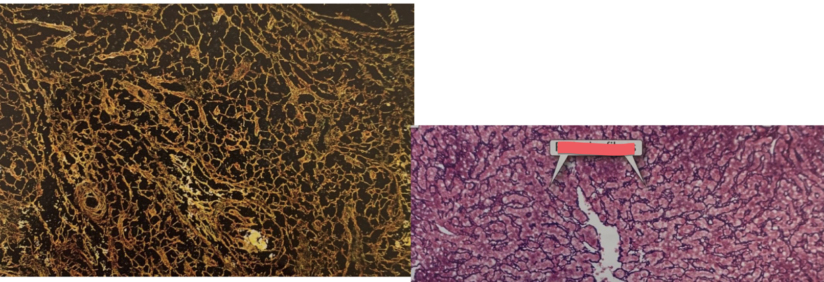
reticular fibers
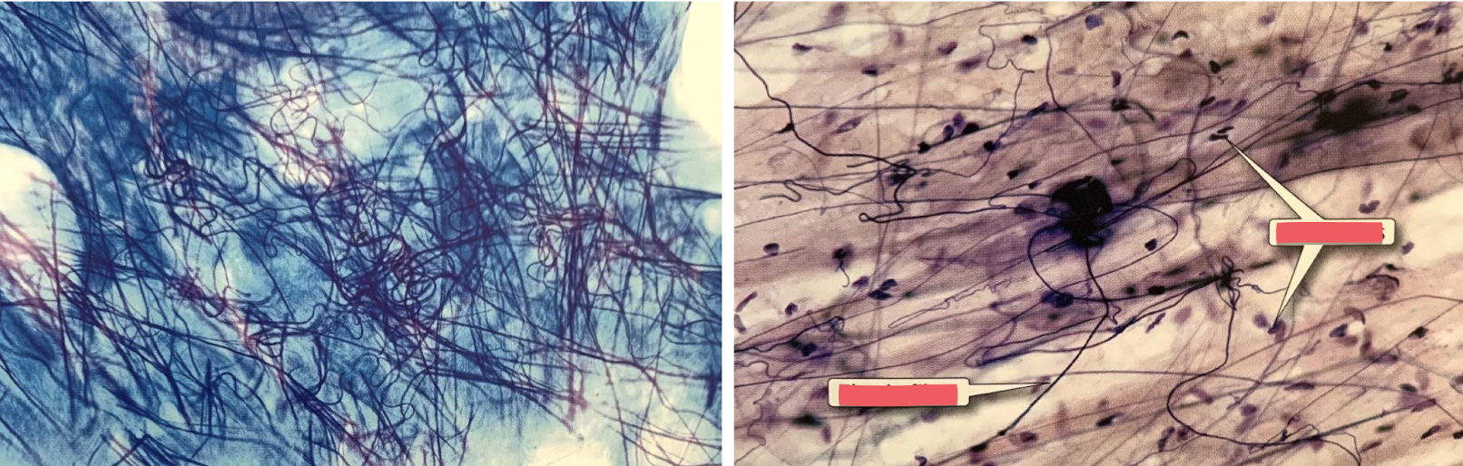
fibroblast (right box), elastic fibers
elastic fibers
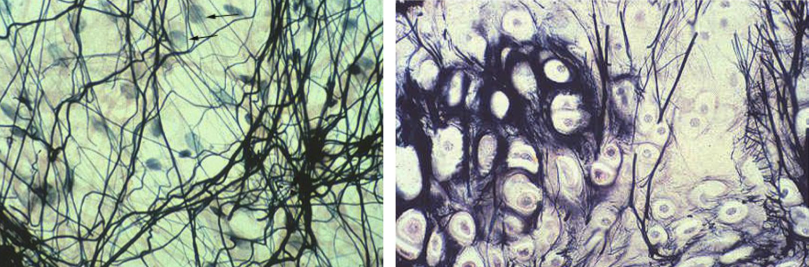
enamel (left box) internal elastic lamina (right box)
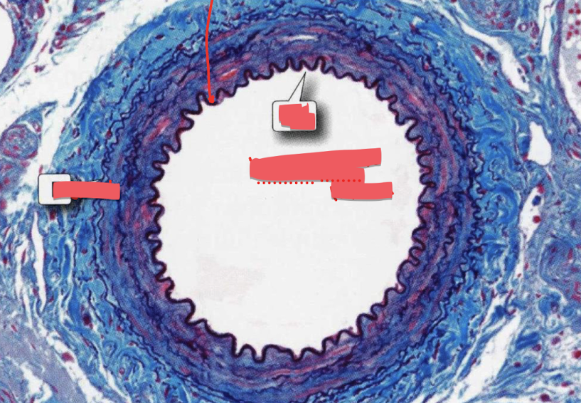
elastic (right box), collagen (left box)
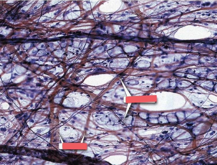
loose connective tissue
identify the type of connective tissue
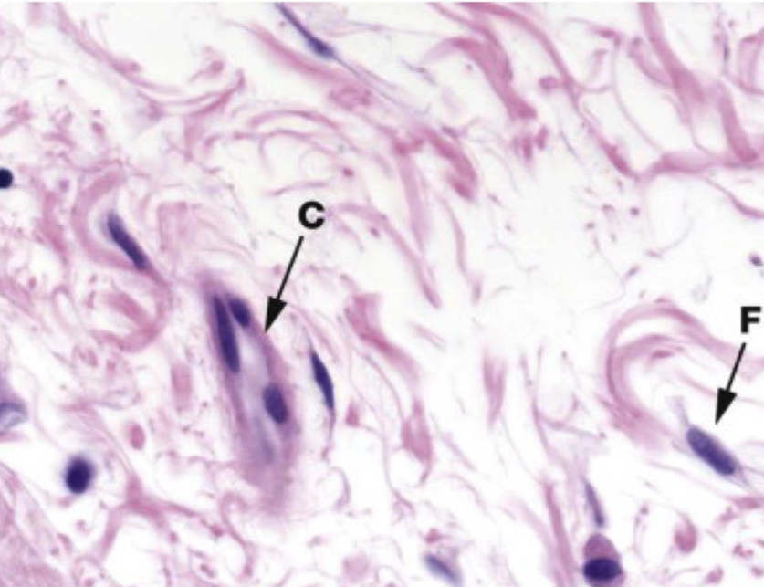
dense conective tissue
identify the type of connective tissue
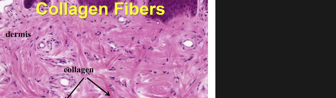
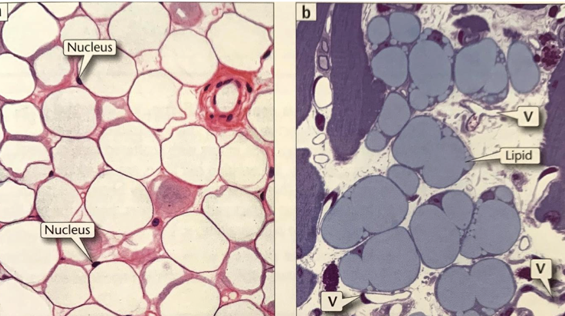
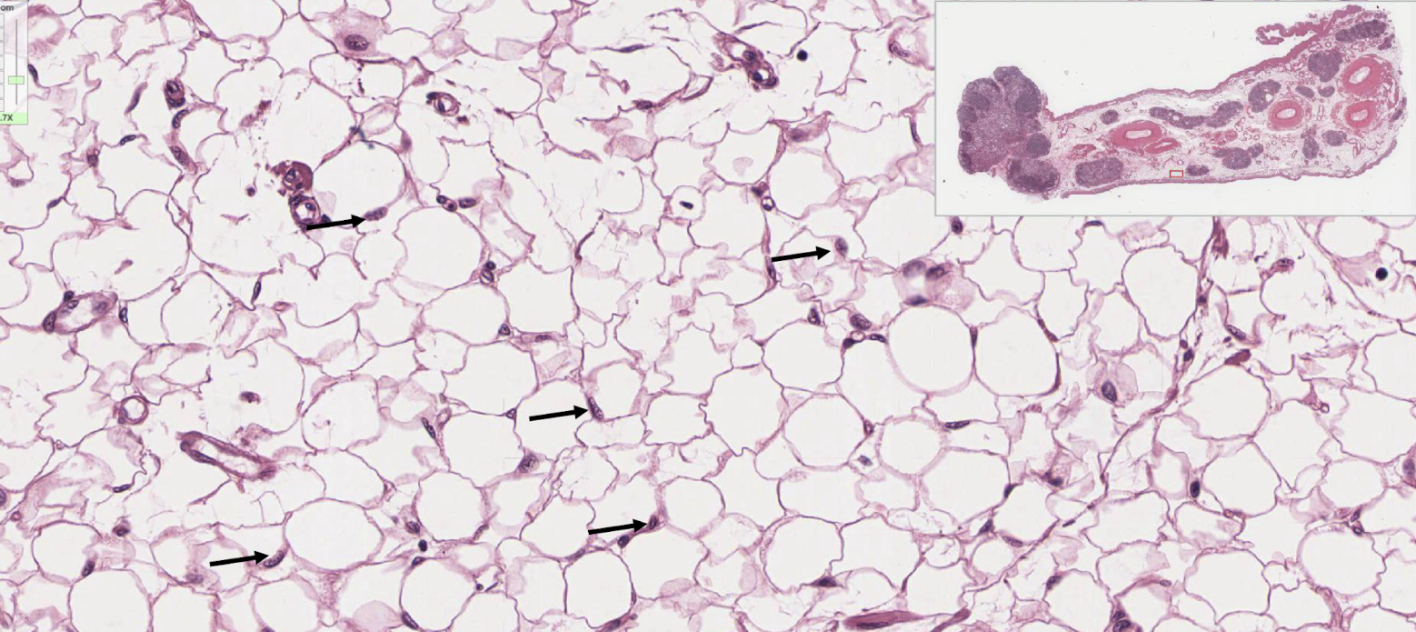
Loose connective tissue: adipose tissue
unilocular adipocyte (A)
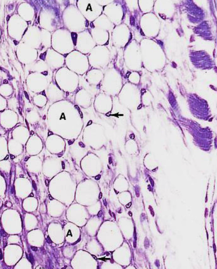
unilocular adipocyte
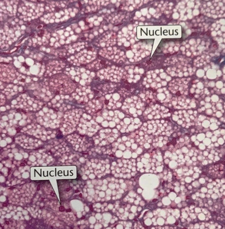
loose connective tissue: brown adipose tissue
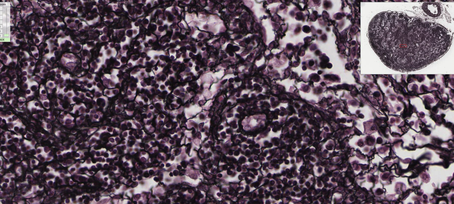
loose connective tissue: reticular tissue
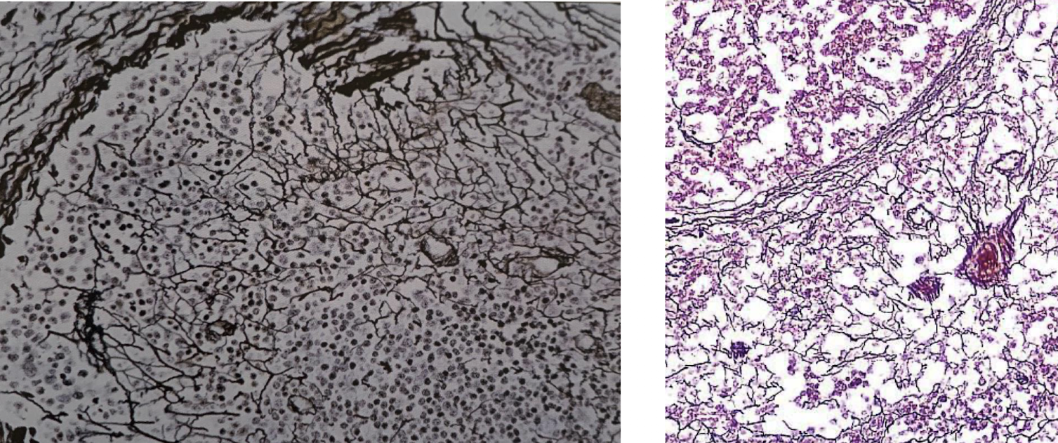
loose connective tissue: reticular tissue
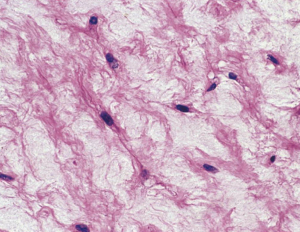
loose connective tissue: mucous connective tissue
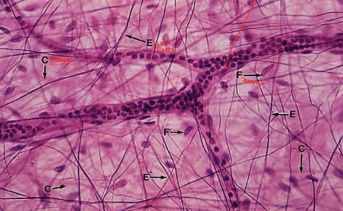
loose connective tissue: mesentery
dense connective tissue: dense irregular connective tissue
top purple is keratinized stratified squamous epithelium
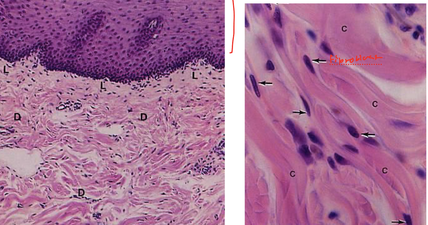
dense connective tissue: dense irregular connective tissue
collagen type 1 going in different direction (salmon pink)
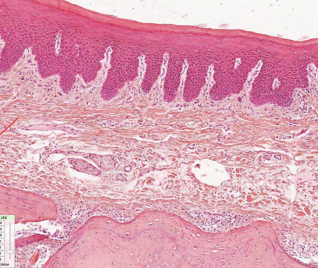
dense connective tissue: dense regular connective tissue
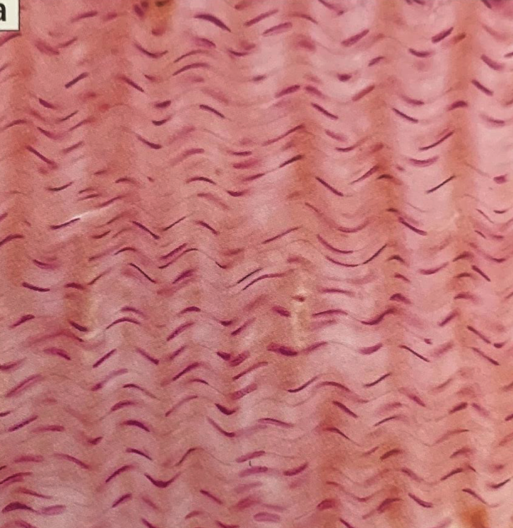
dense connective tissue: dense regular connective tissue
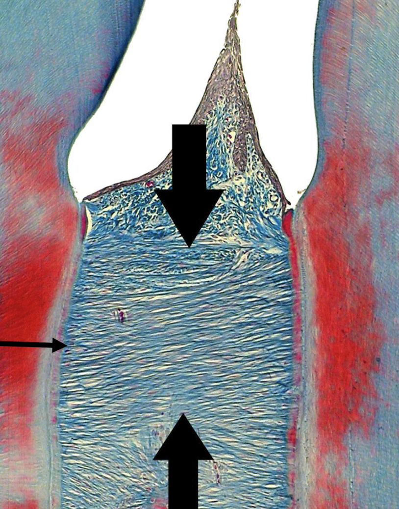
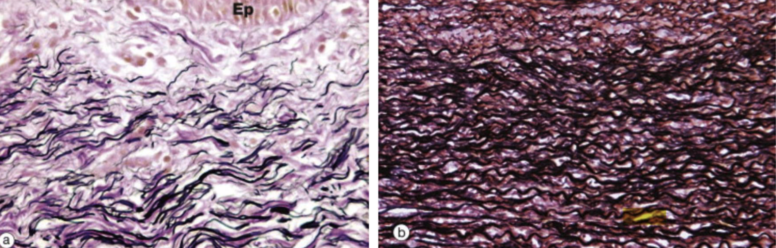
dense connective tissue: elastic connective tissue
elastic fibers in black
collagen fibers in red
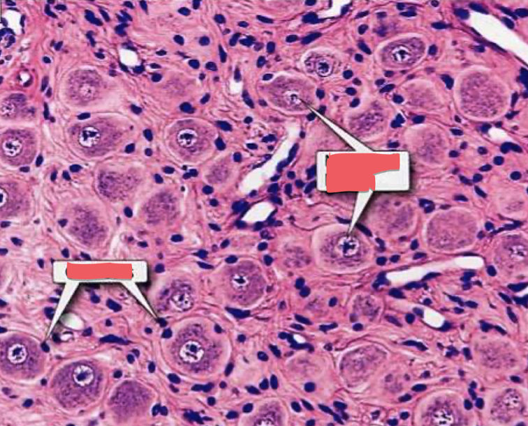
nerve cell (right box), glial cell (left box)
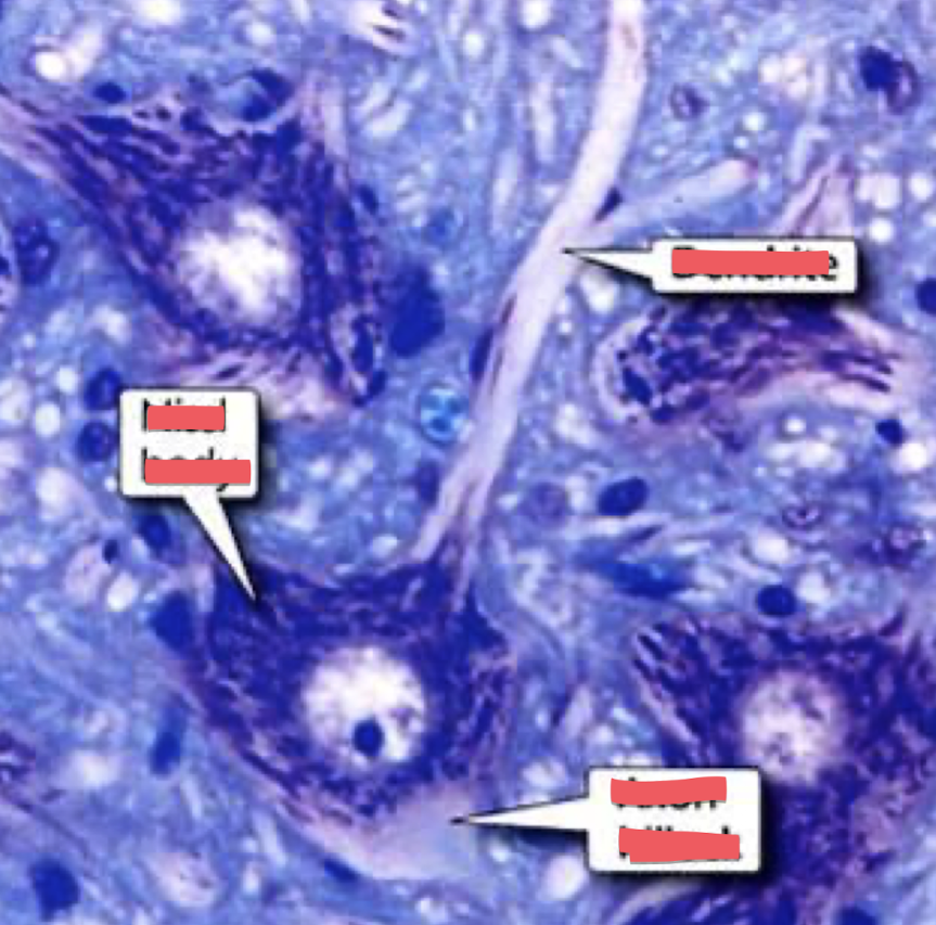
dendrite, nissl body, axon hillock
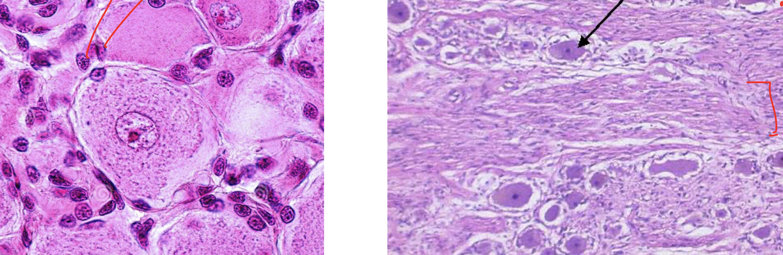
glial cells (red arrow), cell bodies (black arrow), axon sounded by myelin sheath (red segment)
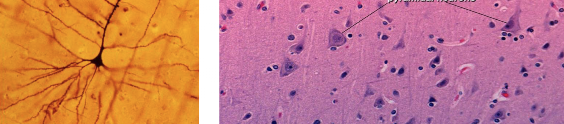
pyramidal neurons
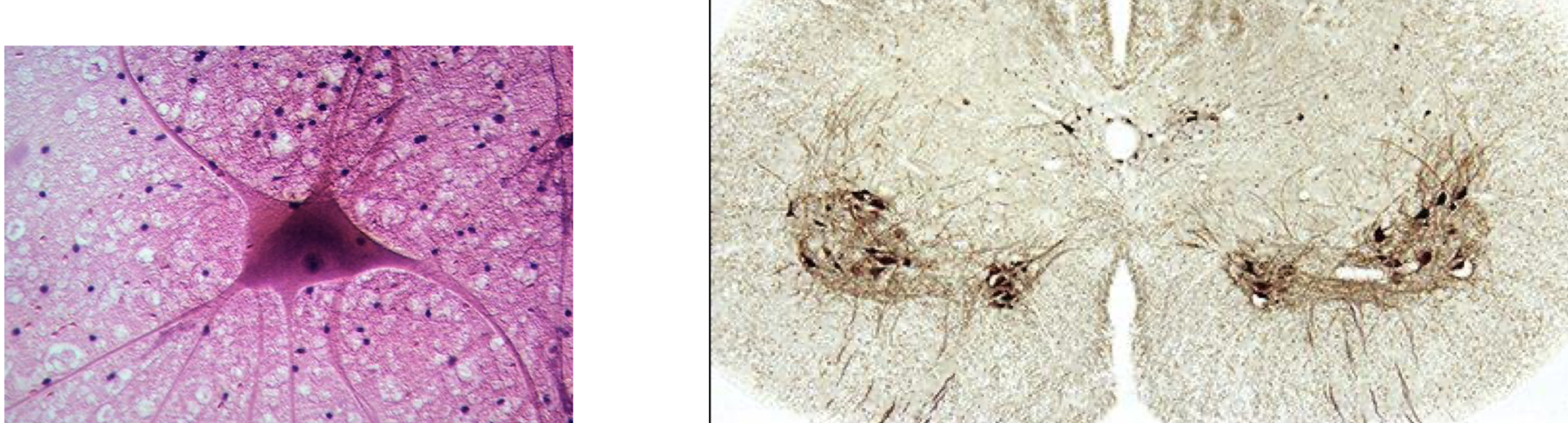
motor neurons
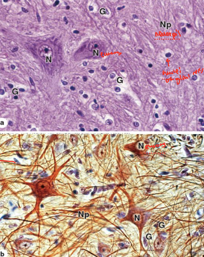
purple (nuclei of glial cells bottom image)
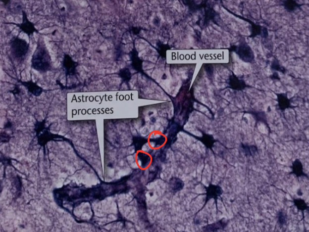
protoplasmic astrocyte (gray matter)
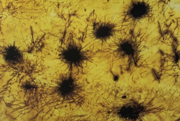
fibrous astrocyte (white matter)
oligodendrocytes
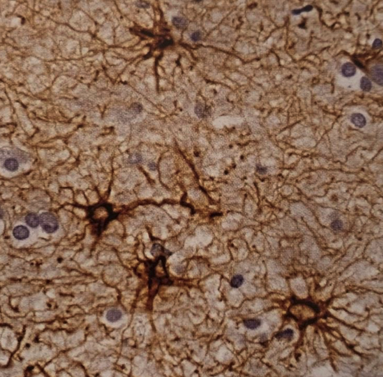
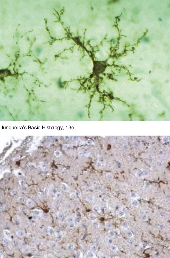
microglia
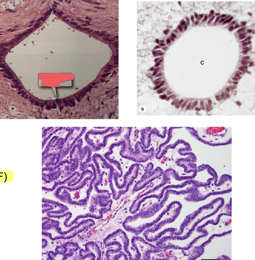
ependymal cells
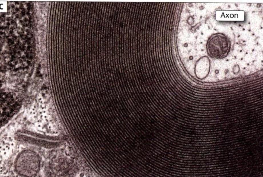
schwann cells (myelinated axon)
schwann cells (unmyelinated axons)
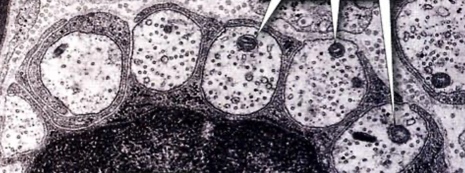
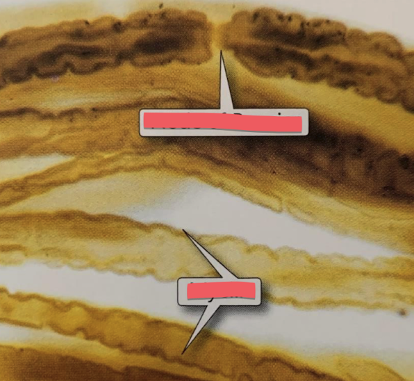
nodes of ranvier (top), myelin (bottom)
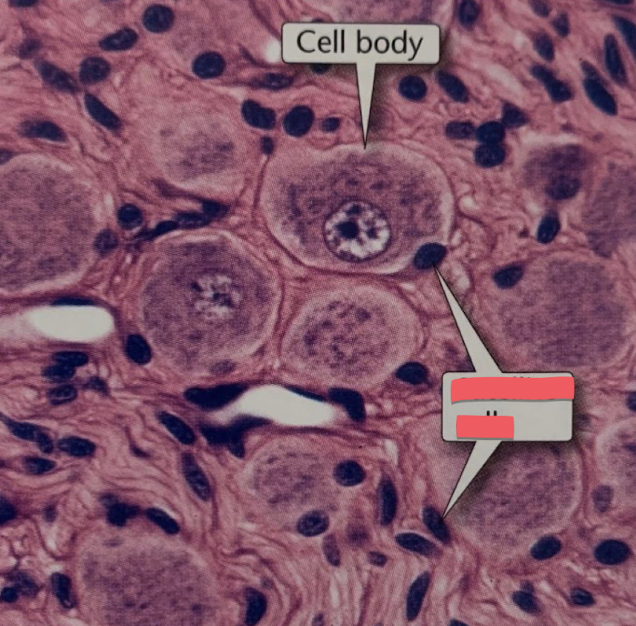
satellite cells
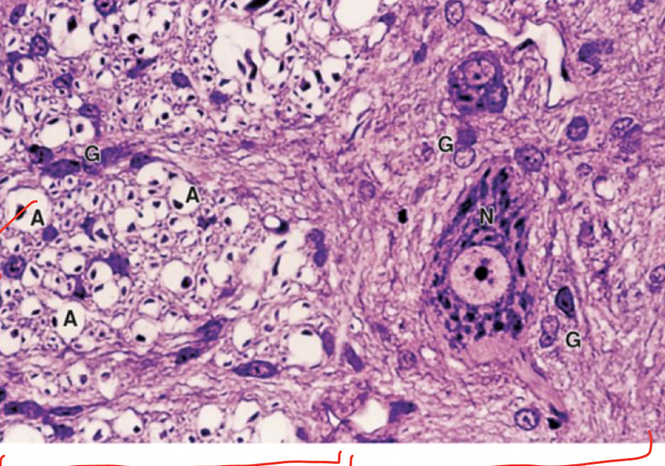
oligodendrocyte (A), white matter (left half), gray matte (right half)
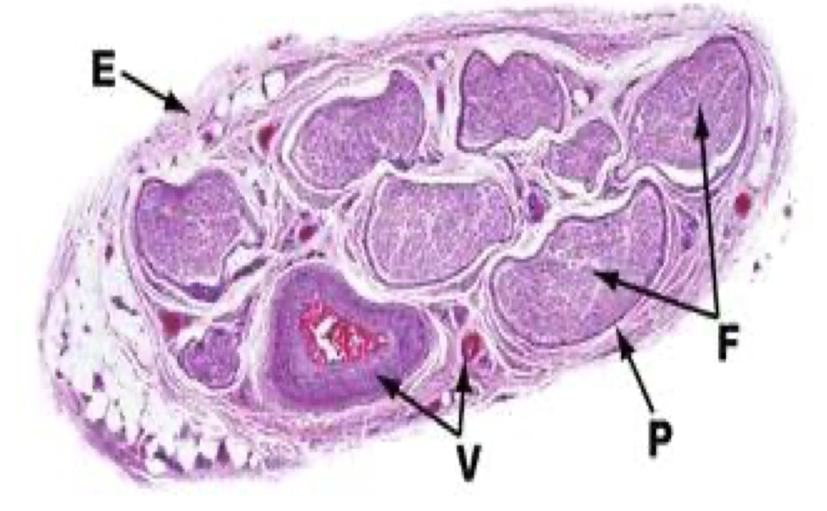
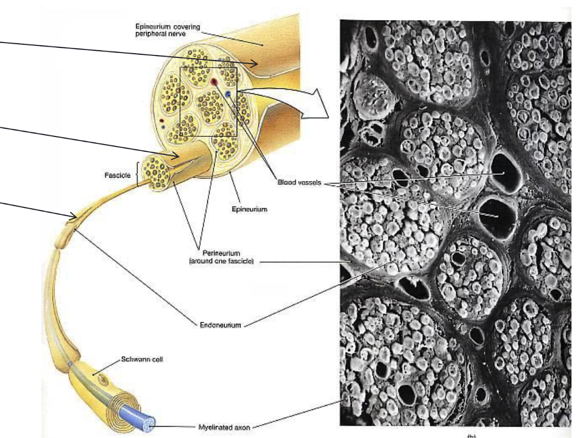
epineurium
perineurum
endoneurium
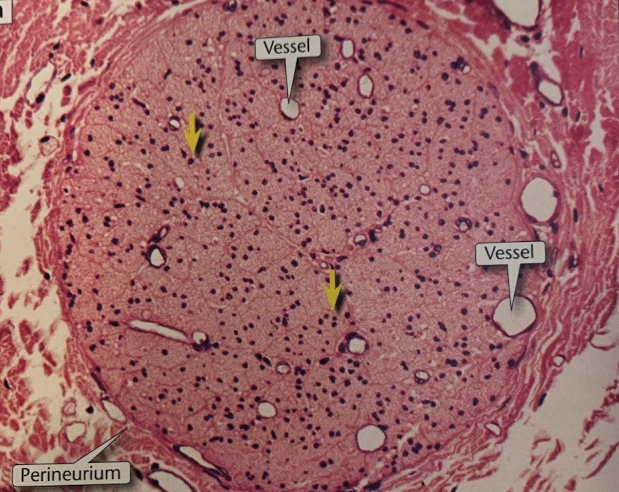
unmyelinated peripheral nerve
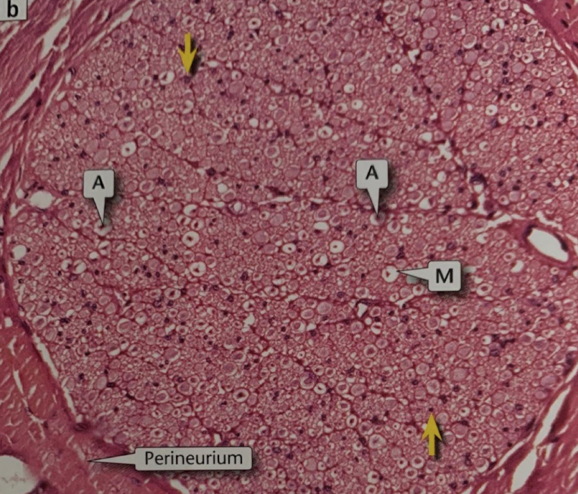
myelinated peripheral nerve
fascicles (F)
F