Mammalian Anatomy Practical 3
1/119
Earn XP
Description and Tags
Neck muscles and skull bone features and some cranial nerves.
Name | Mastery | Learn | Test | Matching | Spaced |
|---|
No study sessions yet.
120 Terms
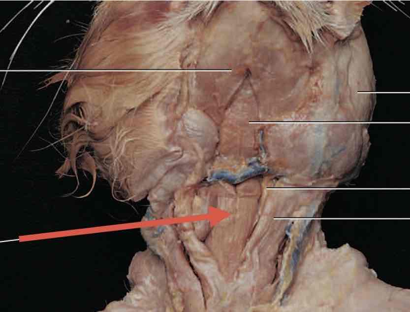
Sternohyoid
Insertion: Lower border of the hyoid bone
Innervation: Ansa Cervicalis
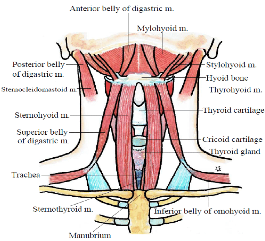
Note that stylohyoid is superior to the digastric muscle, and thin. This image is for a human.
Stylohyoid
Insertion: Body of the hyoid bone
Innervation: Facial Nerve (CN07)
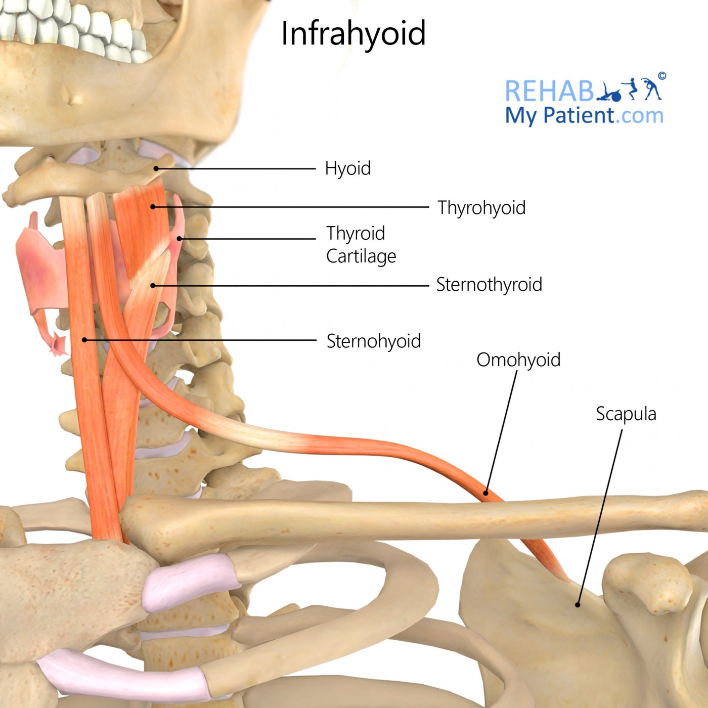
Observe how the omohyoid is directly lateral to the sternohyoid
Omohyoid
Insertion: Lower border of the body of the hyoid bone
Innervation: Branches of spinal nerves (C01-C03)
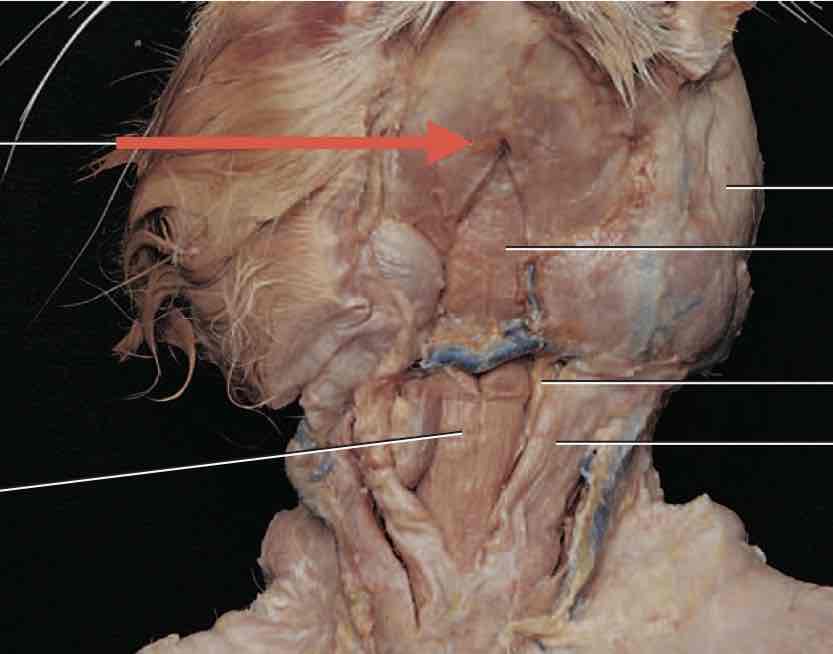
Digastric
Insertion: Common tendon onto the hyoid bone
Innervation: Trigeminal nerve (CN05), mylohyoid branch (anterior belly) + Facial nerve (CN07), digastric branch (posterior belly)
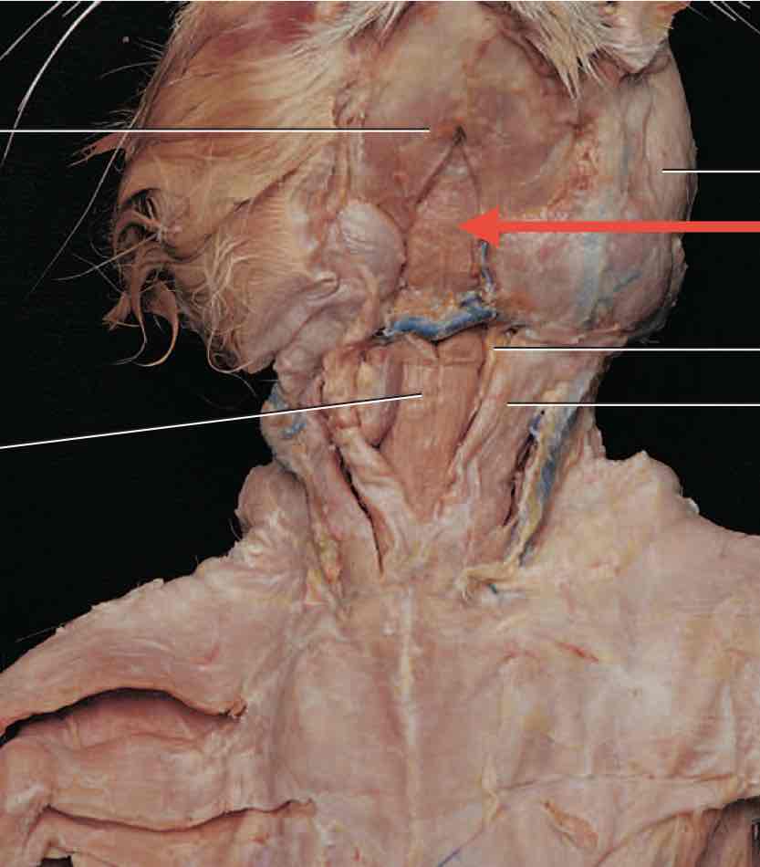
Mylohyoid
Insertion: Body of the hyoid bone
Innervation: Trigeminal nerve (CN05) and mylohyoid branch
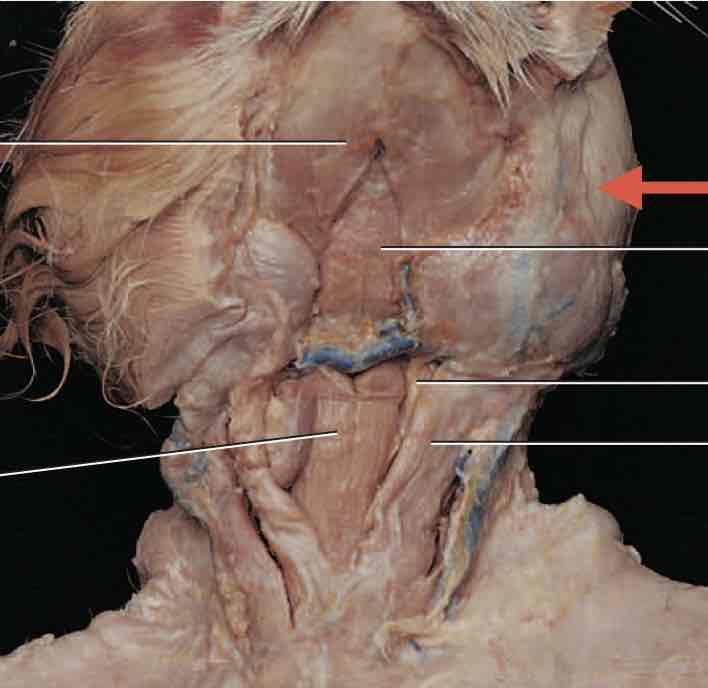
Masseter
Insertion: Ramus of mandible
Innervation: Trigeminal nerve (CN05), mandibular branch (superficial and deep masseter)
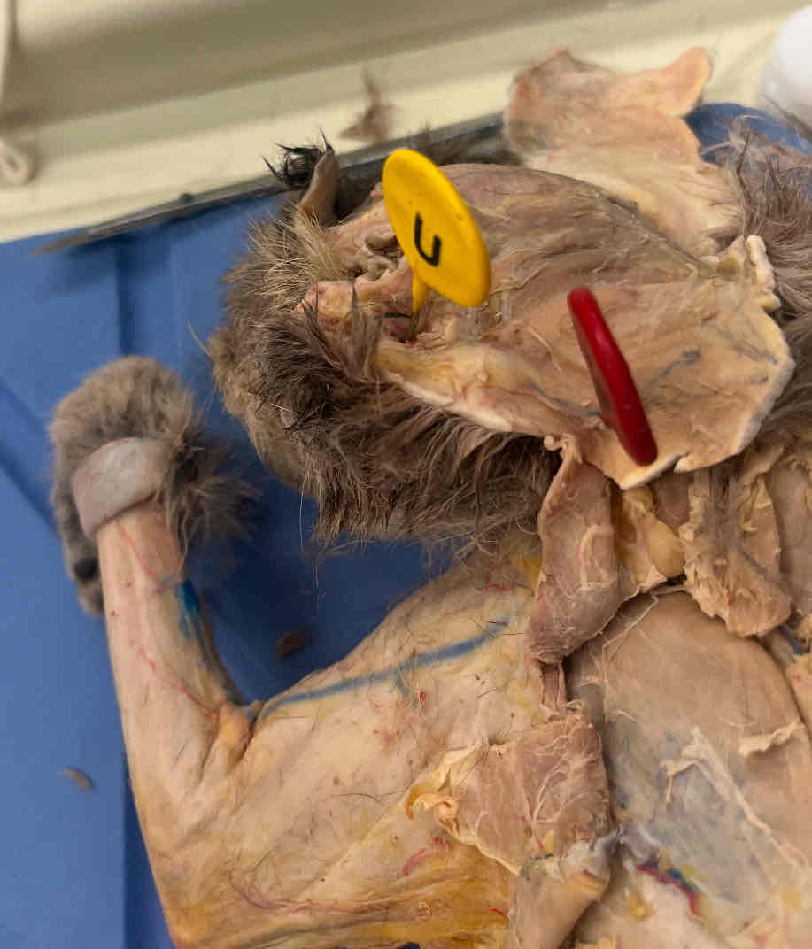
U
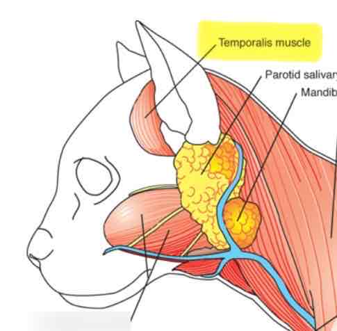
Temporalis
Insertion: Coronoid process and anterior ramus of mandible
Innervation: Trigeminal nerve (V), mandibular branch
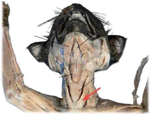
Cleidomastoid
Insertion: Lateral surface of the mastoid process
Innervation: Accessory nerve (CN11); branches from the anterior divisions of nerves C02 and C03
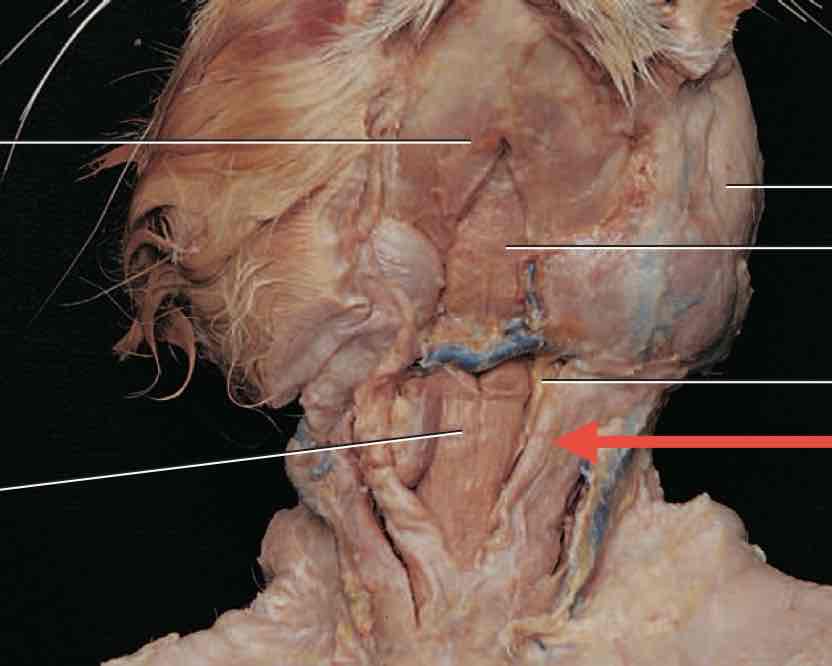
Sternomastoid
Insertion: Lateral surface of the mastoid process
Innervation: Accessory nerve (CN11); branches from the anterior divisions of nerves C02 and C03
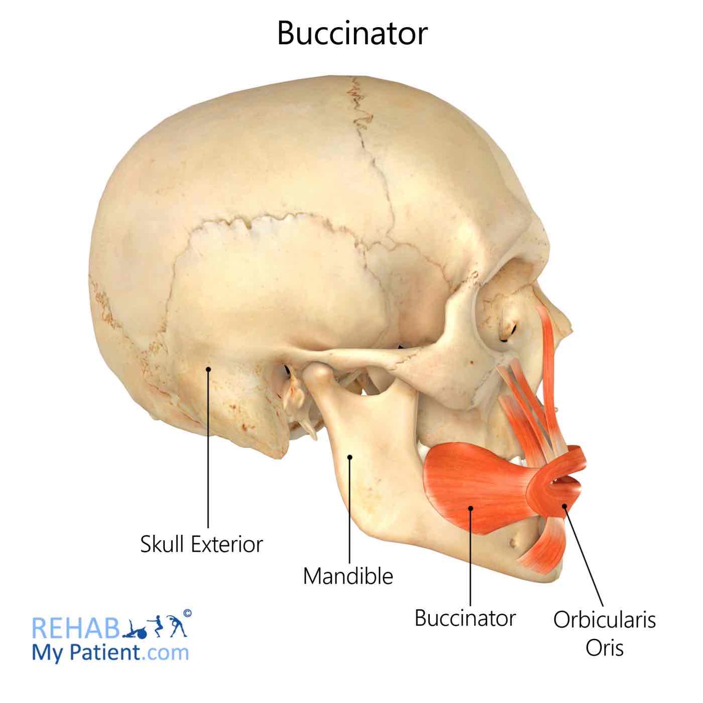
Buccinator
Buccinator
Insertion: Orbicularis oris
Innervation: Facial nerve (VII), buccal branch
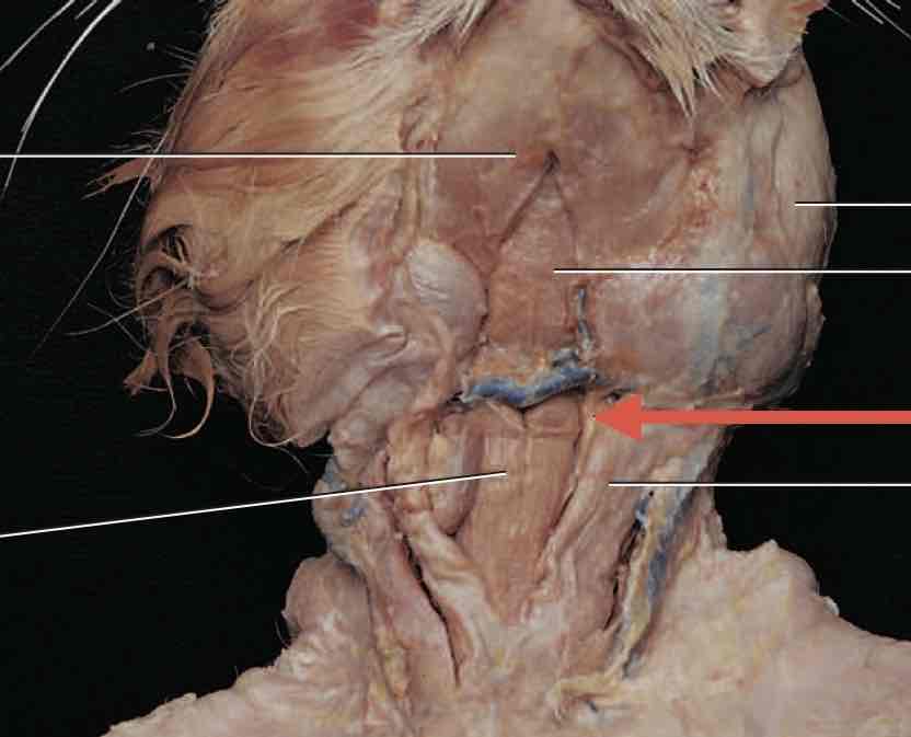
Sternothyroid
Insertion: Oblique line of the thyroid cartilage
Innervation: Ansa cervicalis
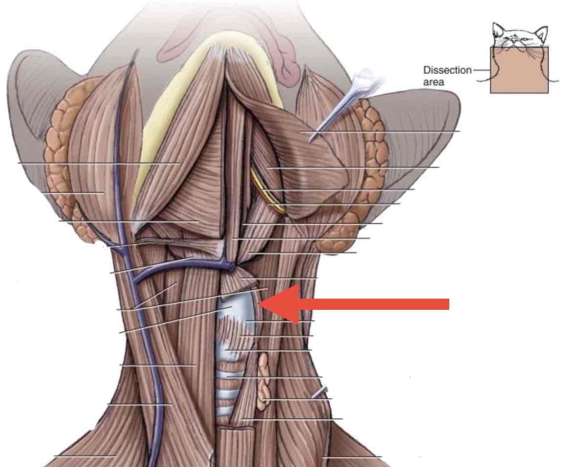
Thyrohyoid
Insertion: Hyoid bone
Innervation: Anterior ramus of C1, via the hypoglossal nerve (CN12)
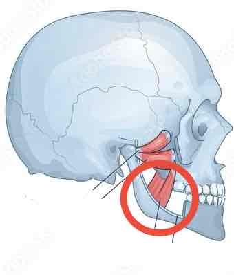
Medial Pterygoid
Insertion: Ramus and angle of the mandible
Innervation: Trigeminal nerve (CN05), mandibular branch
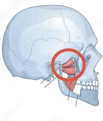
Lateral Pterygoid
Insertion: Condyle of mandible
Innervation: Trigeminal nerve (V), mandibular branch
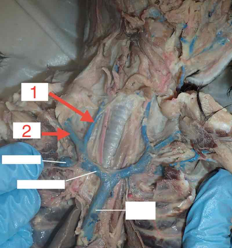
Jugular vein
Internal (1)
External (2)
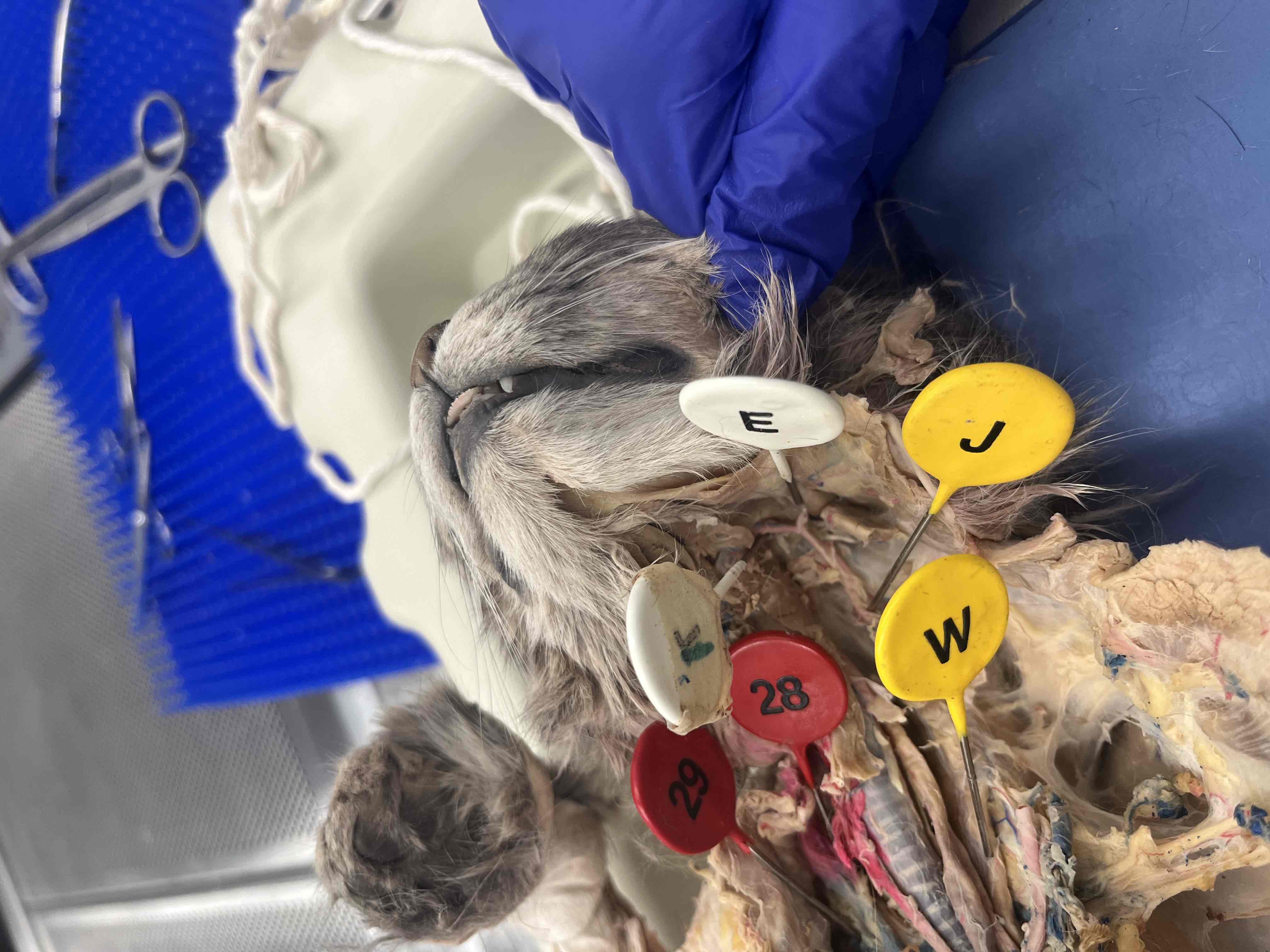
1, E and J
Carotid artery
J - Common
1 - Internal
E - External
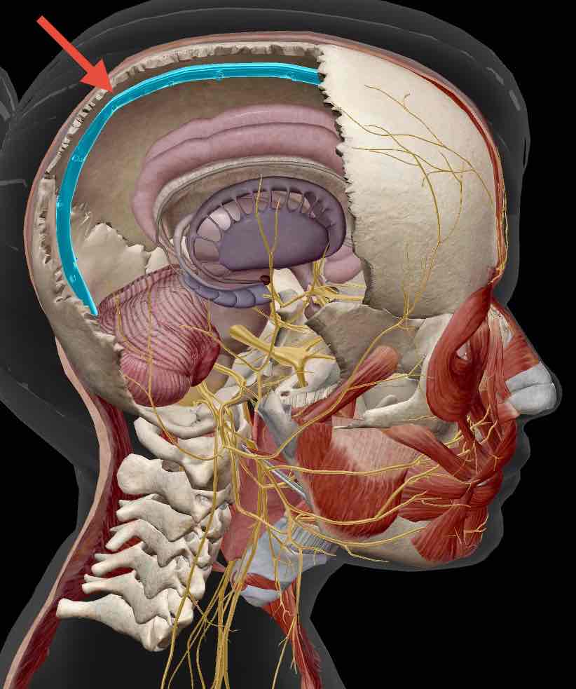
Superior sagittal sinus
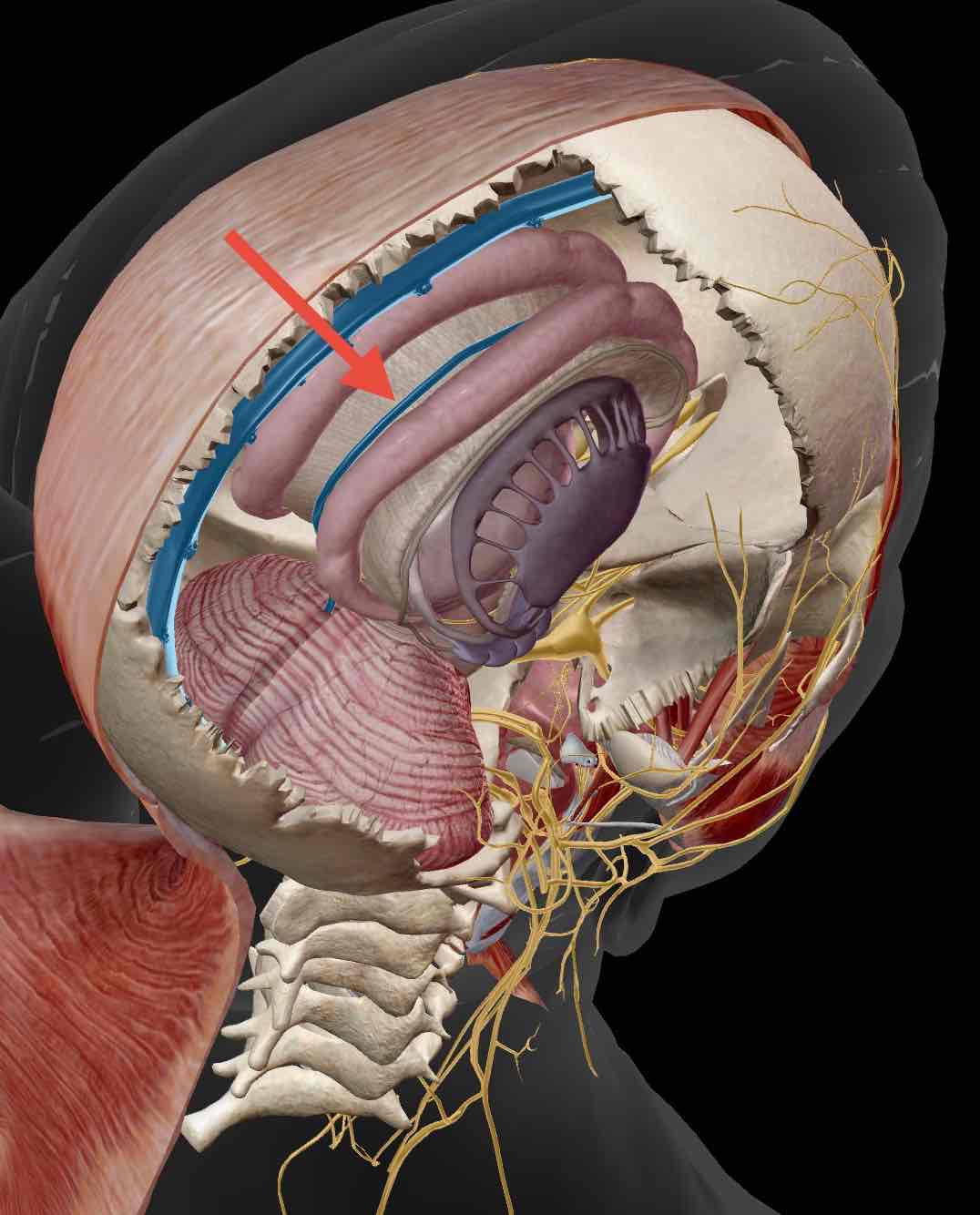
Inferior sagittal sinus
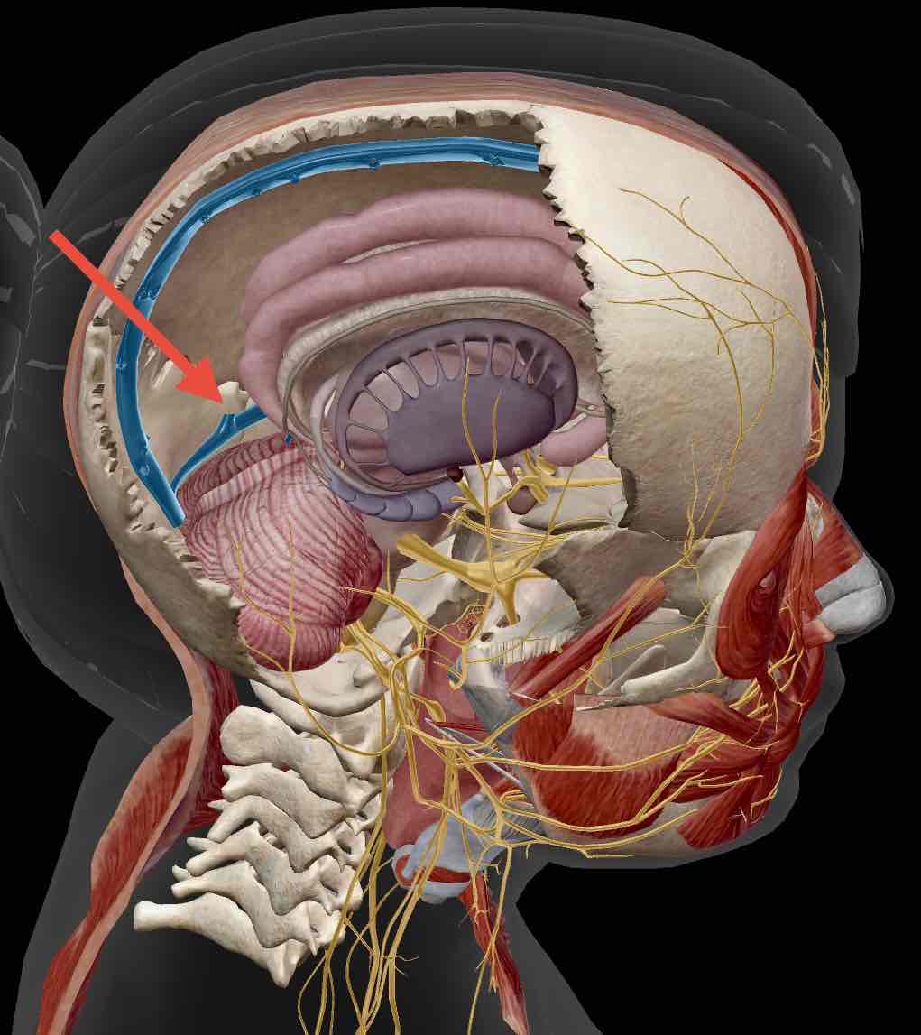
Transverse sinus
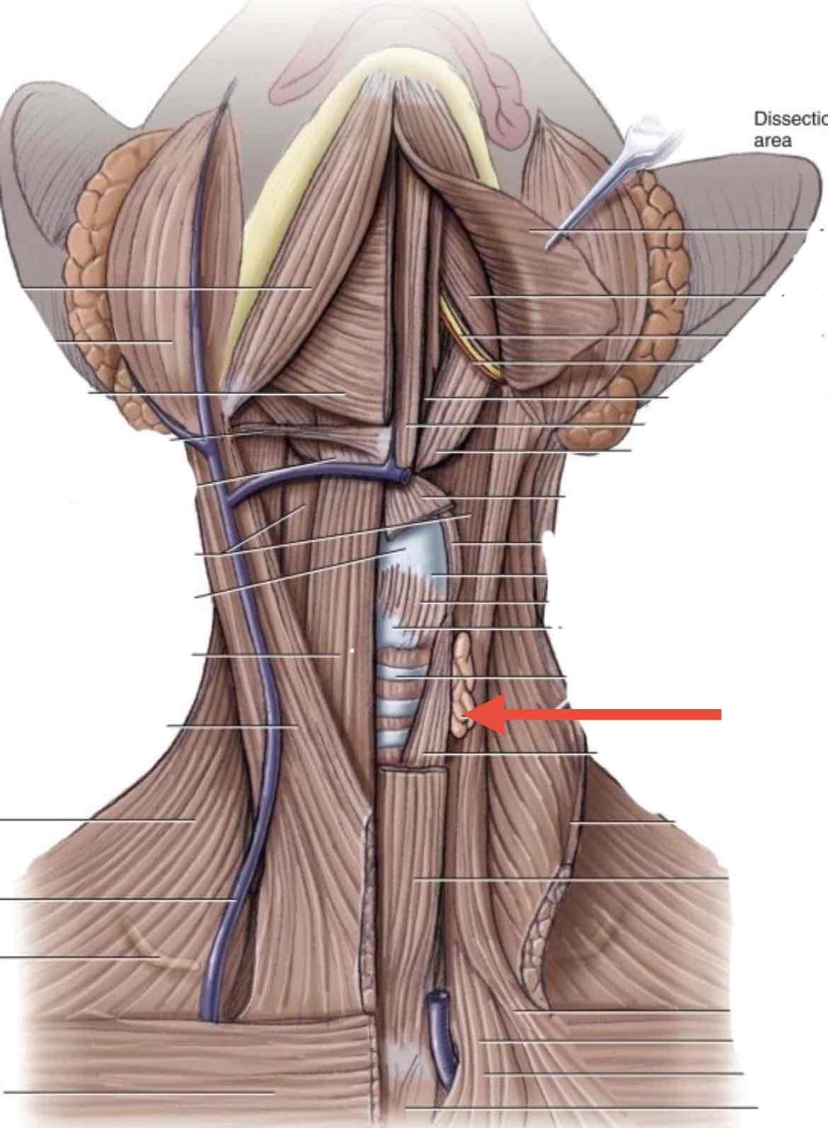
Thyroid gland
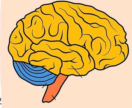
Yellow Region
Cerebrum (The largest portion of the brain, there are still lobes within the cerebrum)
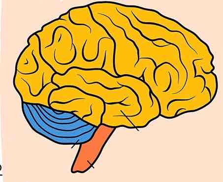
Blue region
Cerebellum
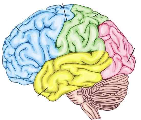
Blue lobe
Frontal lobe
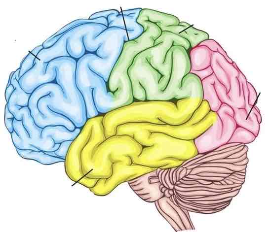
Green lobe
Parietal lobe
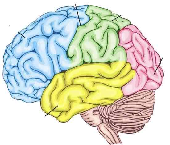
Yellow lobe
Temporal lobe
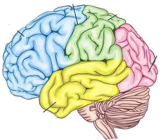
Pink lobe
Occipital lobe
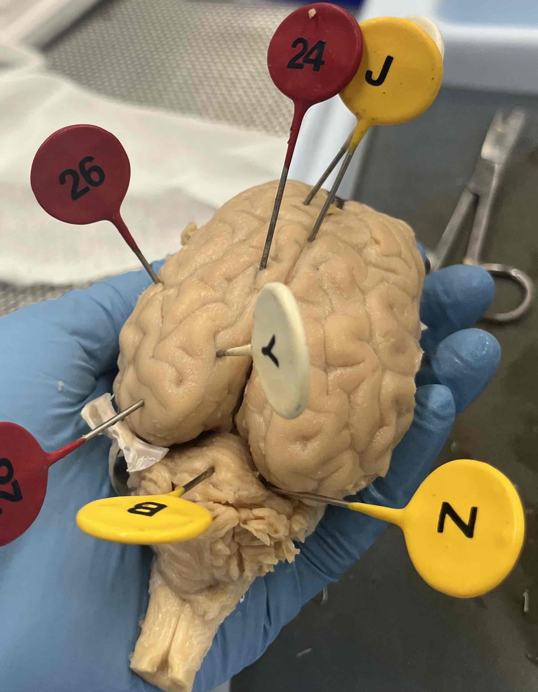
J
Longitudinal fissure
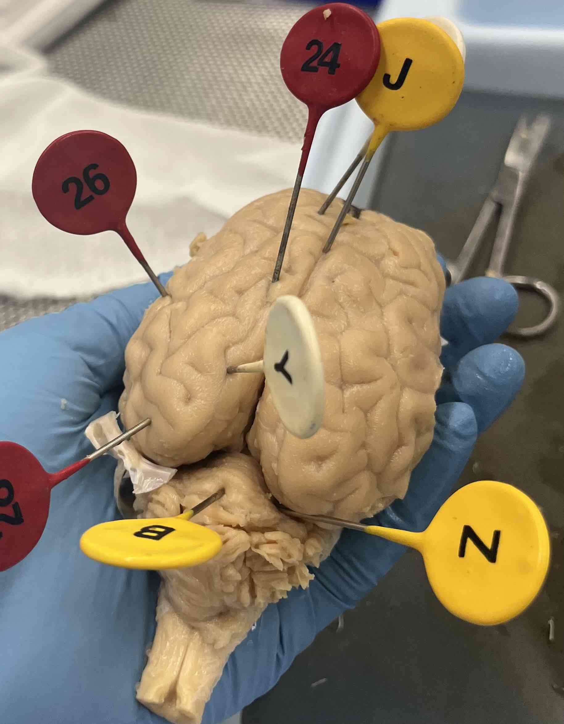
Z
Transverse fissure
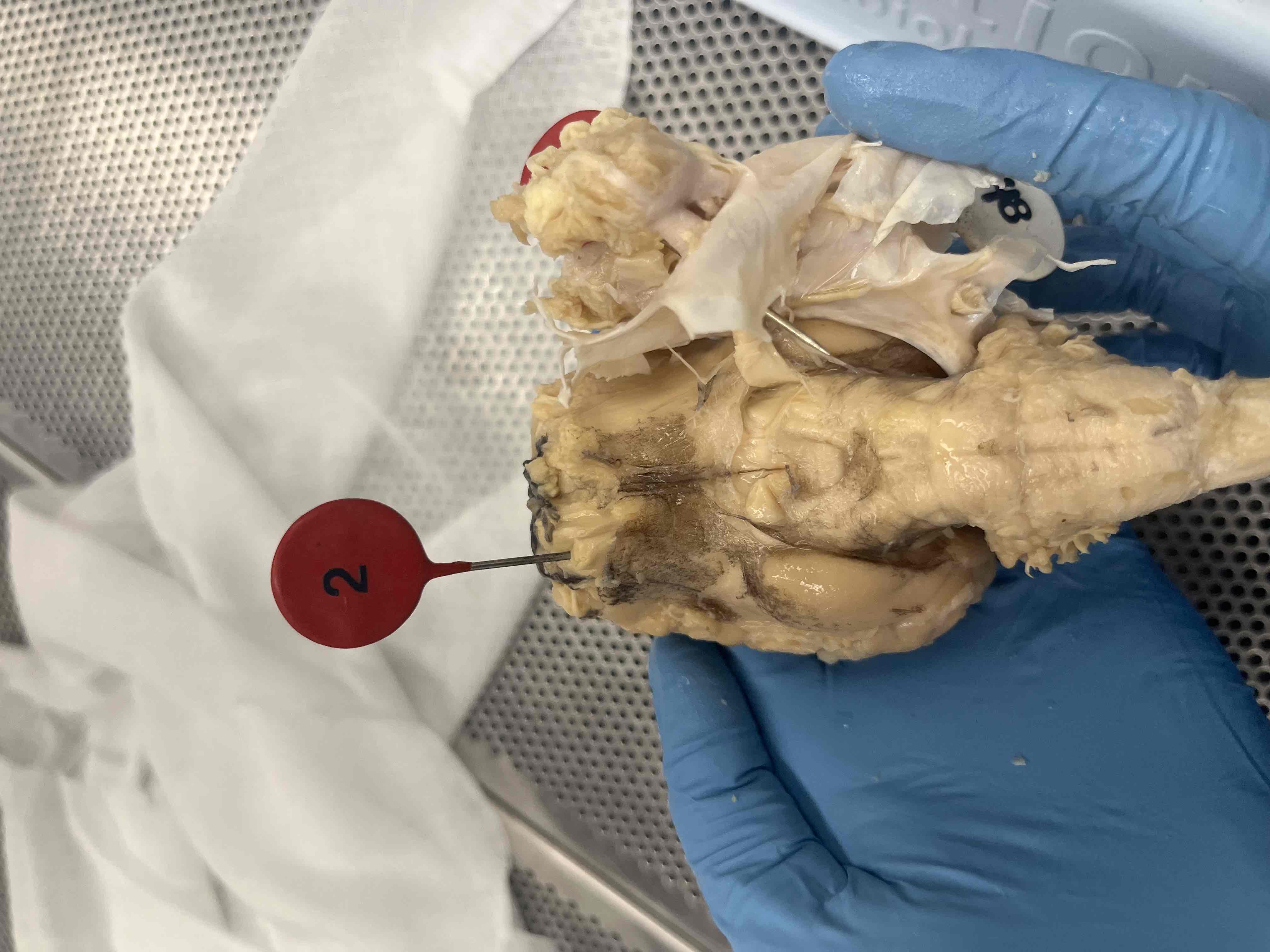
2
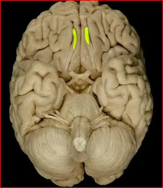
Olfactory bulbs
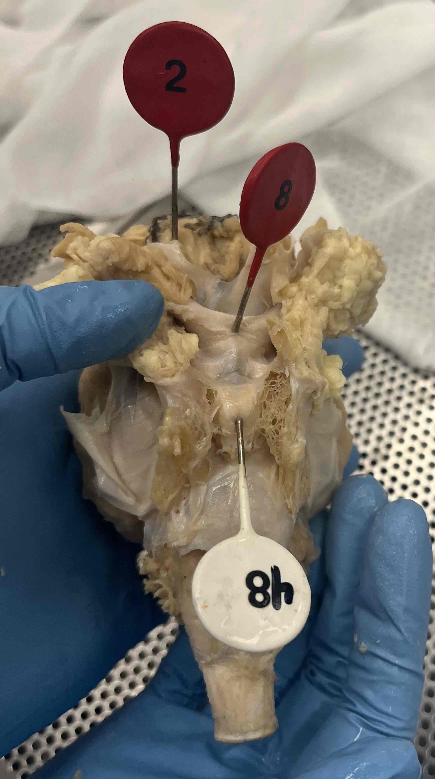
8
Optic Chiasm
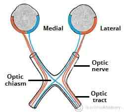
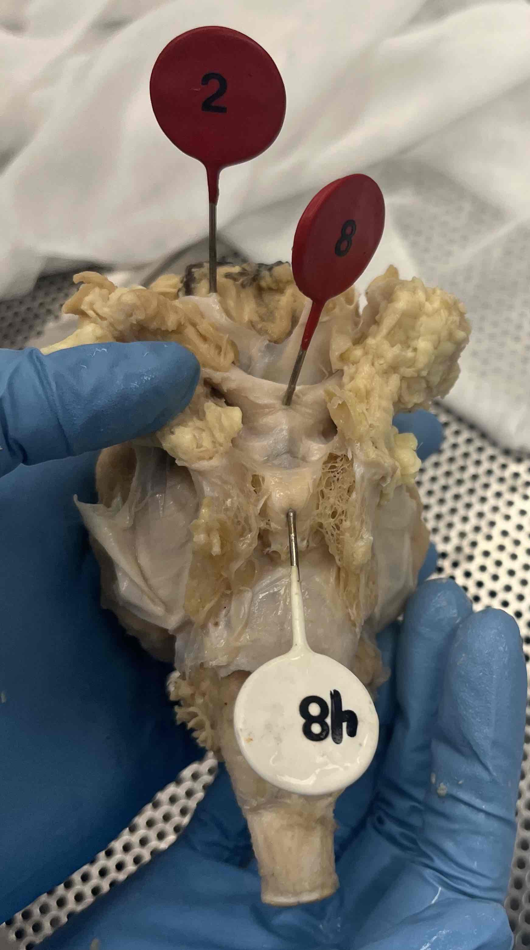
48
Pituitary gland
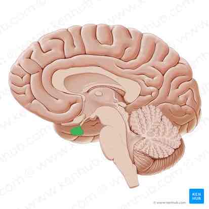
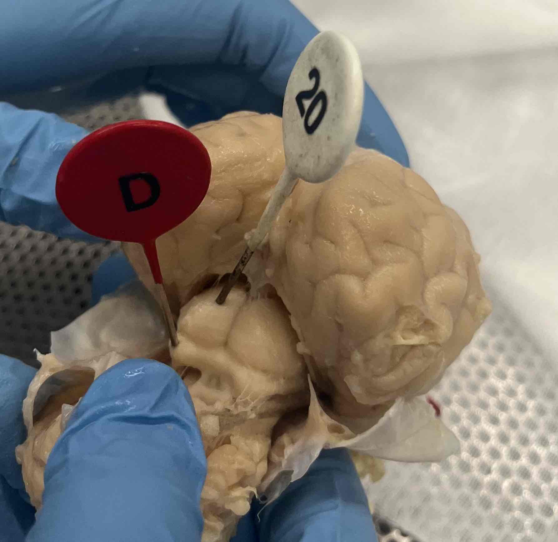
20
Corpora quadrigemina - superior colliculus
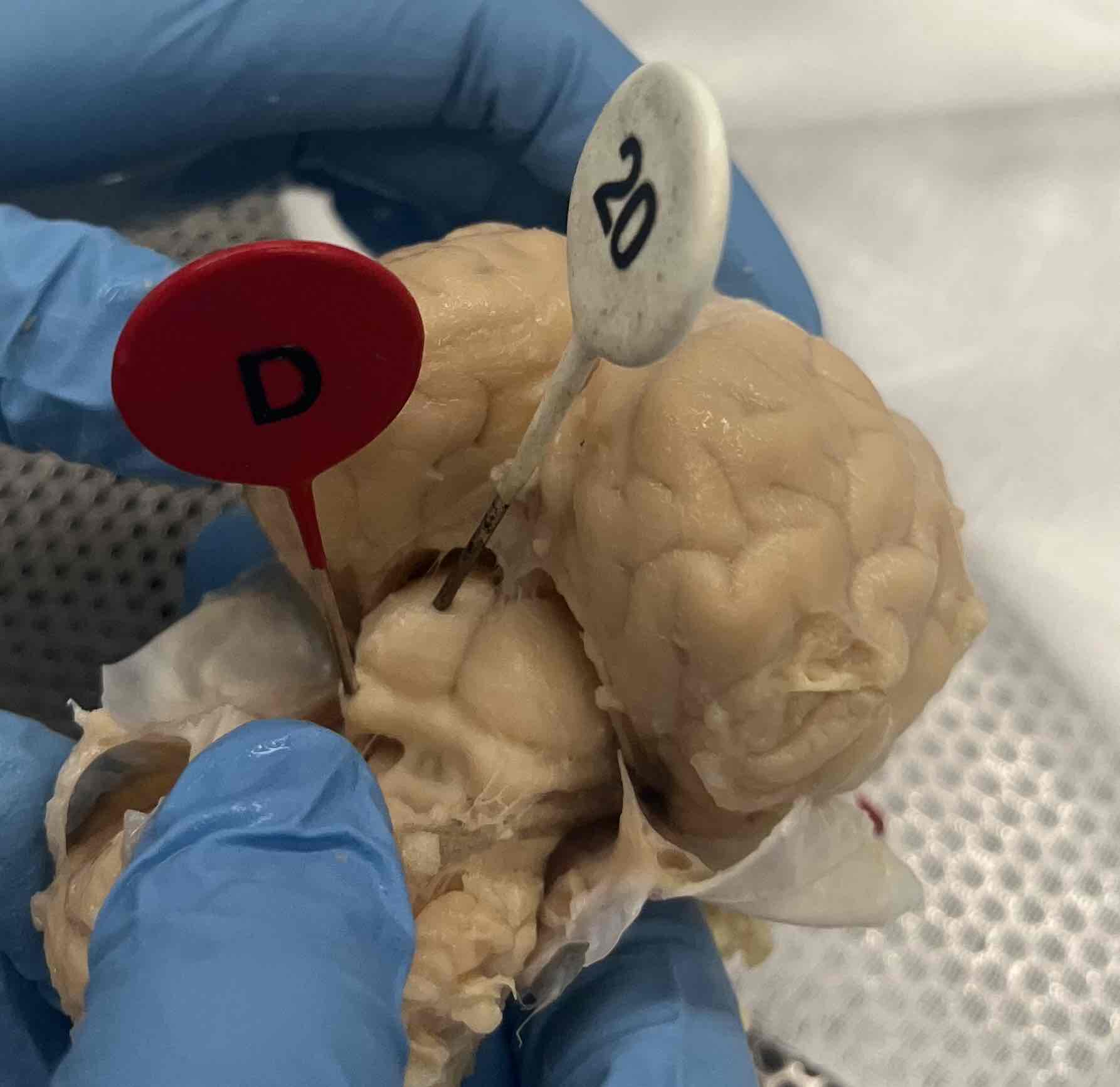
D
Corpora quadrigemina - inferior colliculus
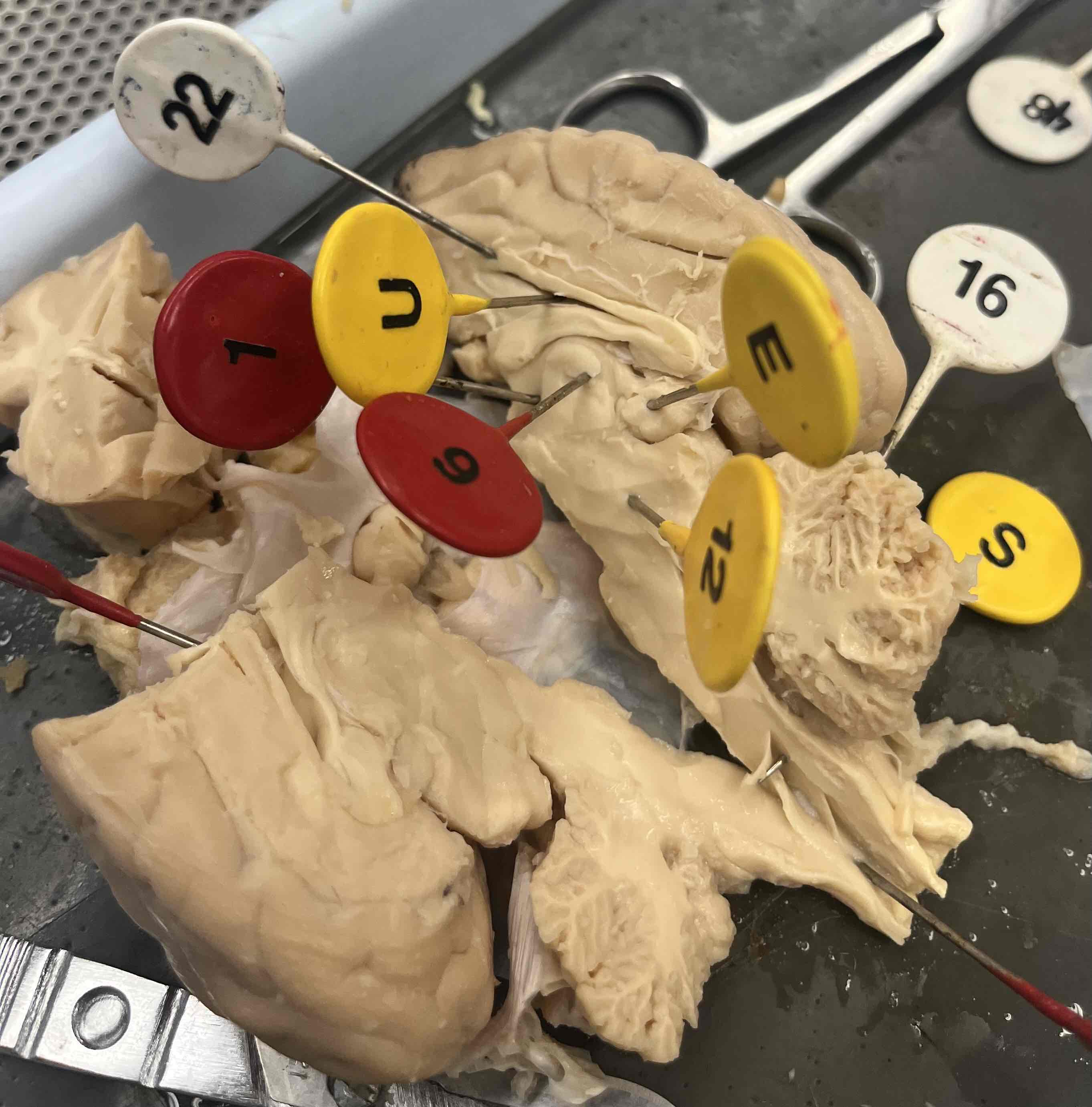
22
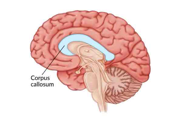
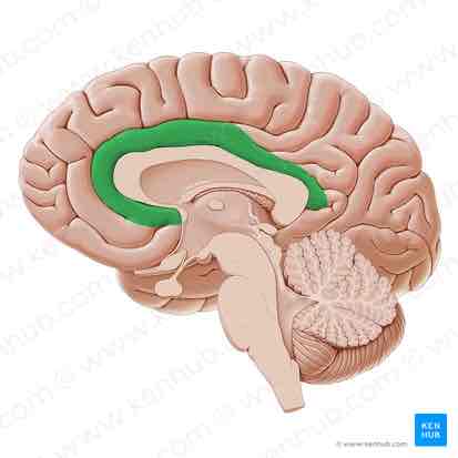
Corpus callosum
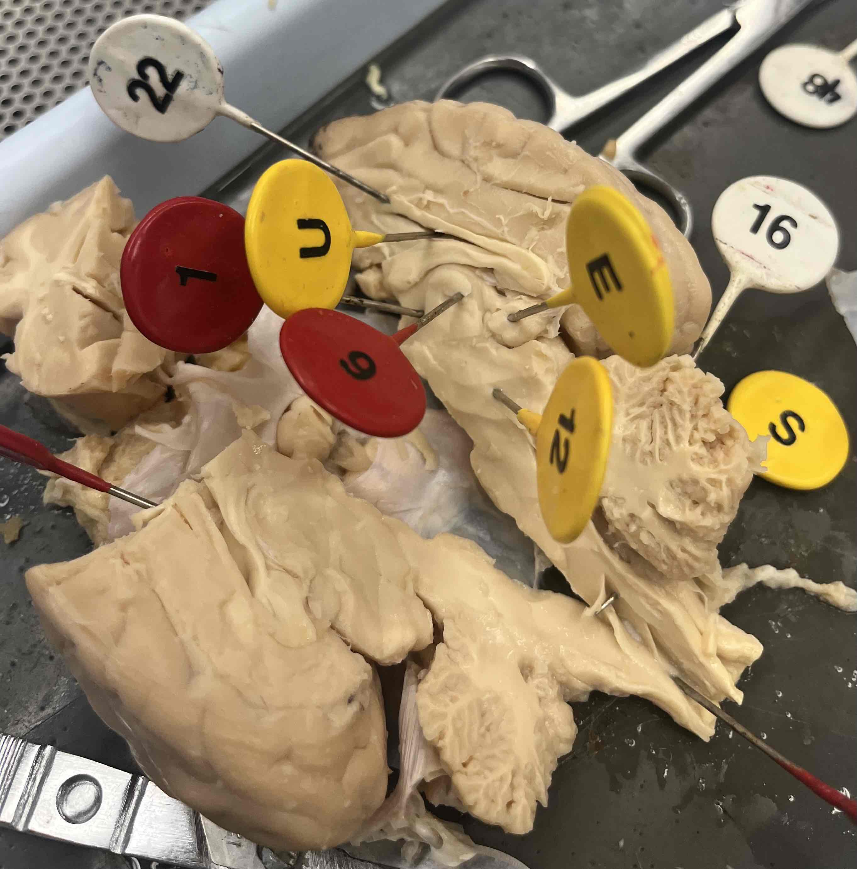
U
Fornix
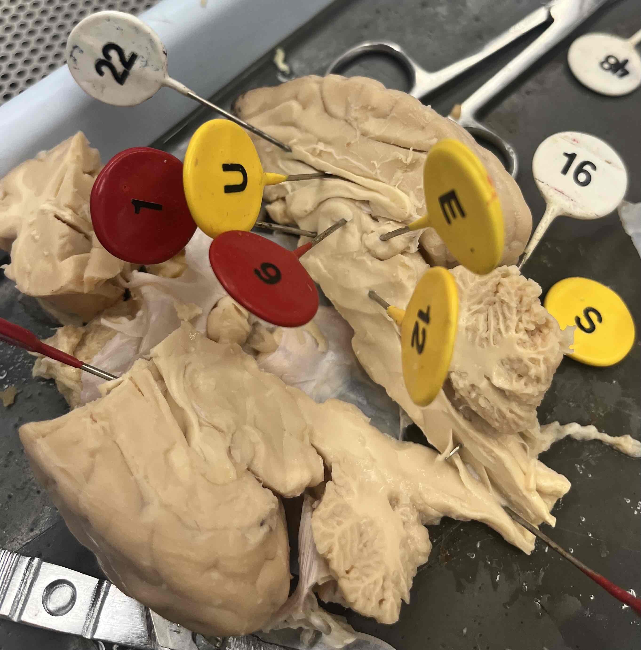
E
Pineal body
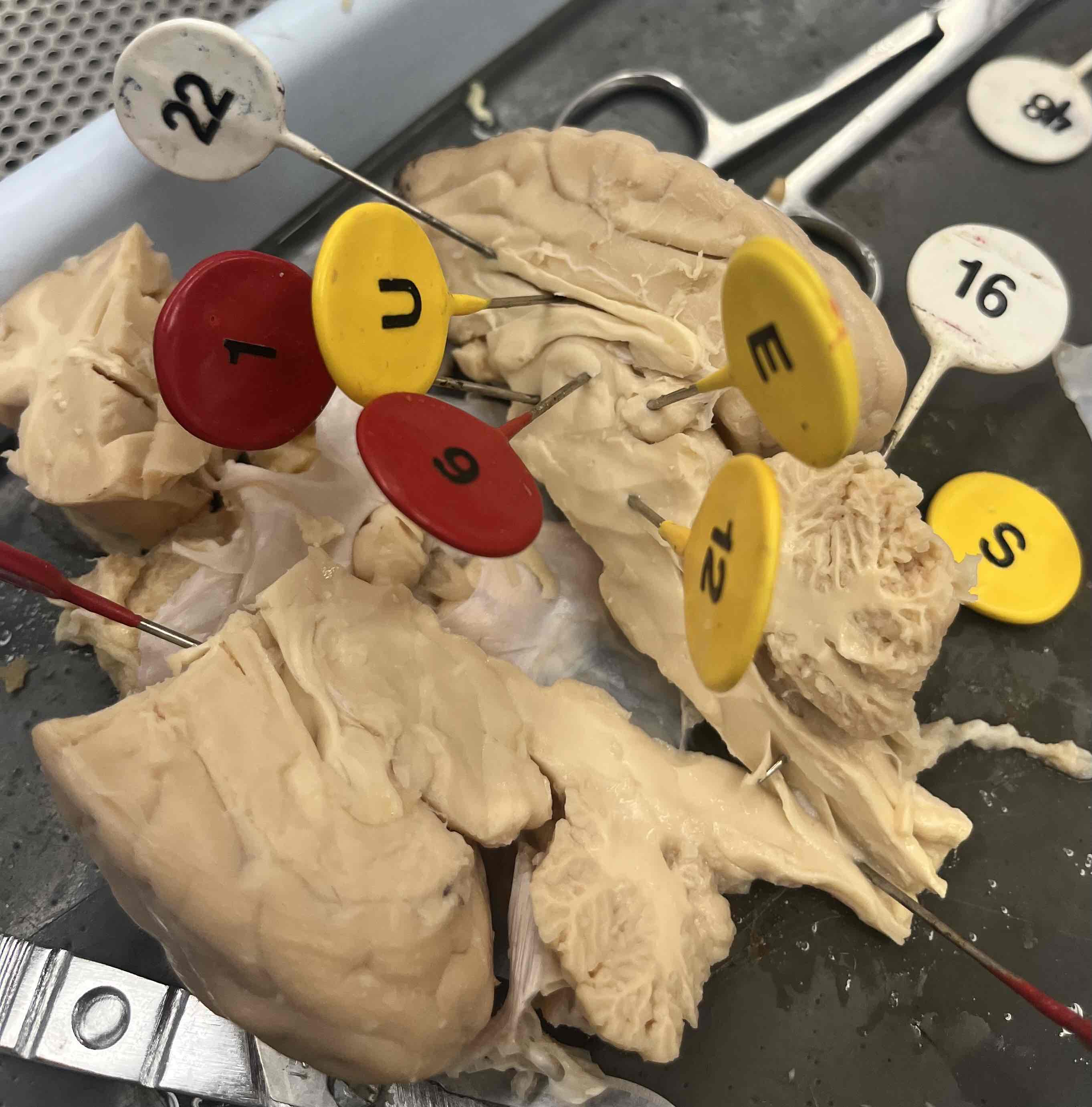
6 (number is upside down, below U)
Thalamus
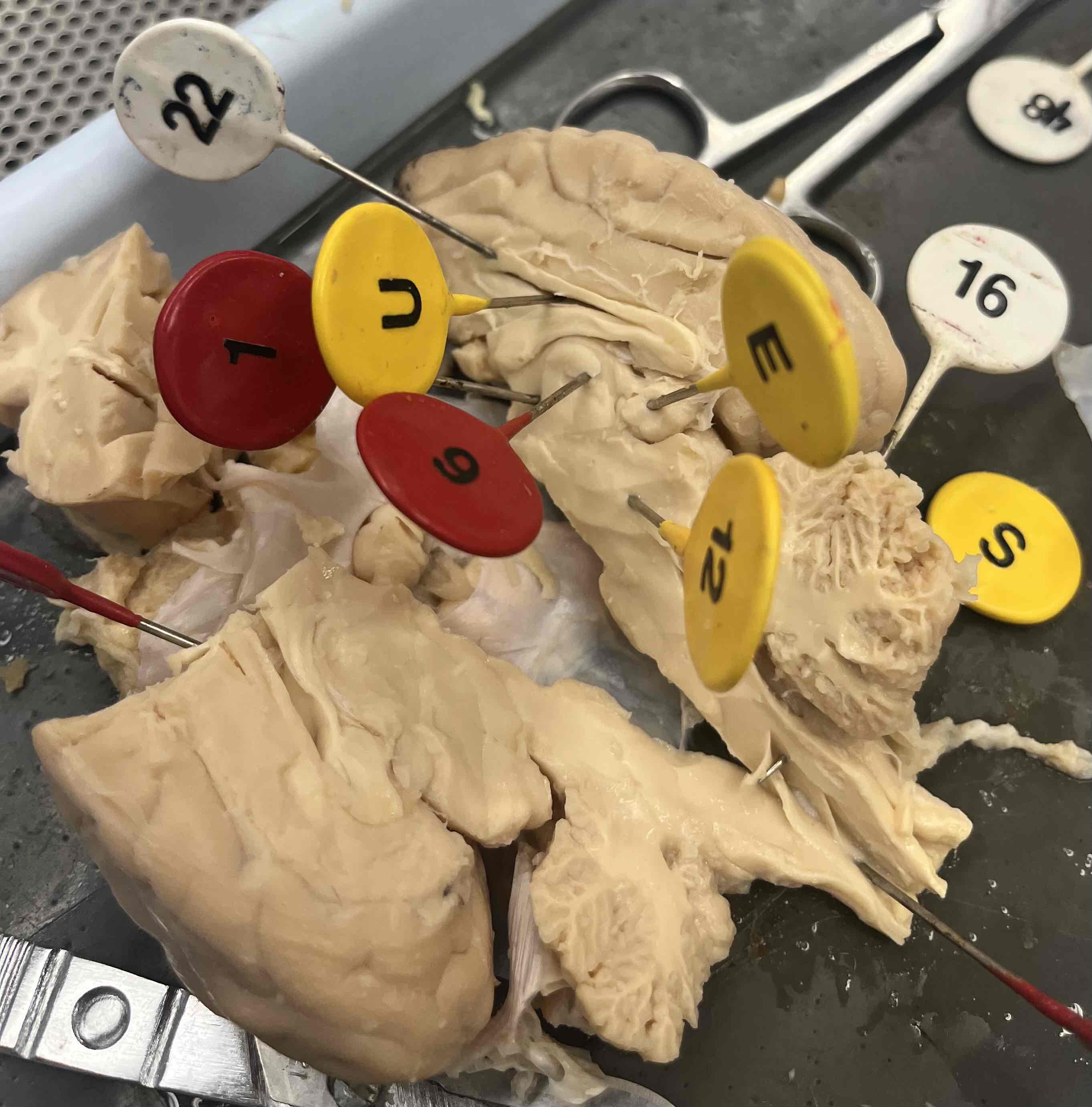
1 (on left of U)
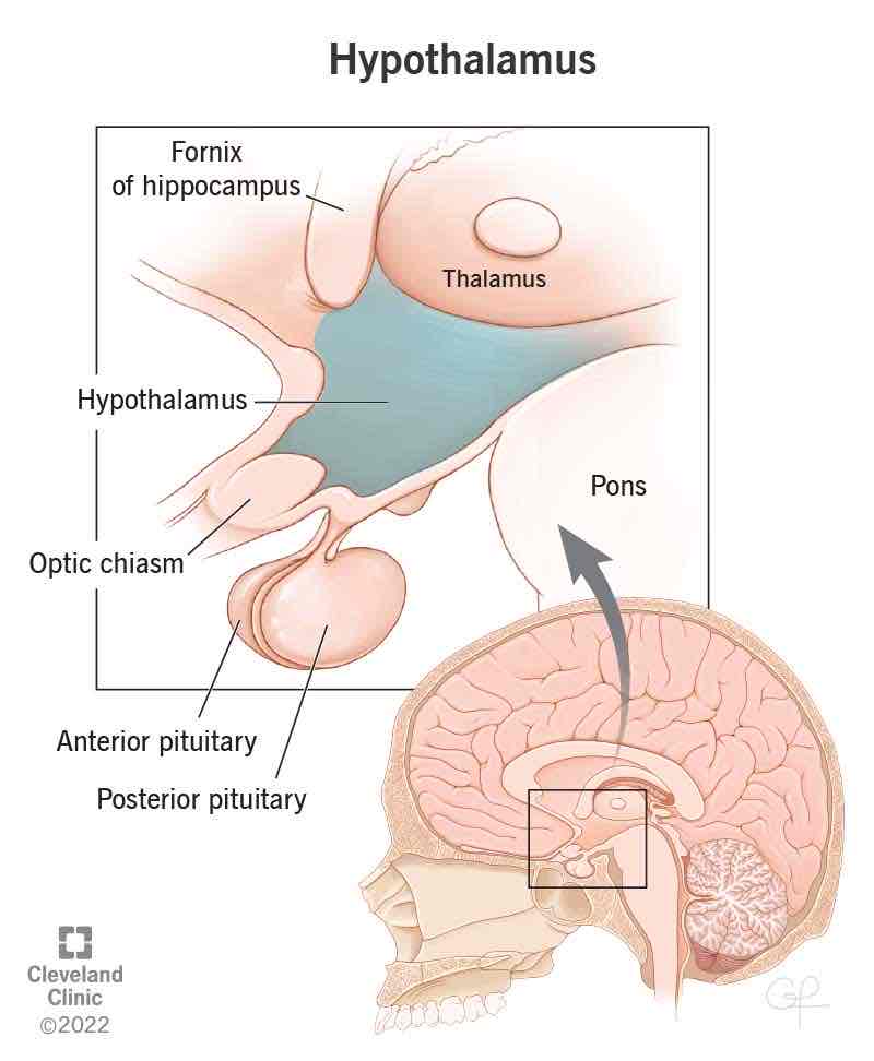
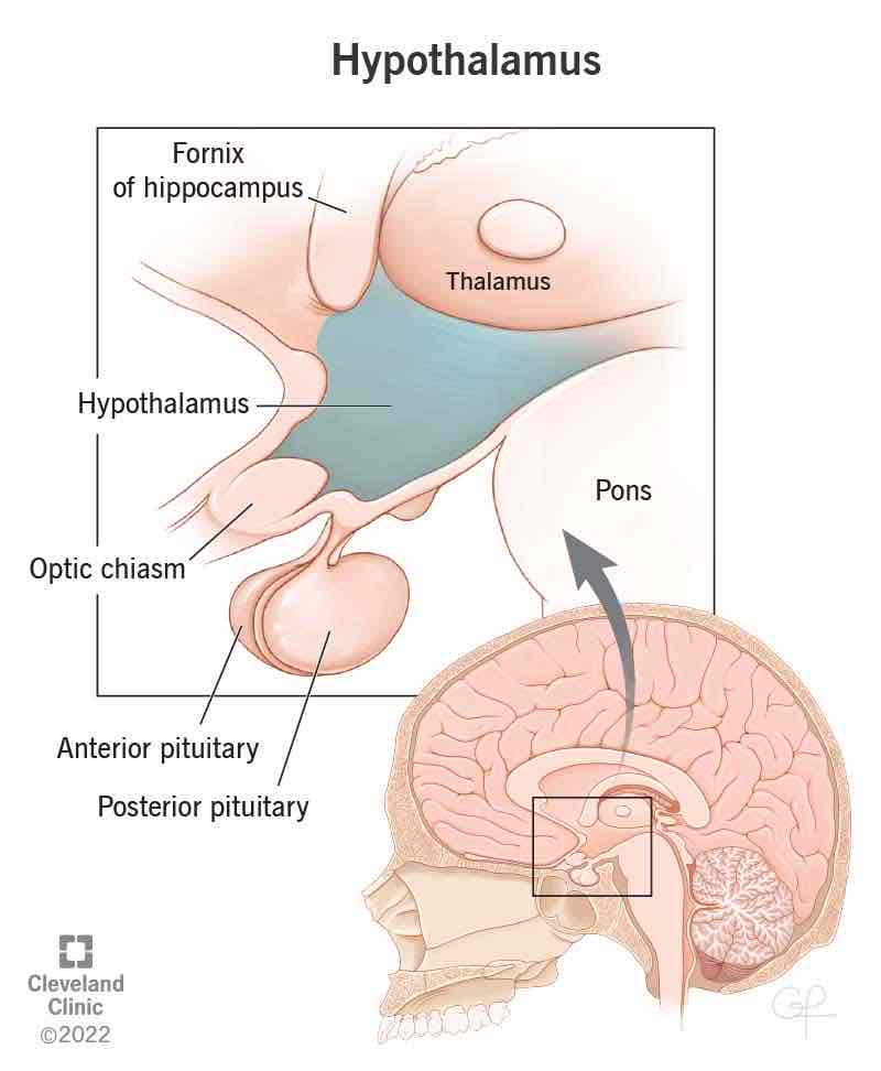
Hypothalamus
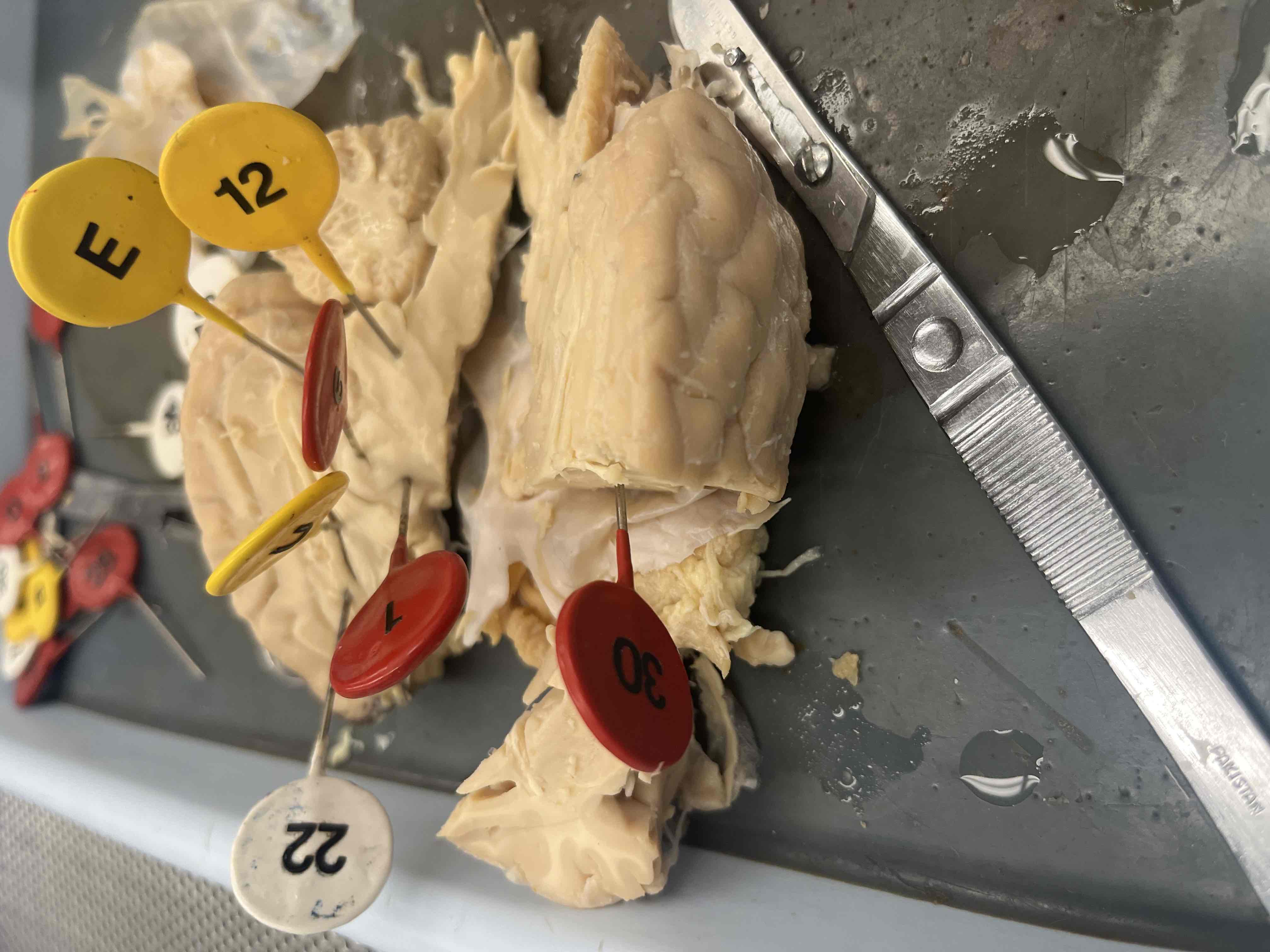
30 (bottom right corner of image)
Septum pellucidum
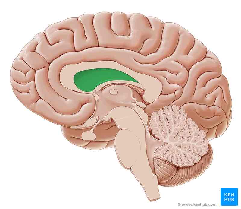
Brain ventricles to know:
1st, 2nd, 3rd, 4th ventricles
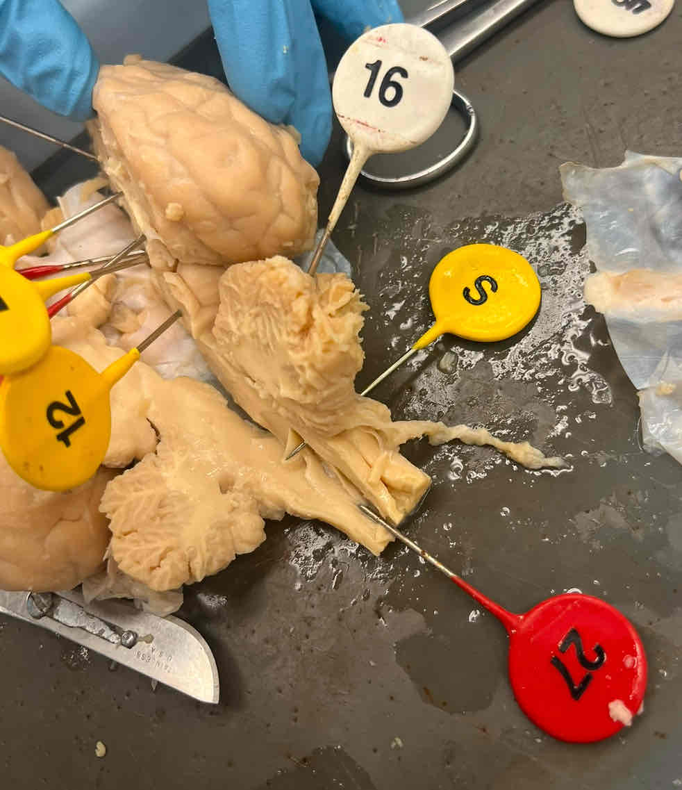
12, 16, and S
Brain stem:
1) Midbrain (12)
2) Pons (16)
3) Medulla oblongata (S)
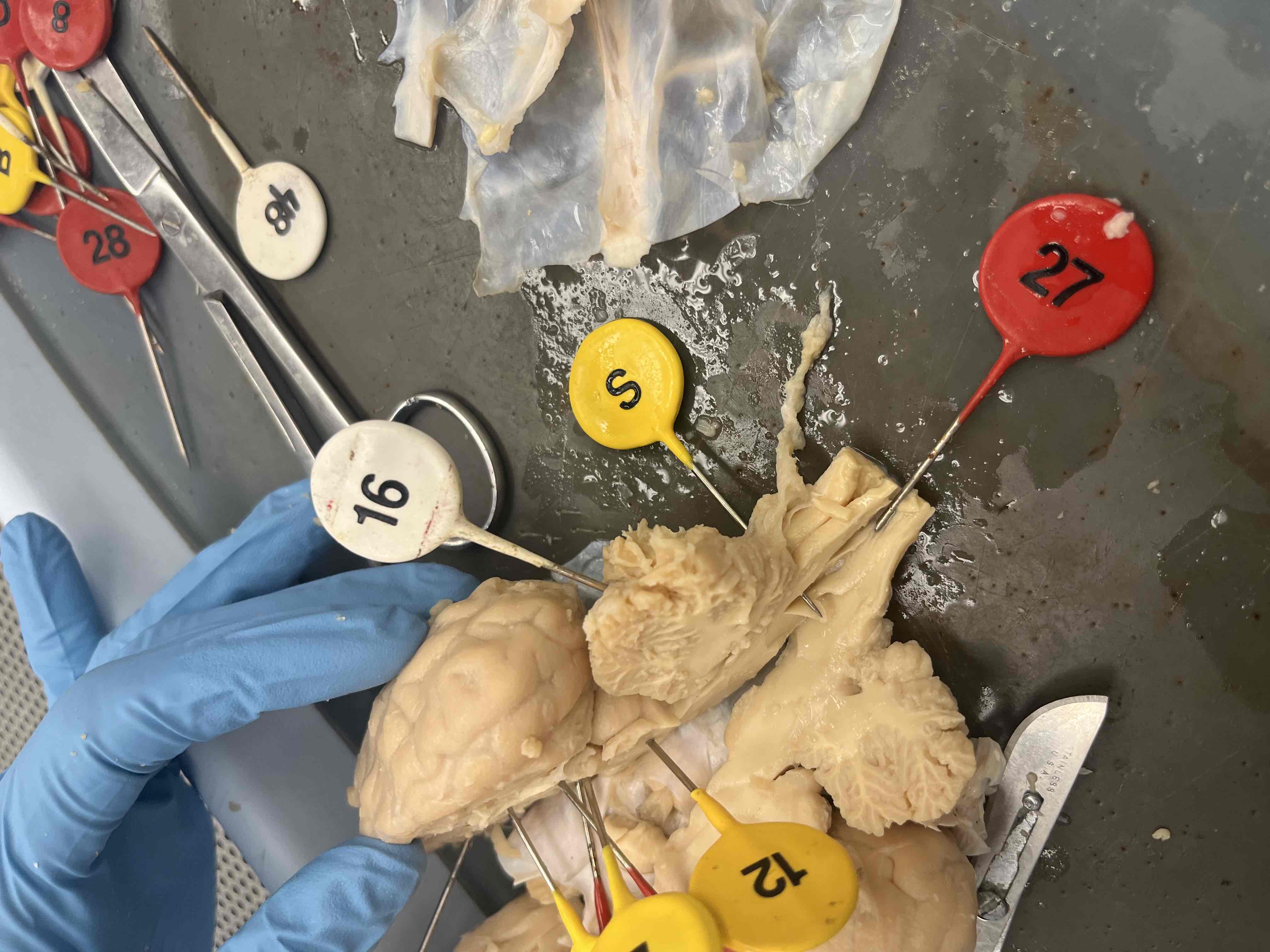
27
Spinal cord
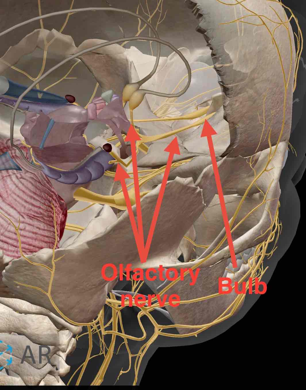
I (Olfactory + bulbs)
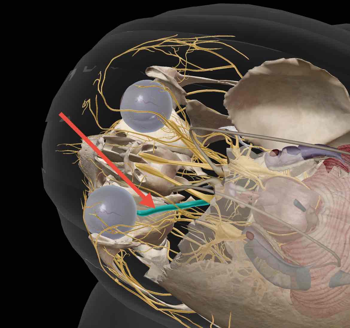
II (Optic)
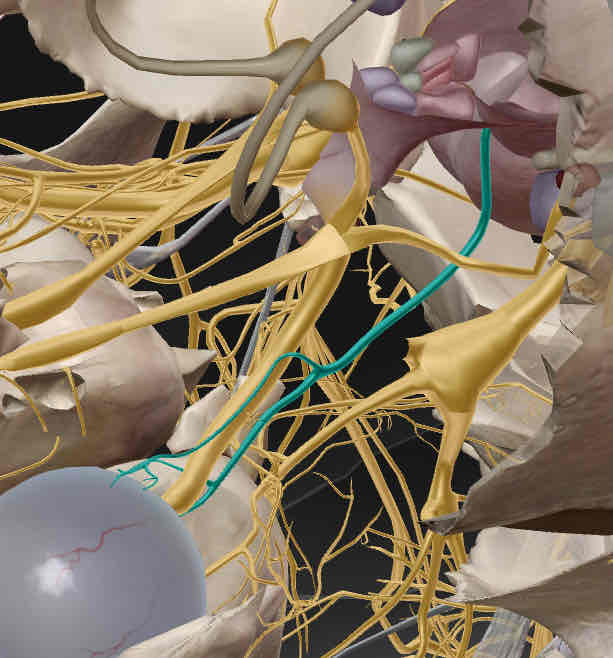
III (Oculomotor)
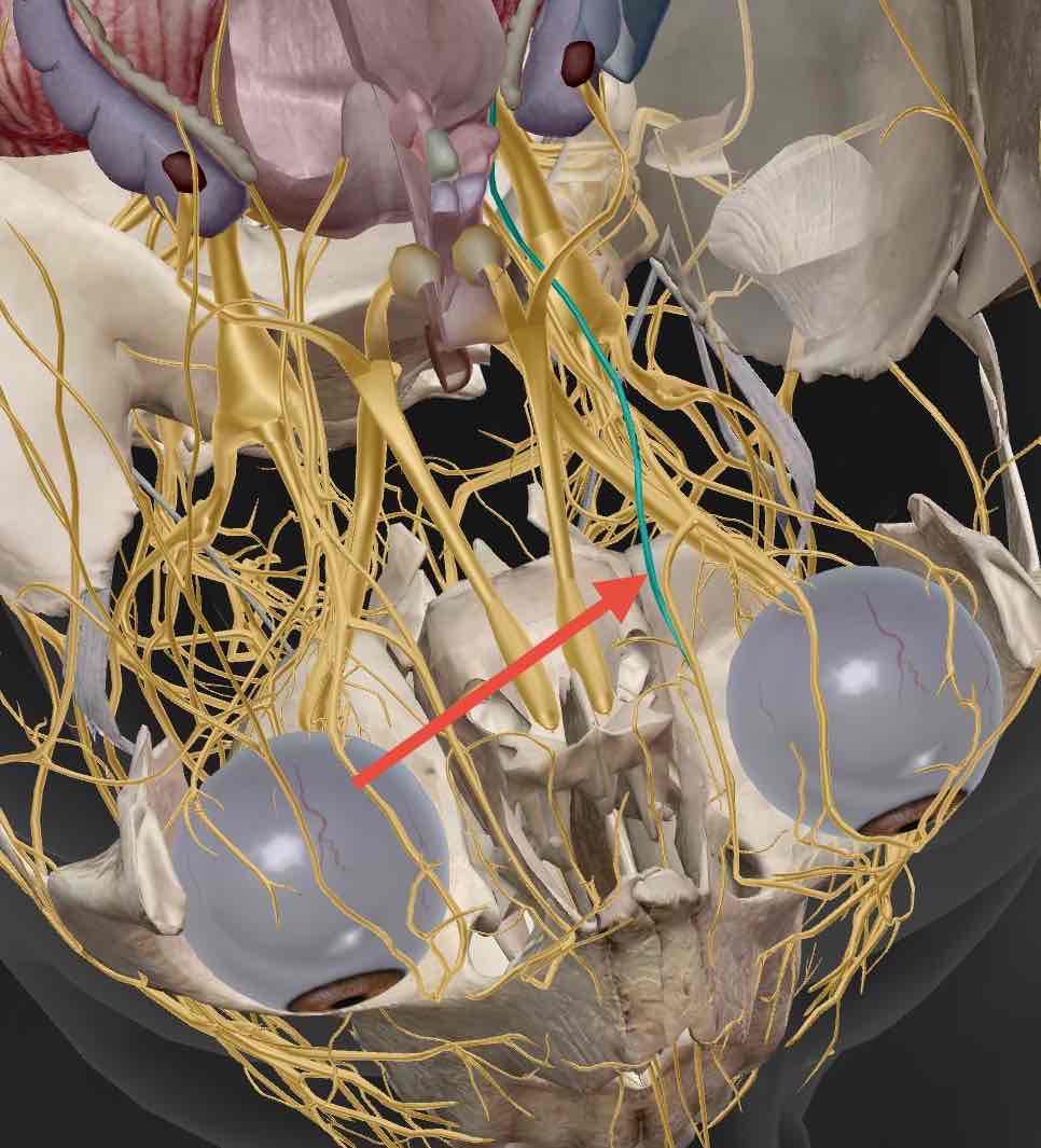
IV (Trochlear)
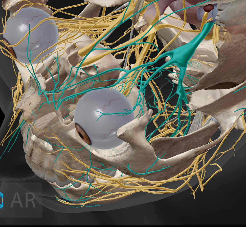
V (Trigeminal)
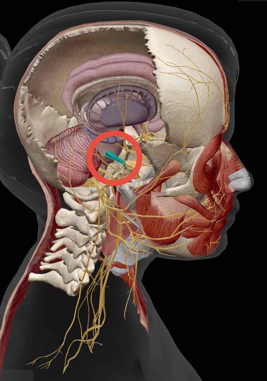
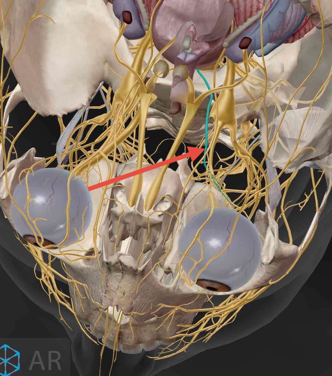
VI (Abducens)
(The Trochlear (IV) crosses over this nerve)

VII (Facial)
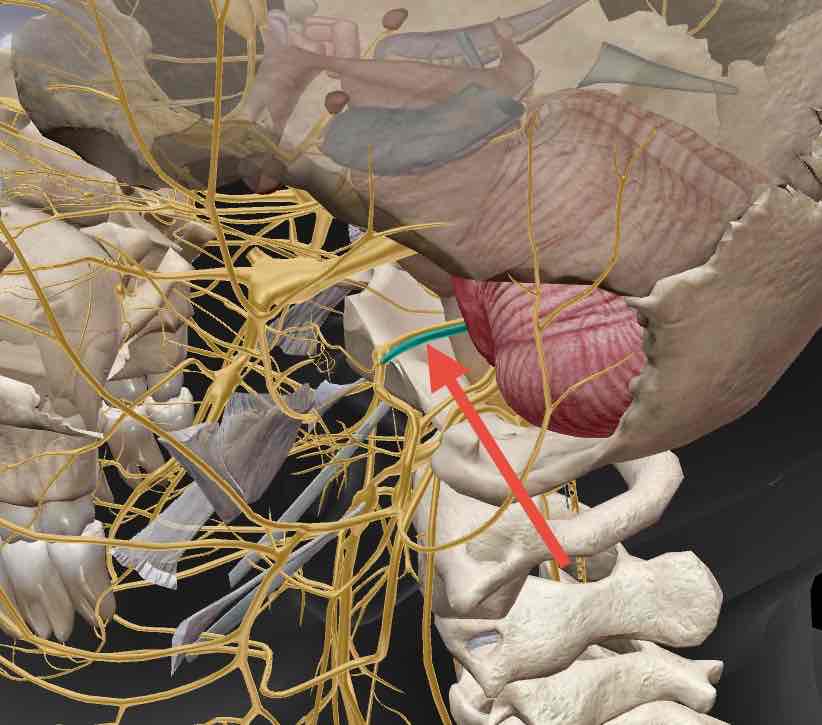
VIII (Vestibulocochlear)
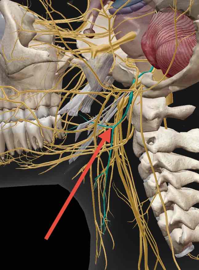
IX (Glossopharyngeal)
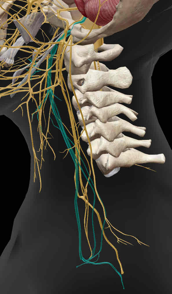
X (Vagus)
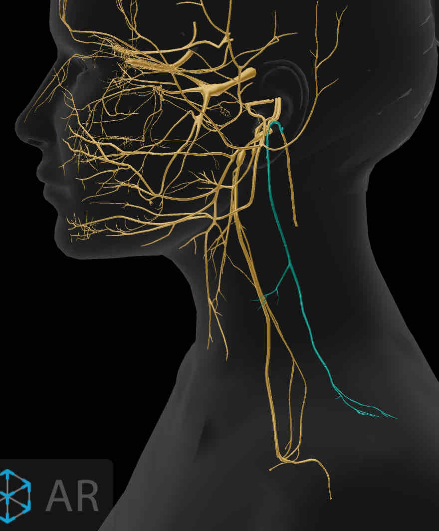
XI (Accessory)
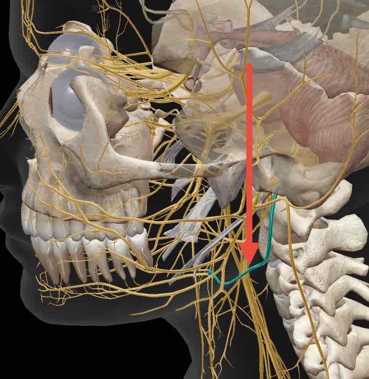
XII (Hypoglossal)
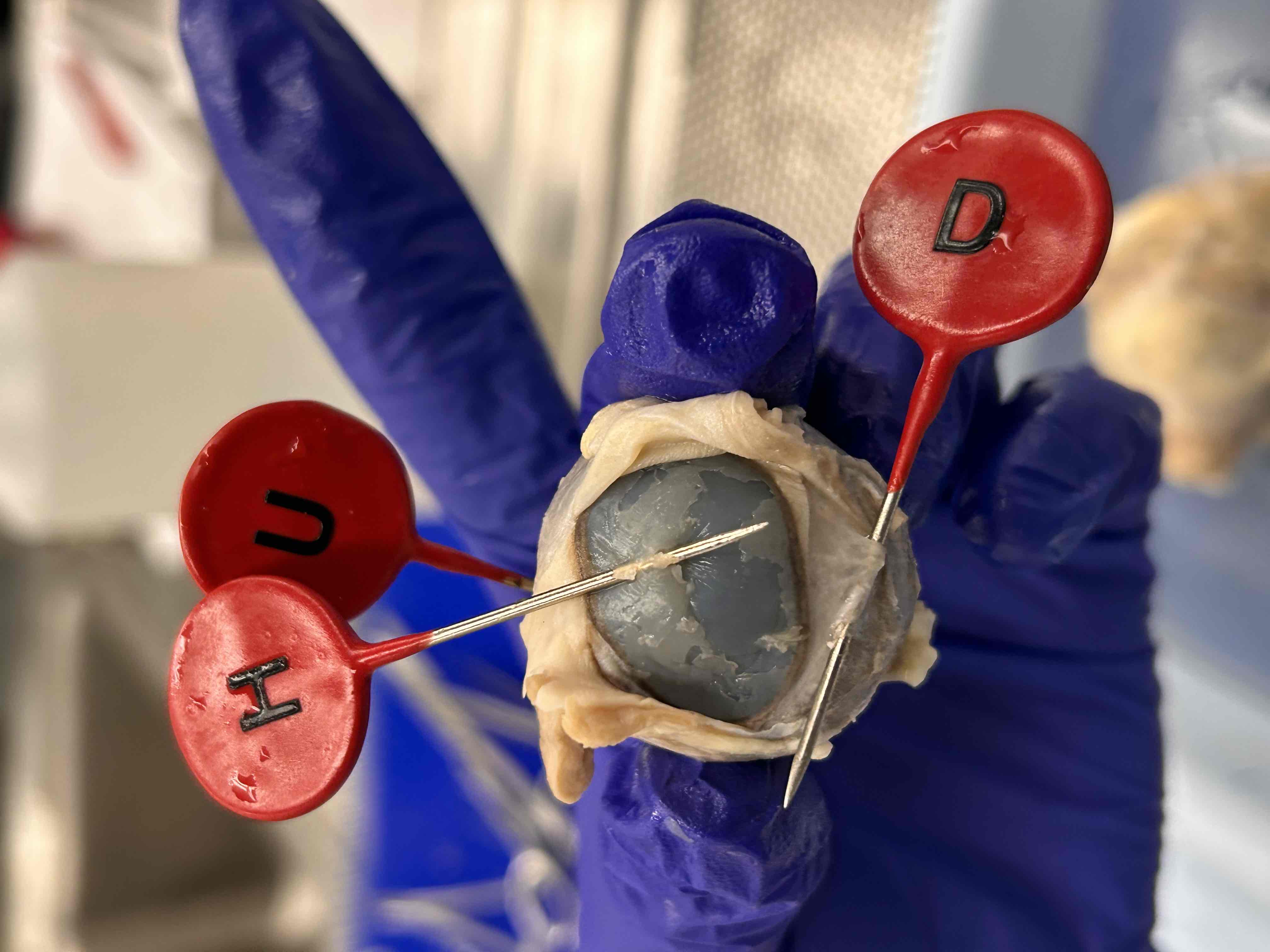
D
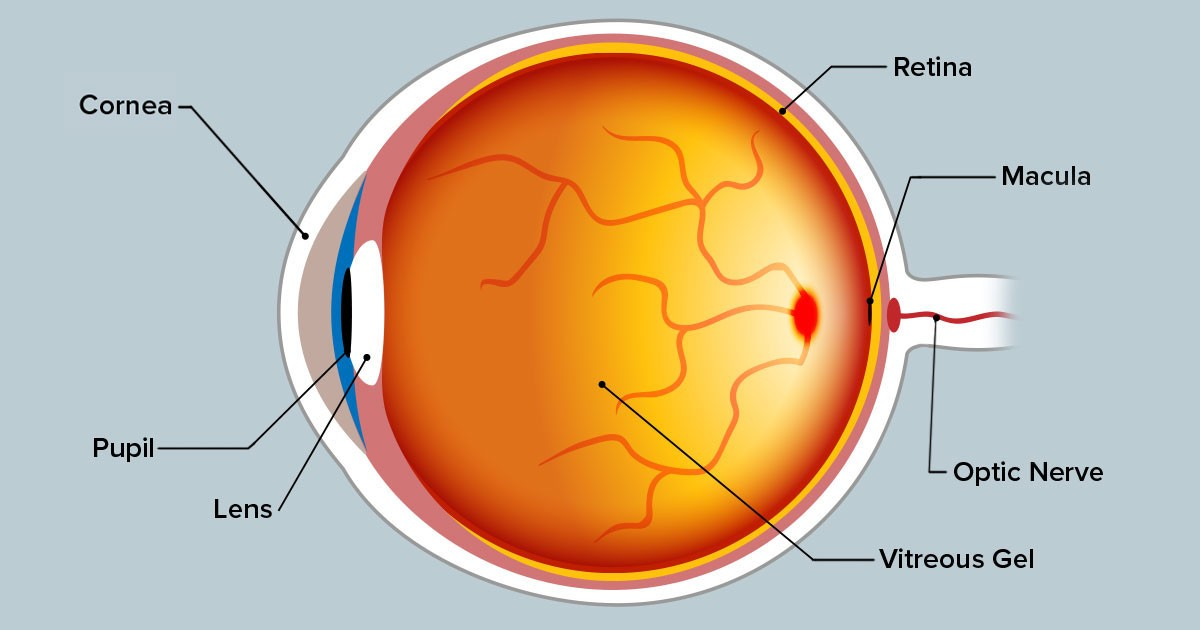
Sclera
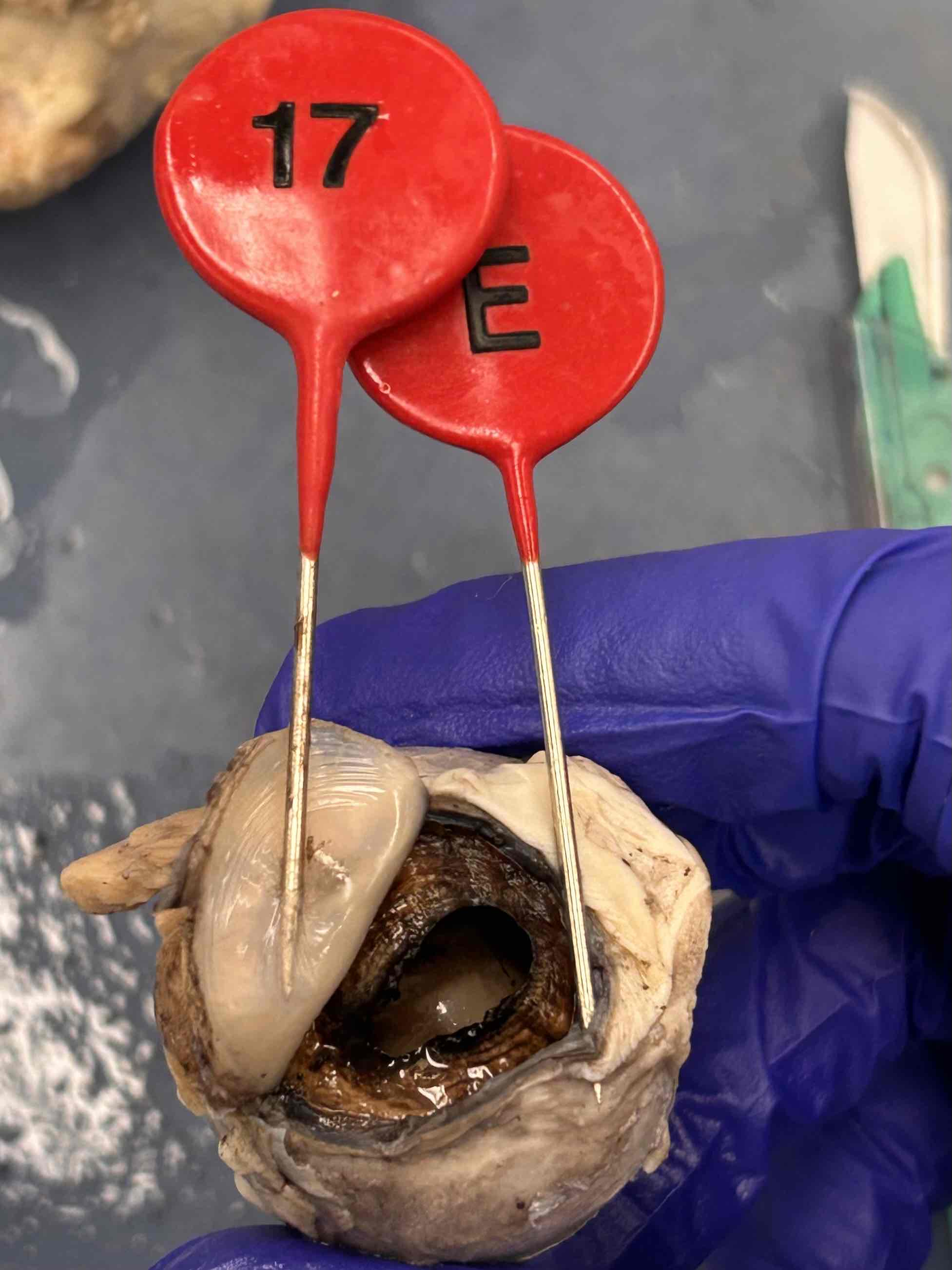
E
Choroid
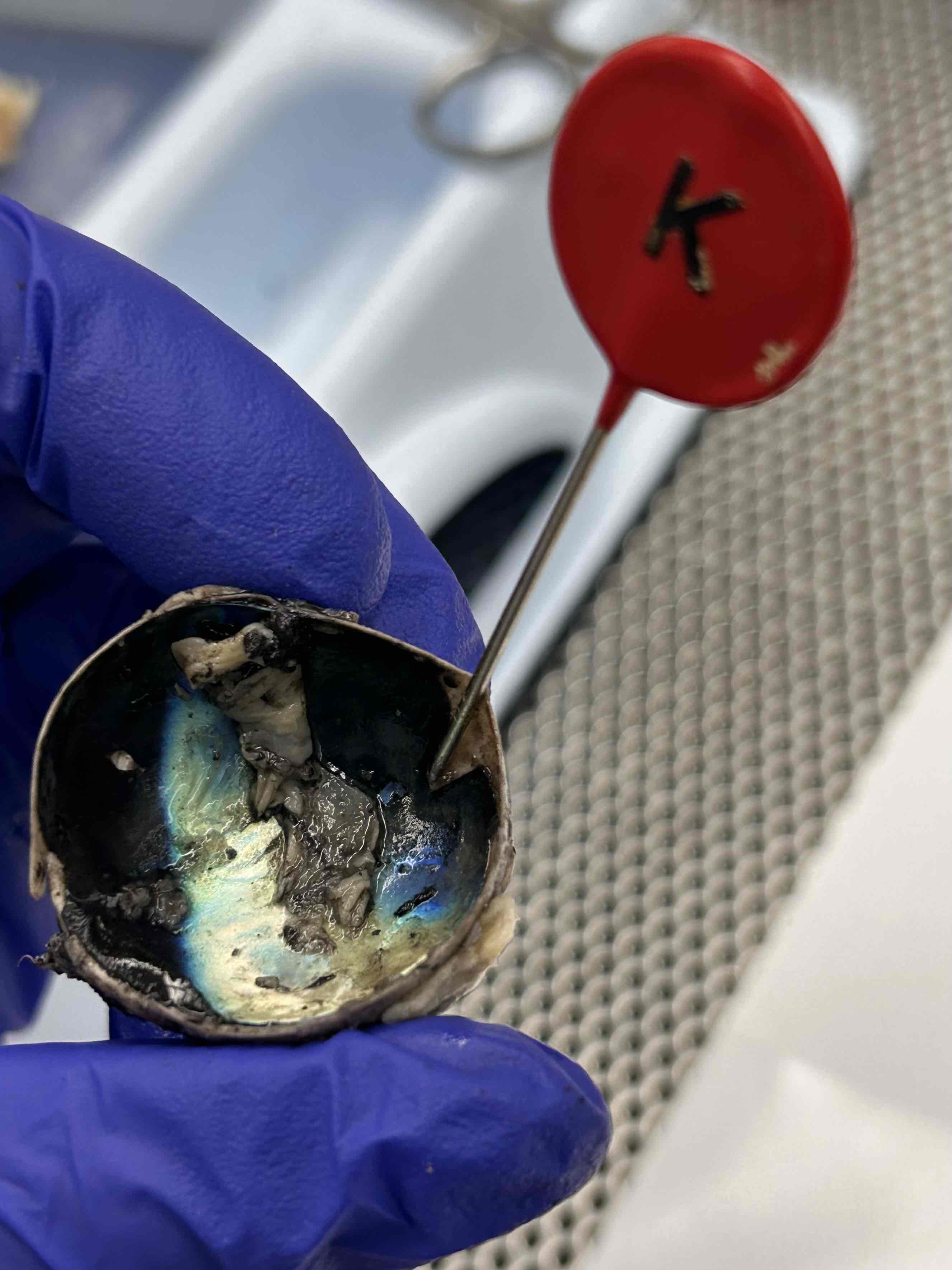
K
Retina
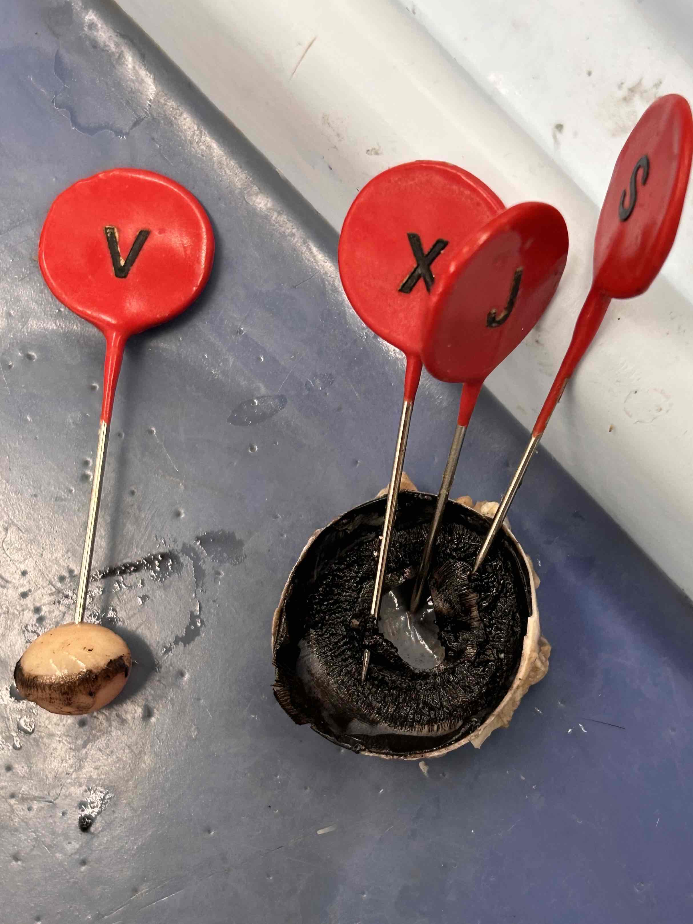
S
Ciliary Body
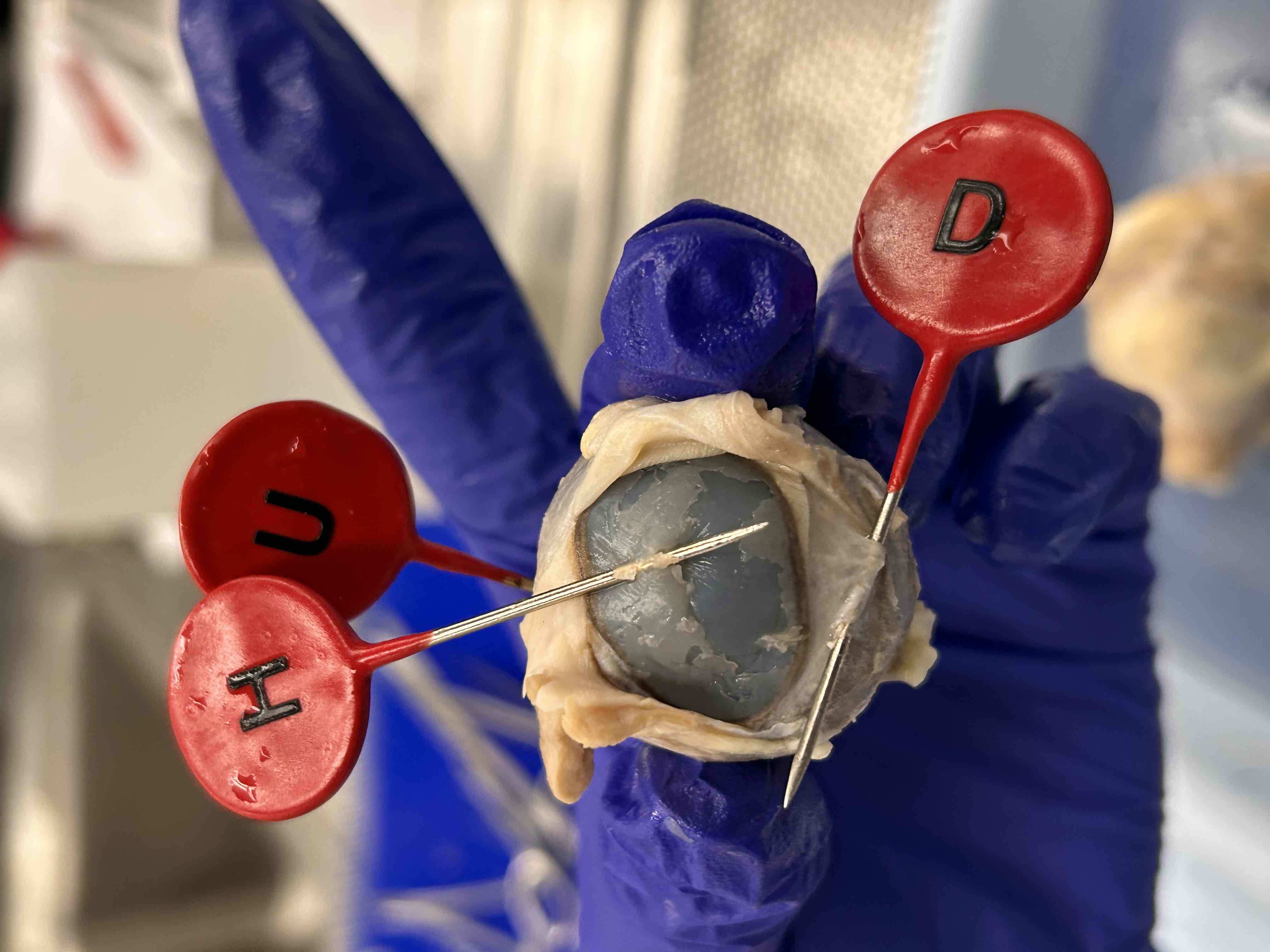
H
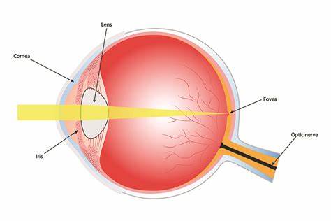
Cornea
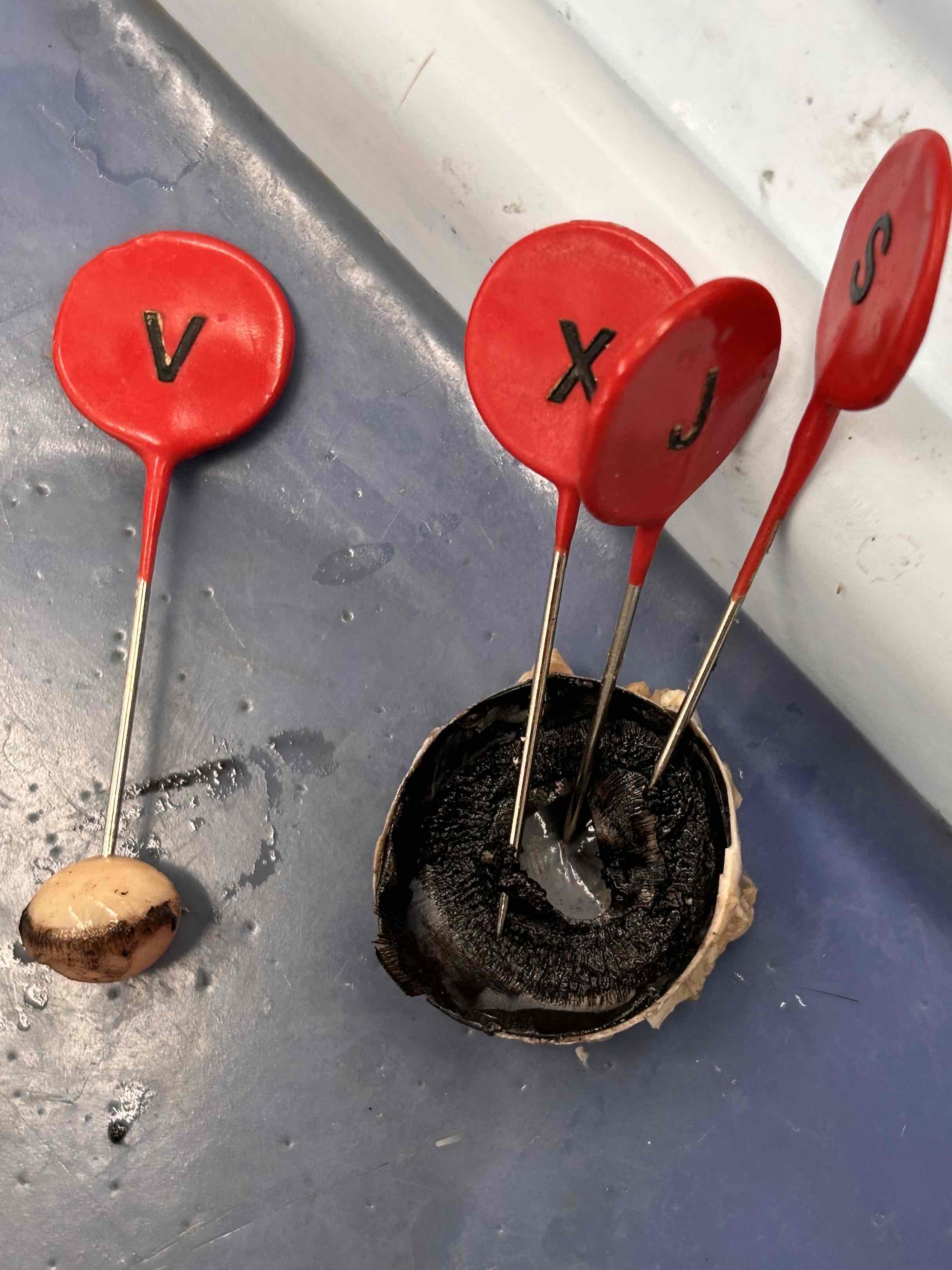
X
Iris
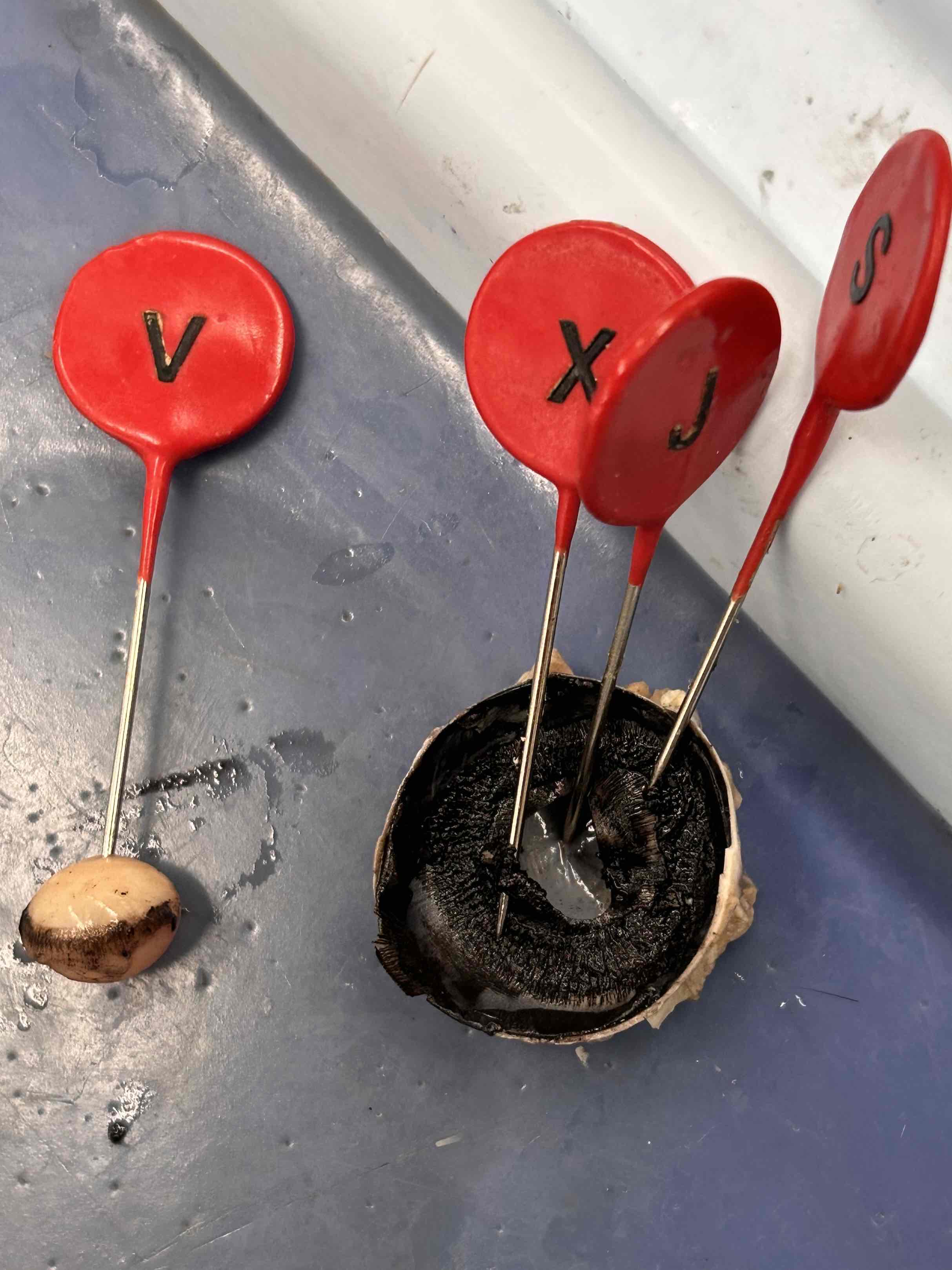
V
Lens
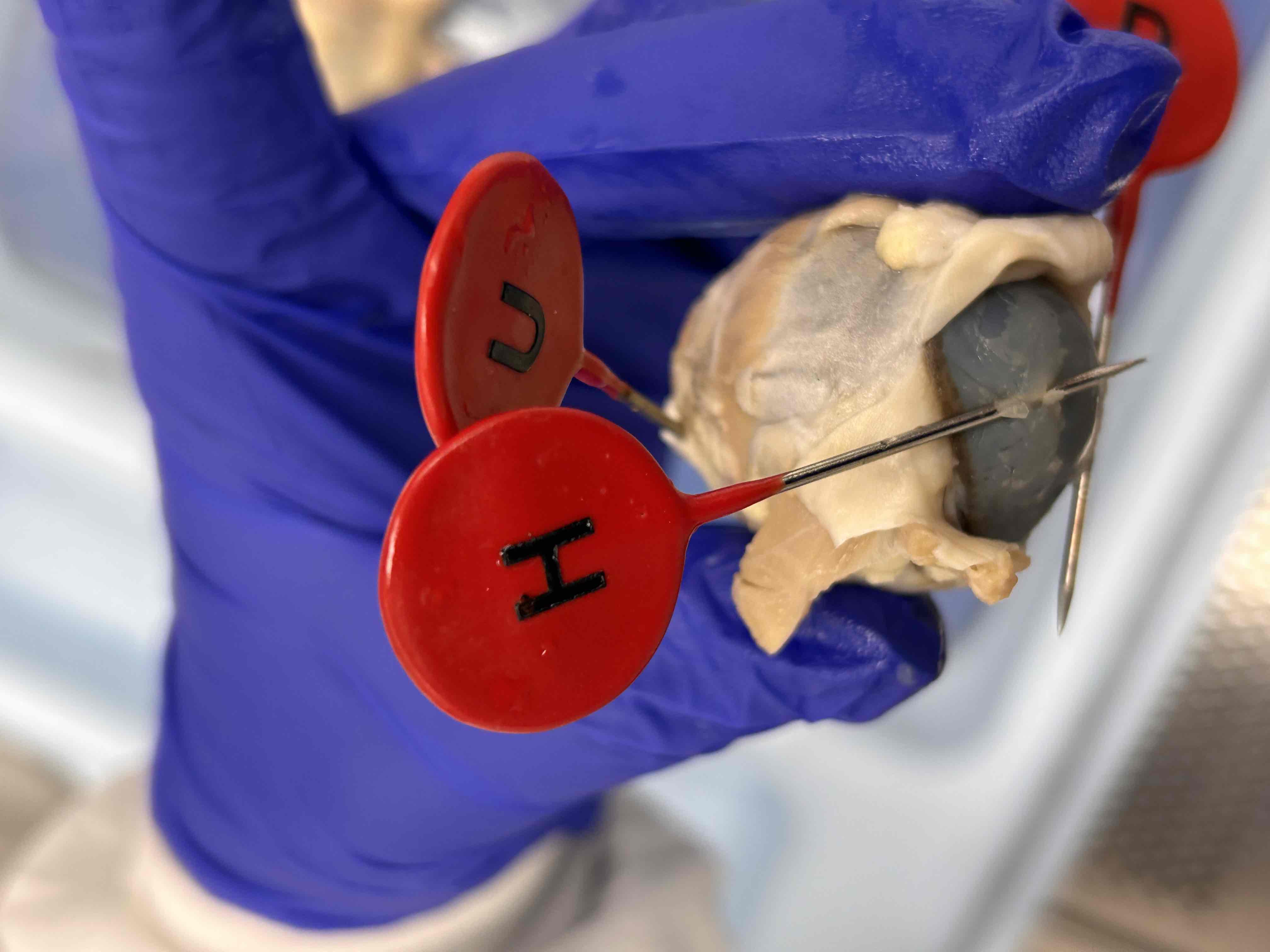
U
Optic nerve
Macula
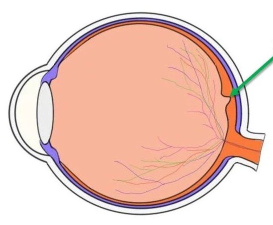
Fovea
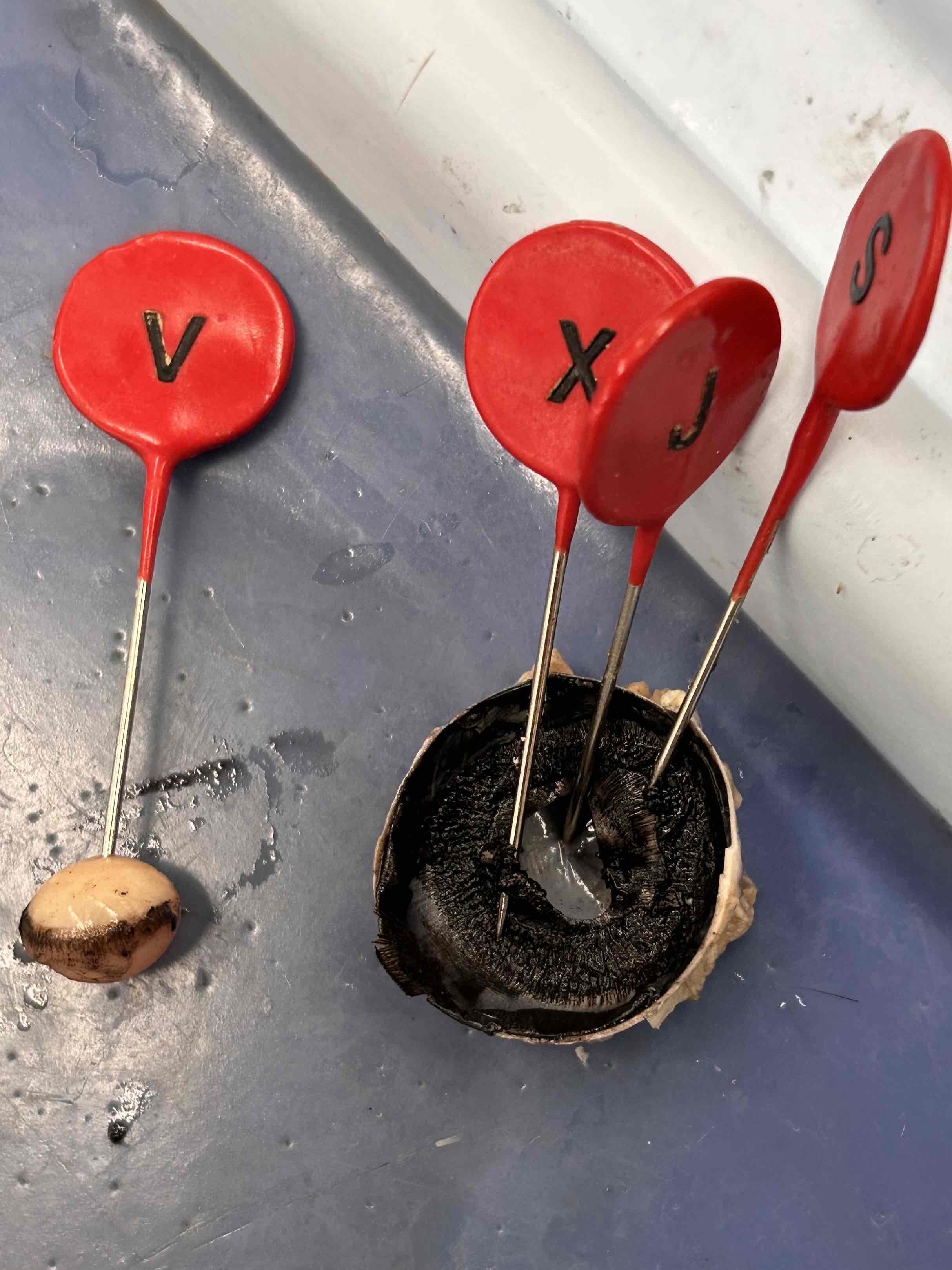
J
Pupil
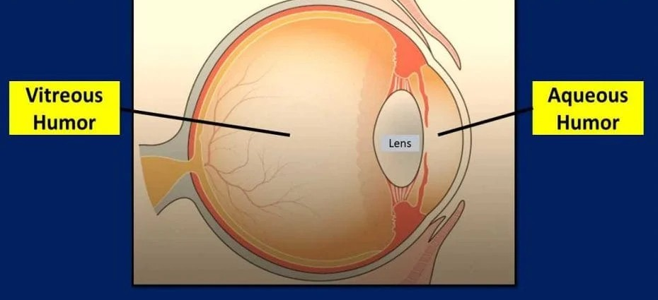
Vitreous Humor;
The gel-like substance filling the space between the lens and the retina in the eye, providing support and maintaining the shape of the eyeball.
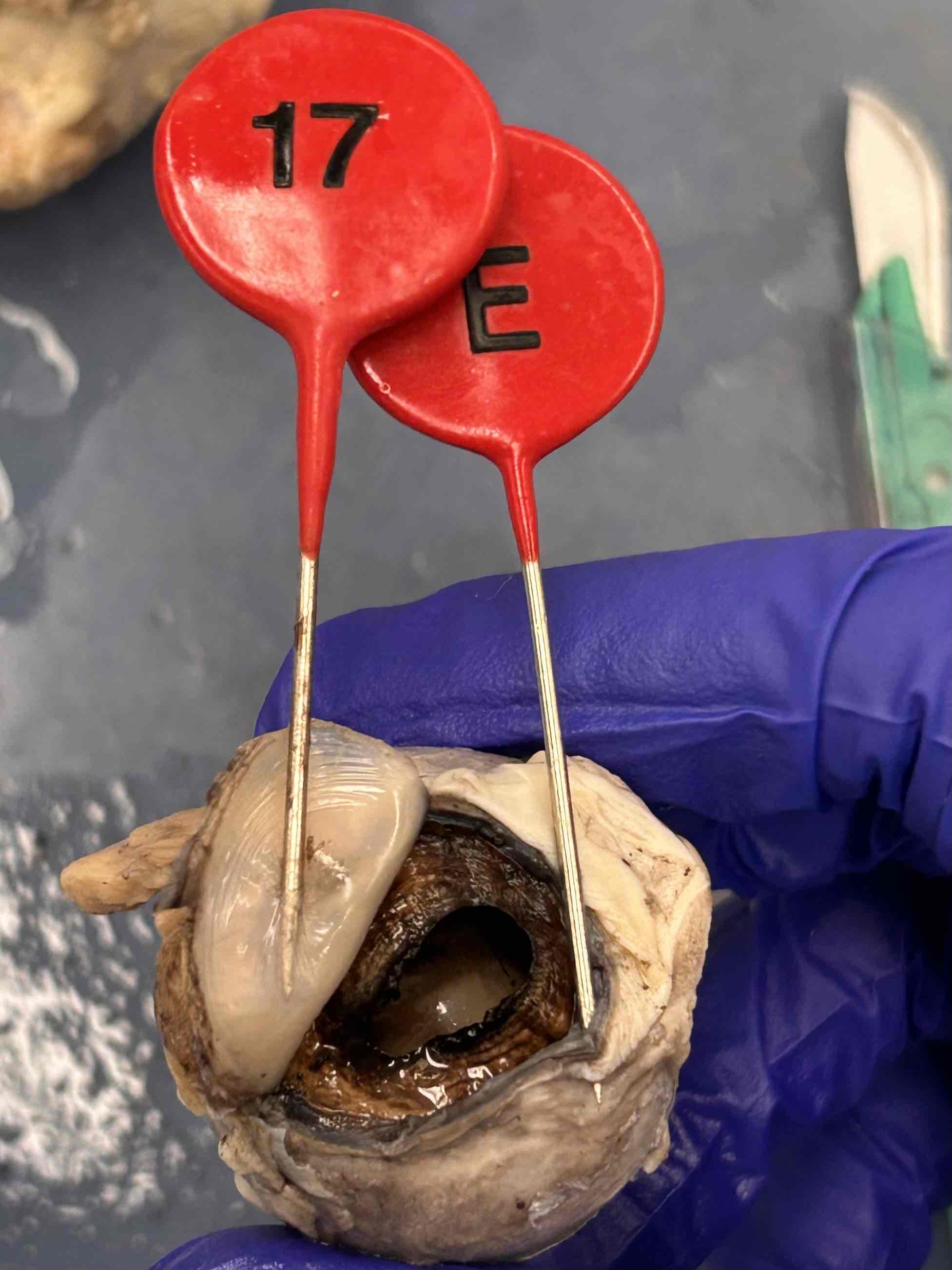
17
Anterior Chamber-Aqueous Humor;
The fluid-filled space between the cornea and the iris, containing aqueous humor that nourishes and maintains intraocular pressure.
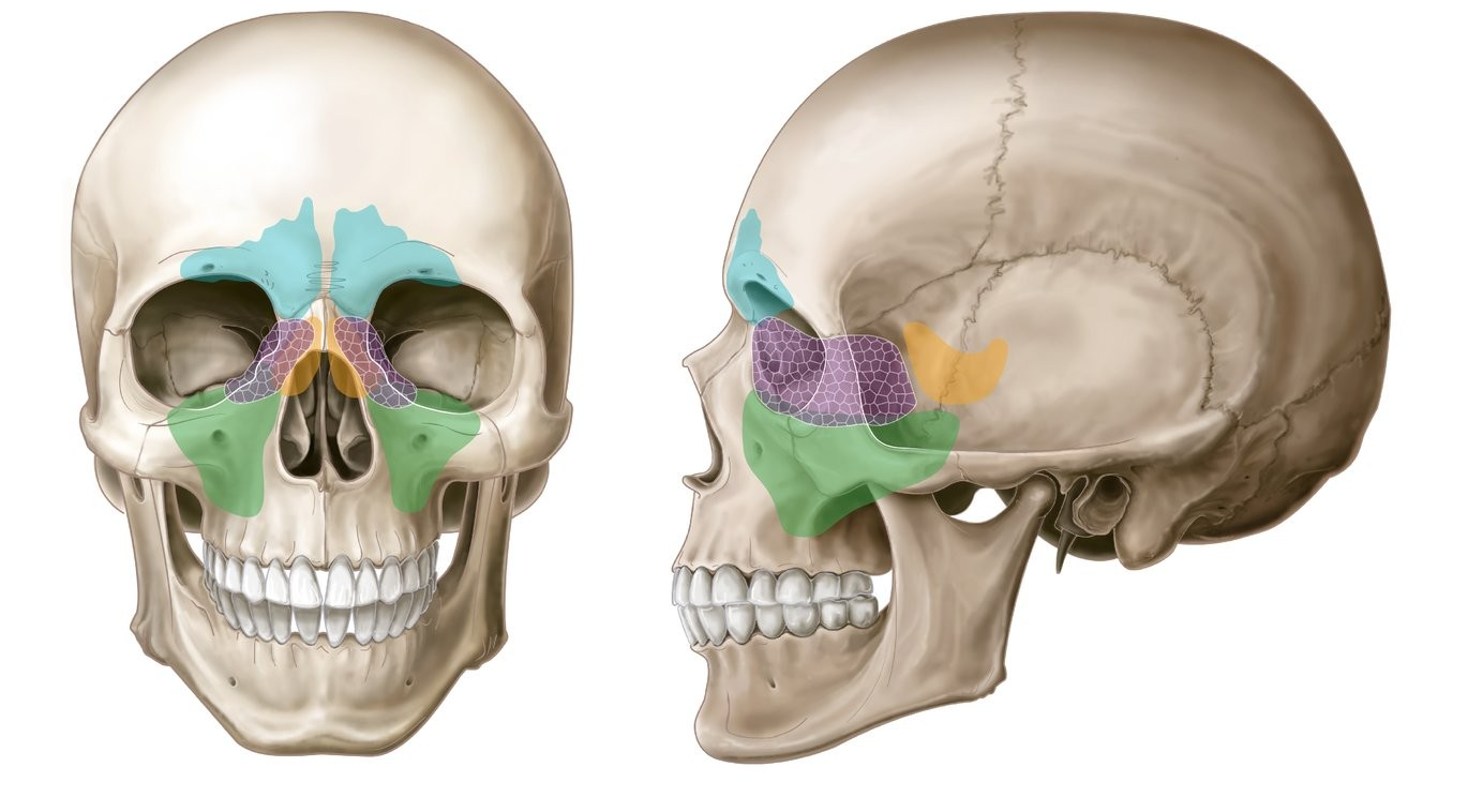
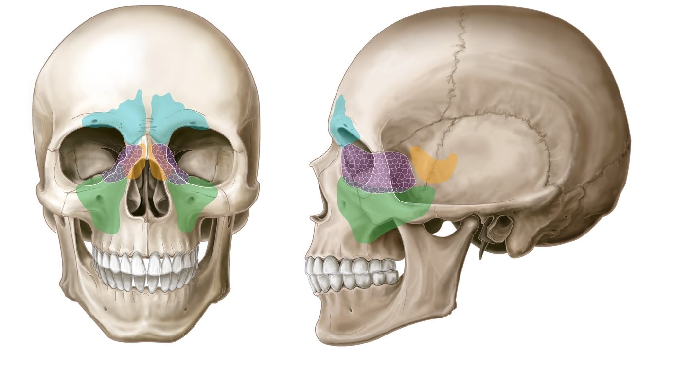
Green region
Maxillary Sinuses
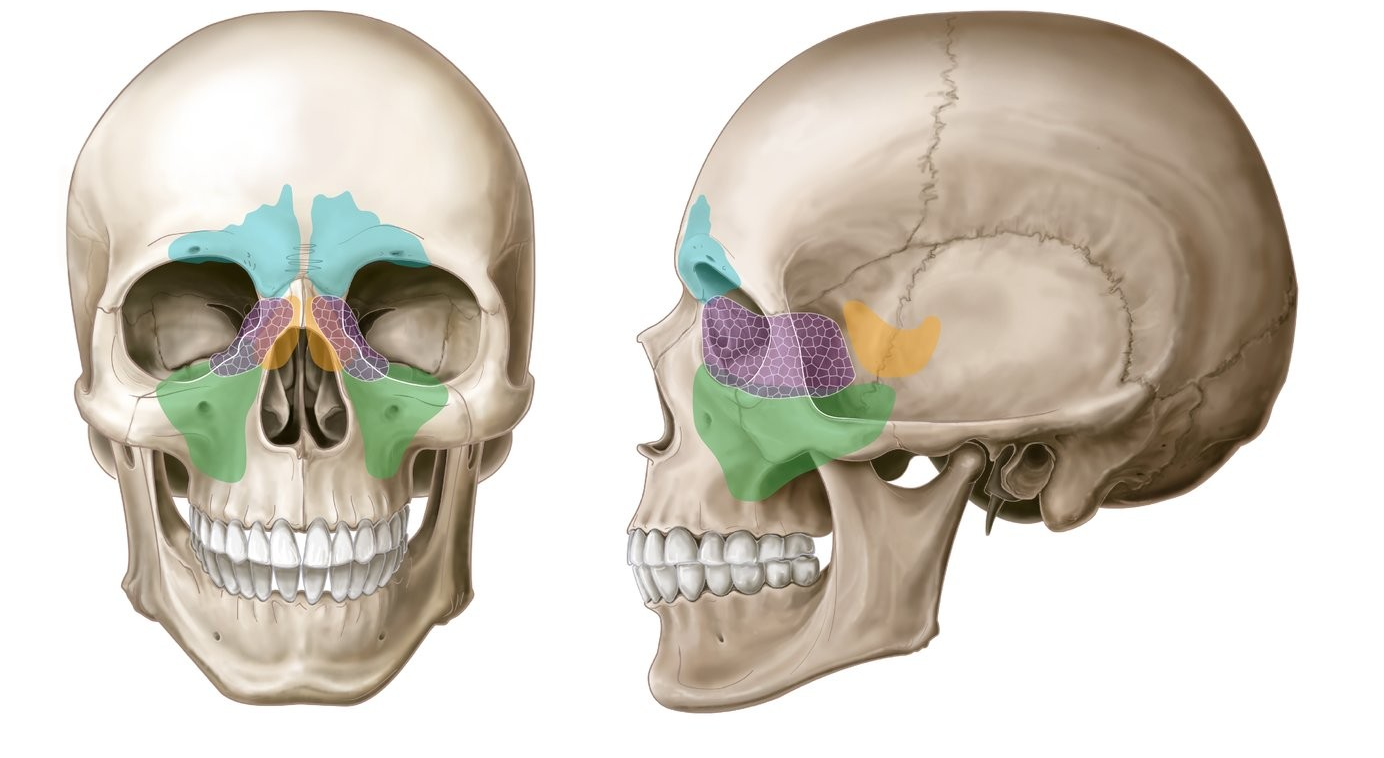
Blue region
Frontal sinuses
Purple region
Ethmoid sinuses
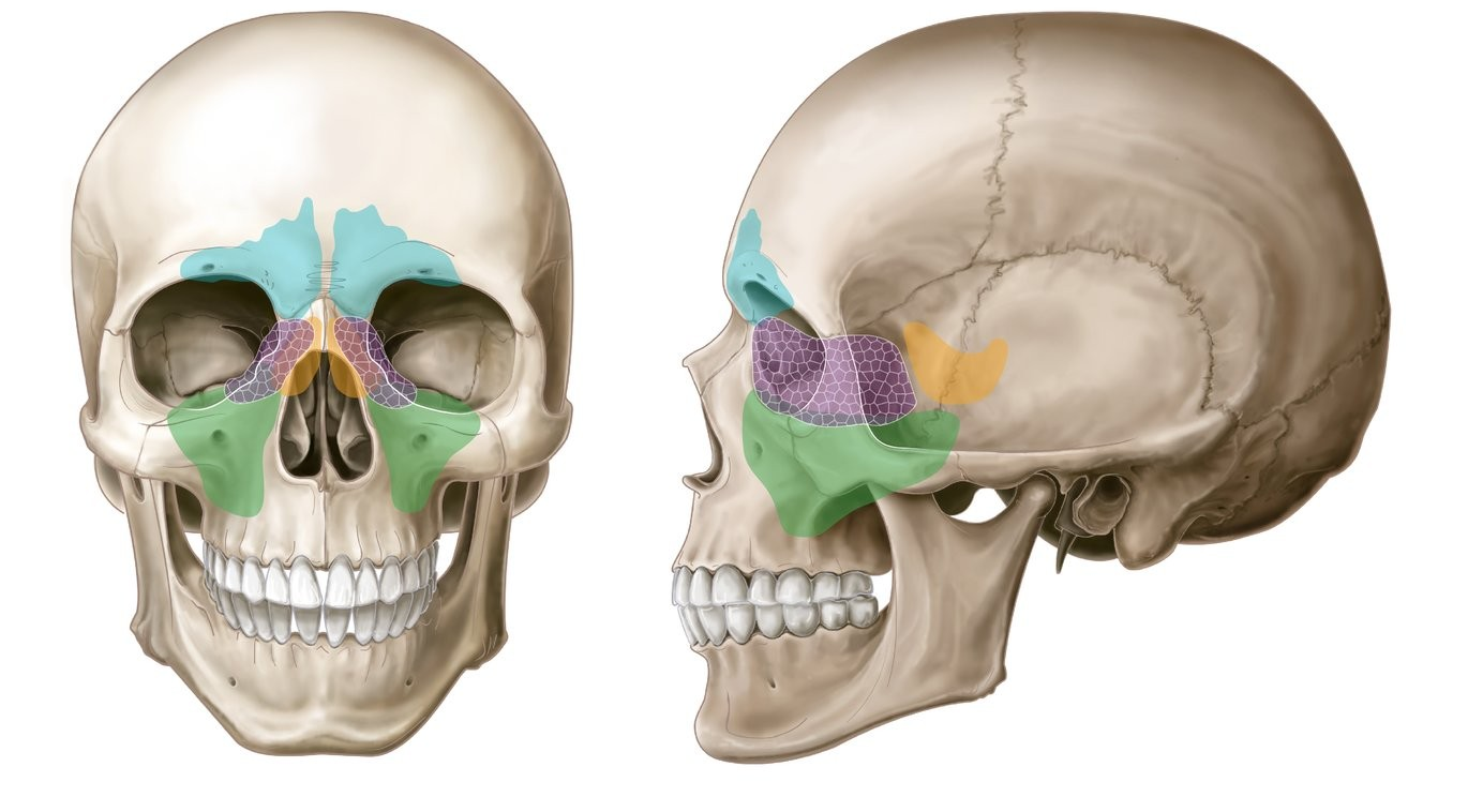
Orange region
Sphenoid sinuses
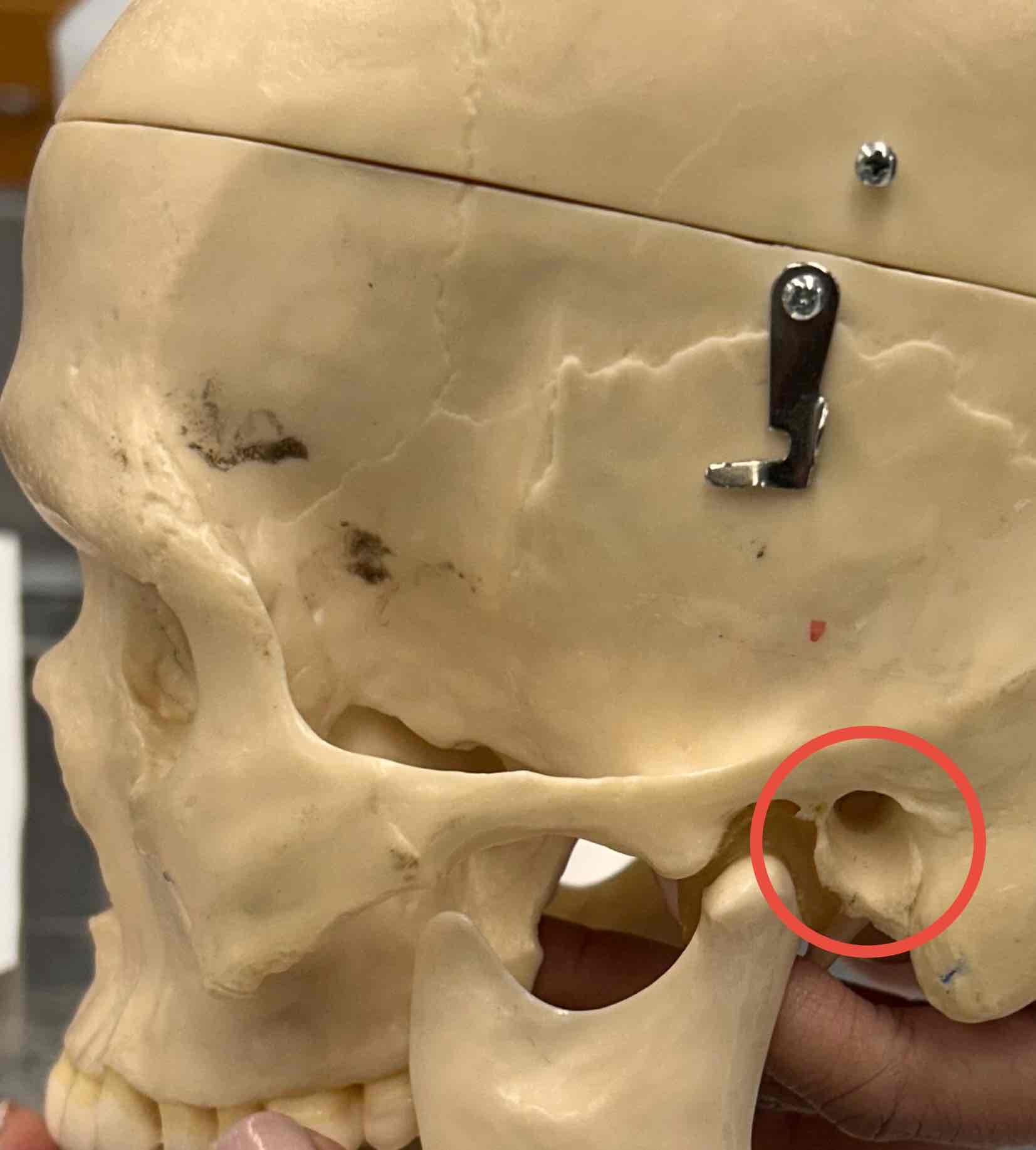
External acoustic meatus
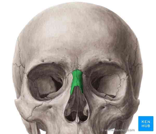
Nasal bone
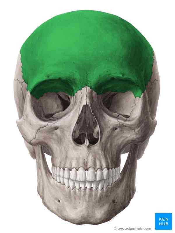
Frontal bone
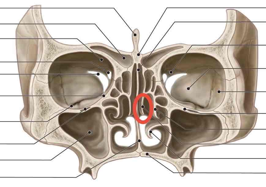
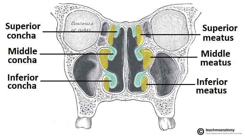
Superior nasal conchae
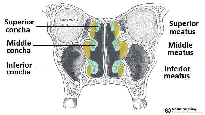
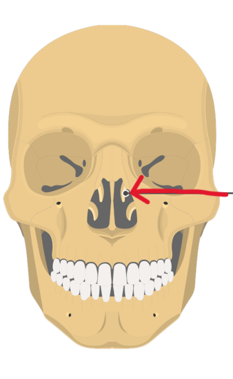
Middle nasal conchae
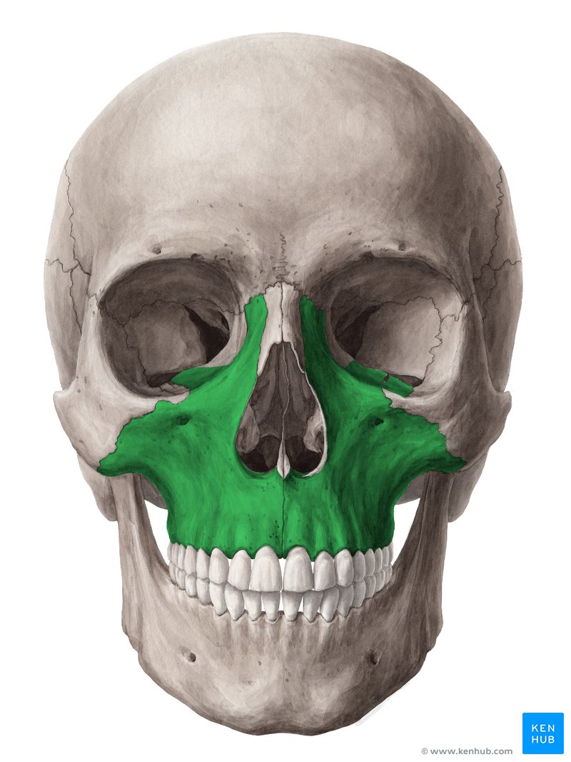
Maxilla
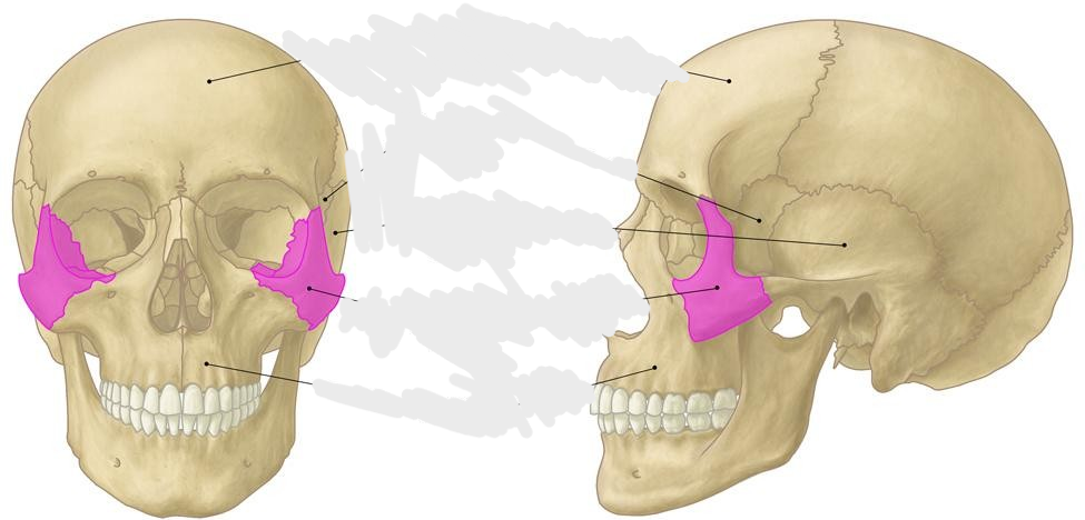
Zygomatic bone
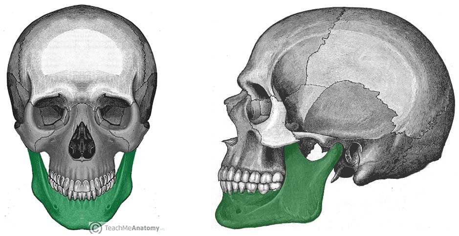
Mandible
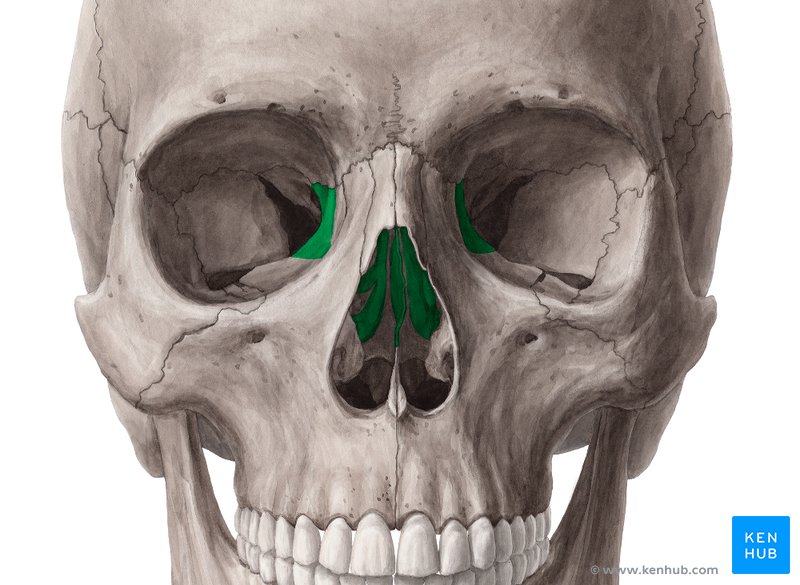
Ethmoid bone
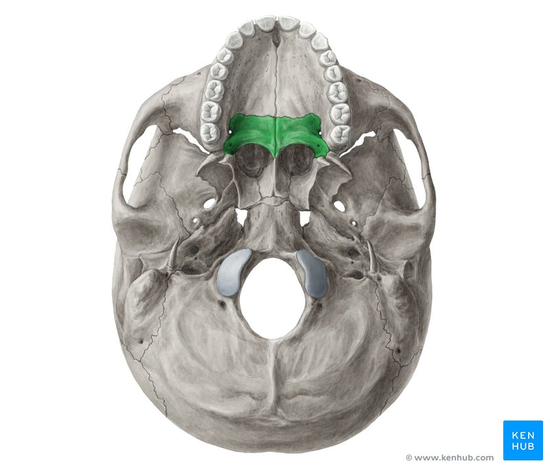
Palatine bone
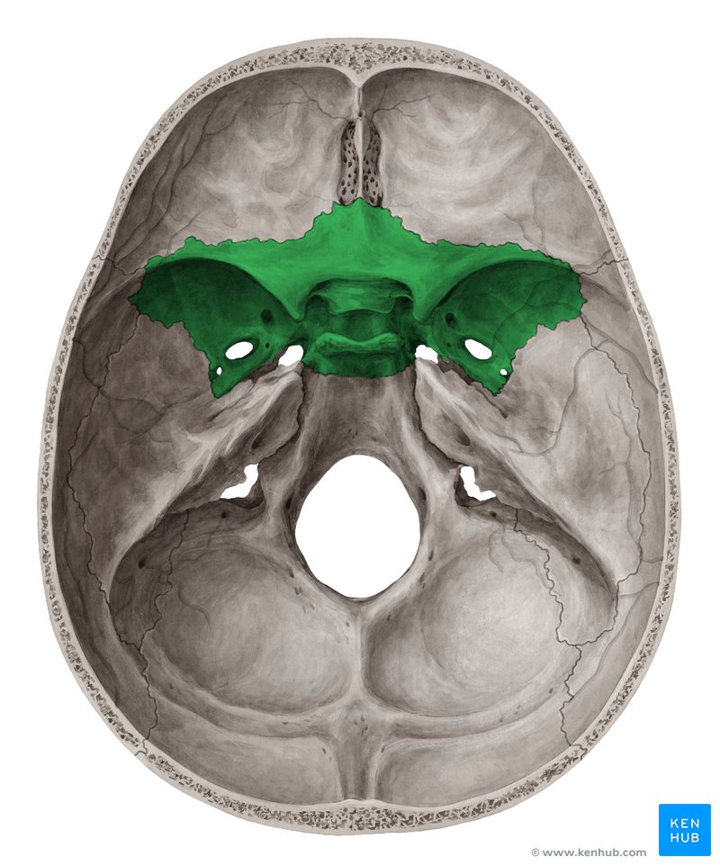
Sphenoid bone
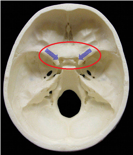
Name (of these holes) and Cranial nerve passing through?
Optic canal- optic nerve (CN II)
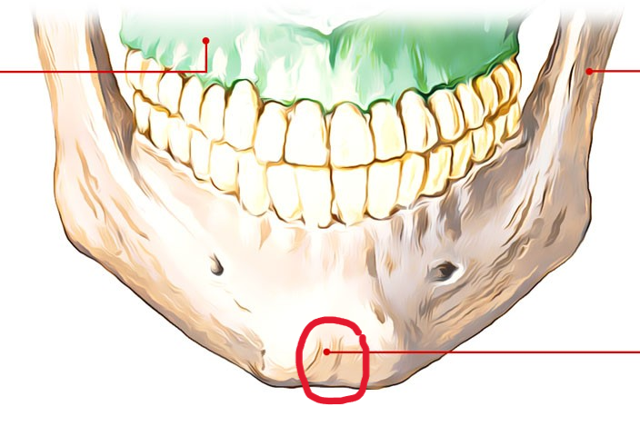
Mental protuberance
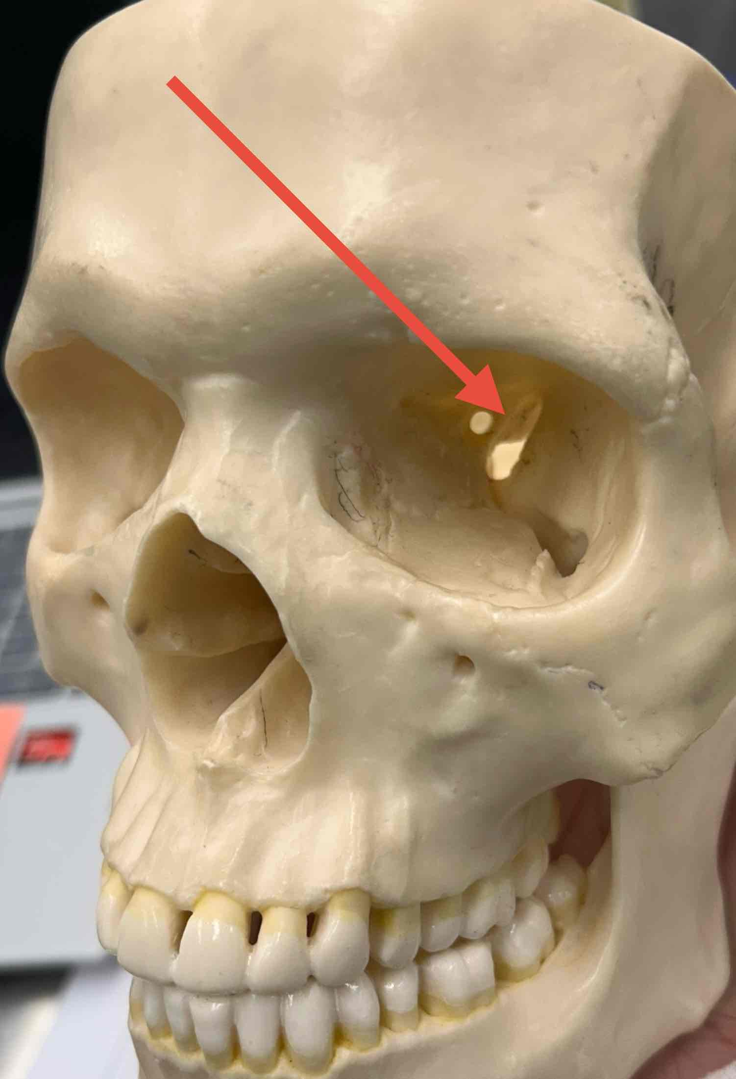
Name and Cranial nerve passing through?
Superior orbital fissure- oculomotor nerve (CN III), trochlear nerve (CN IV), trigeminal nerve (V), and abducens nerve (CN VI)
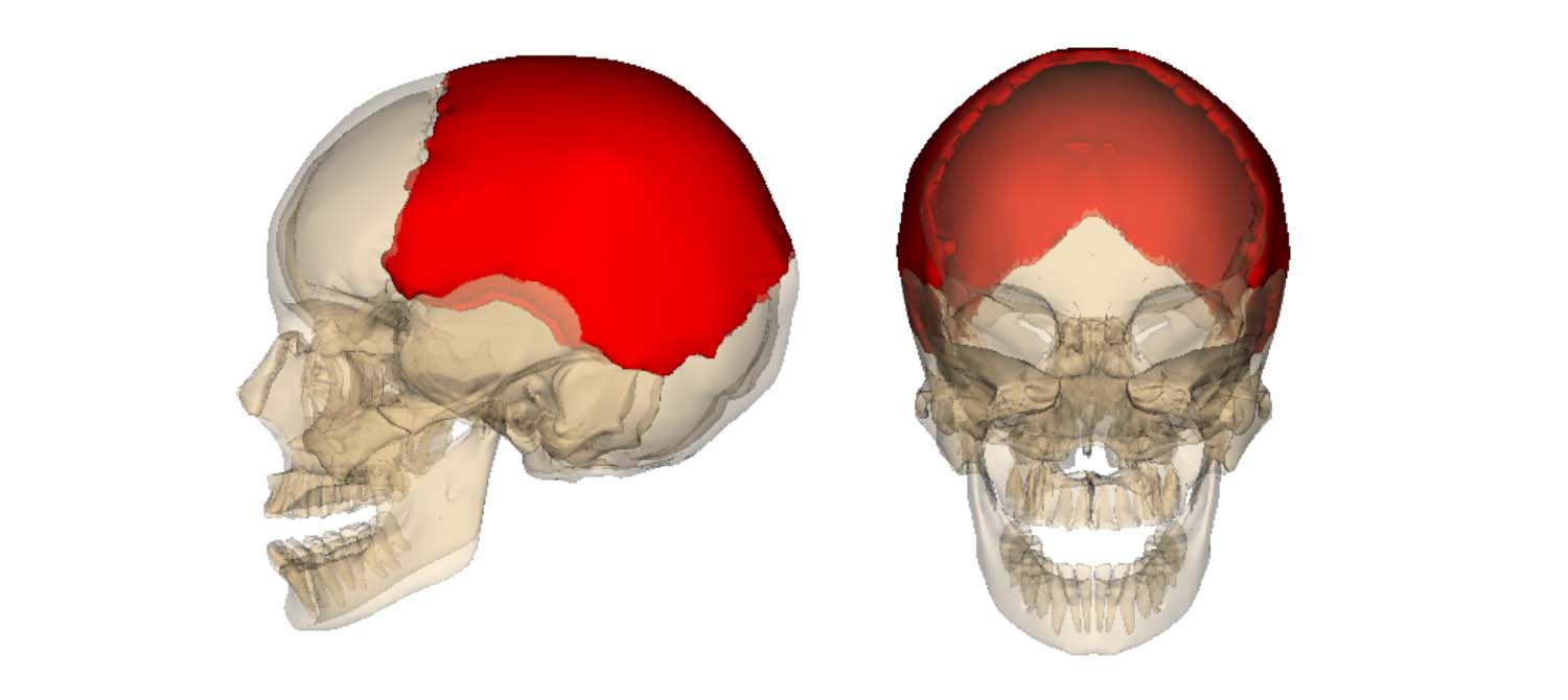
Parietal bones
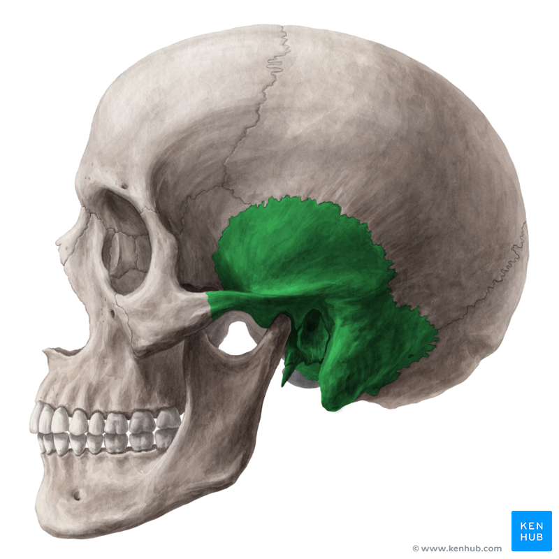
Temporal bone
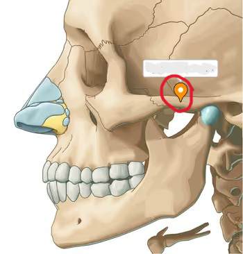
Zygomatic arch
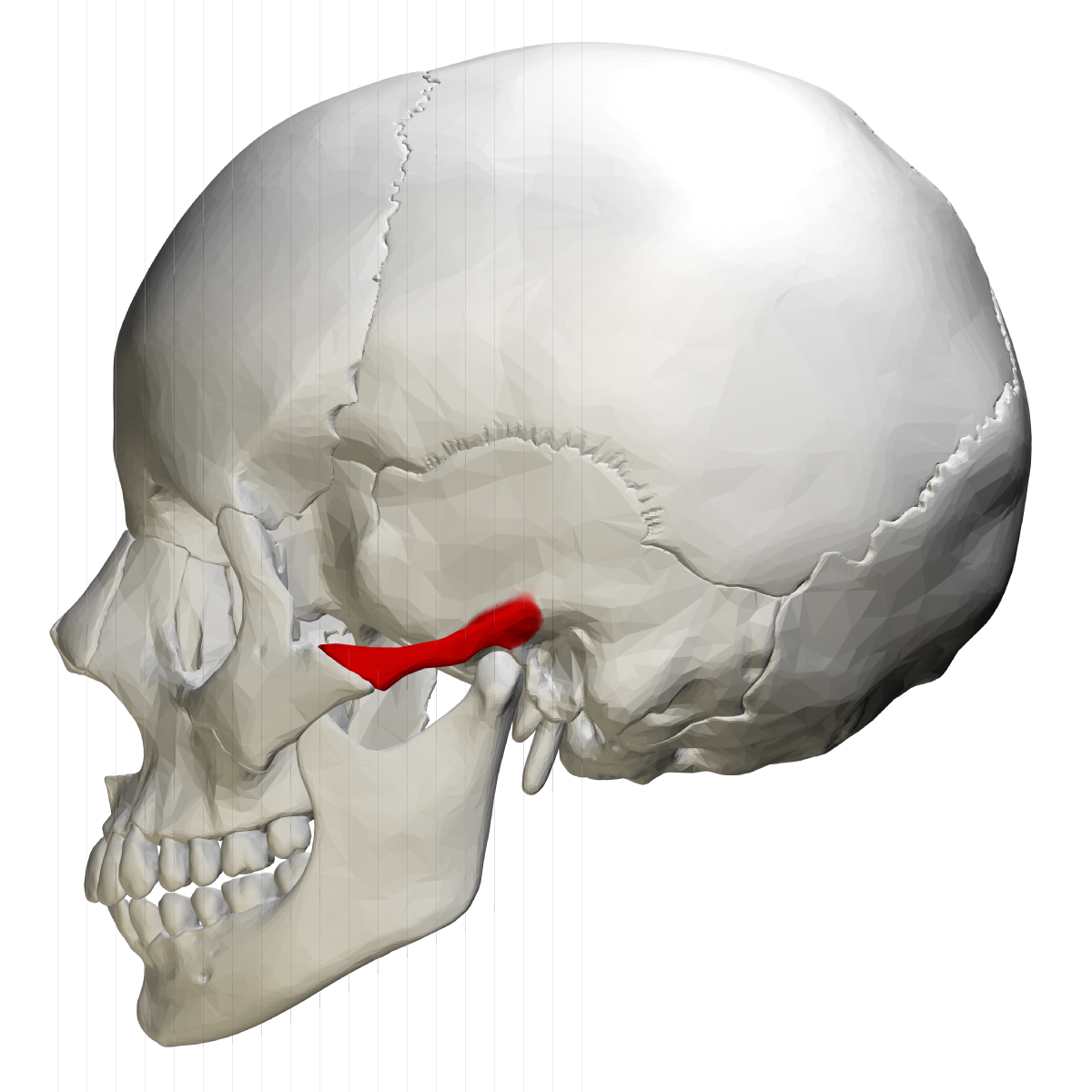
Zygomatic process
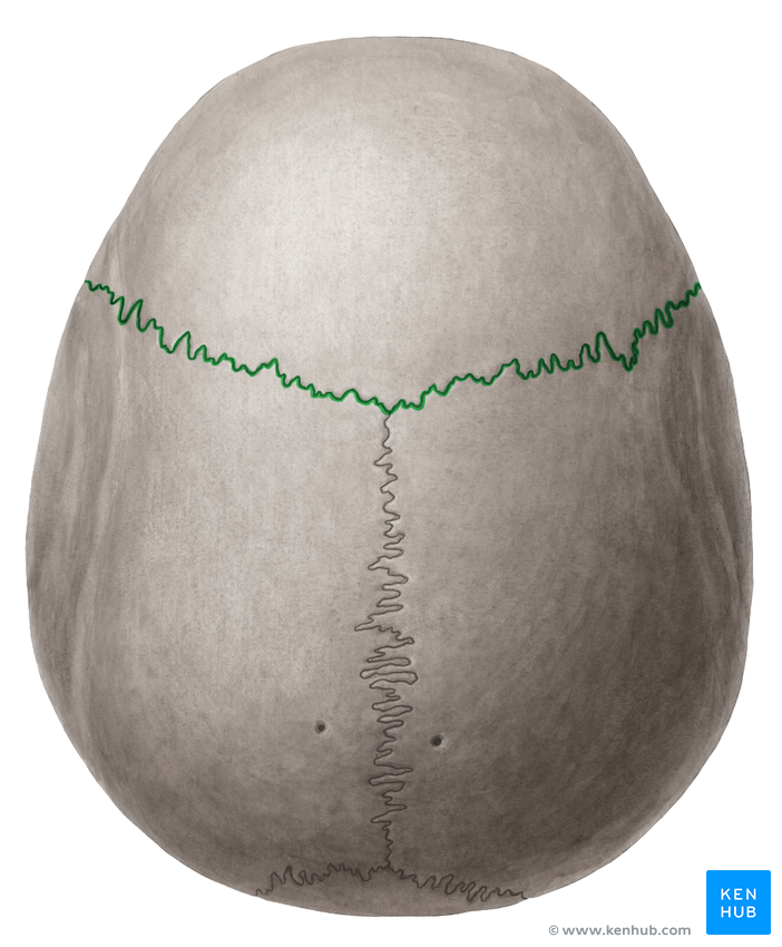
Outlined in green
Coronal suture
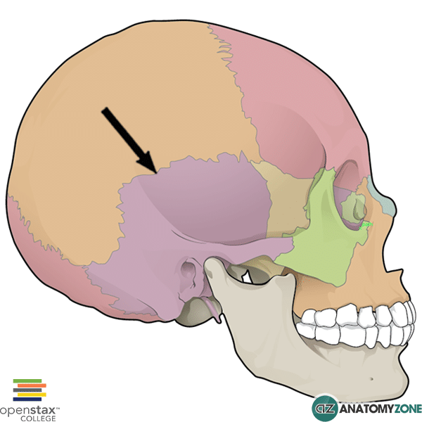
Not the bone but the suture
Squamous suture
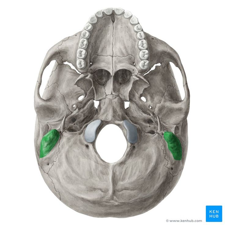
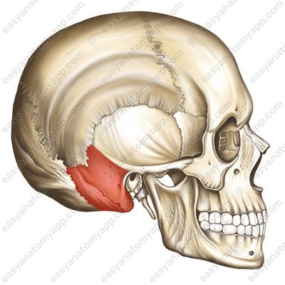
Mastoid process
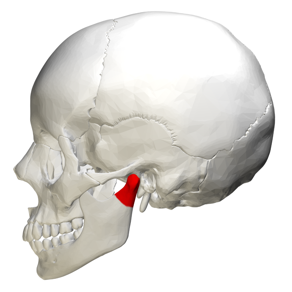
Condylar process
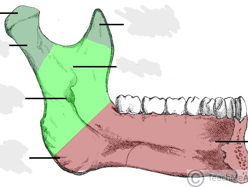
Green region
Ramus of mandible
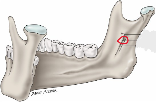
Mandibular foramen
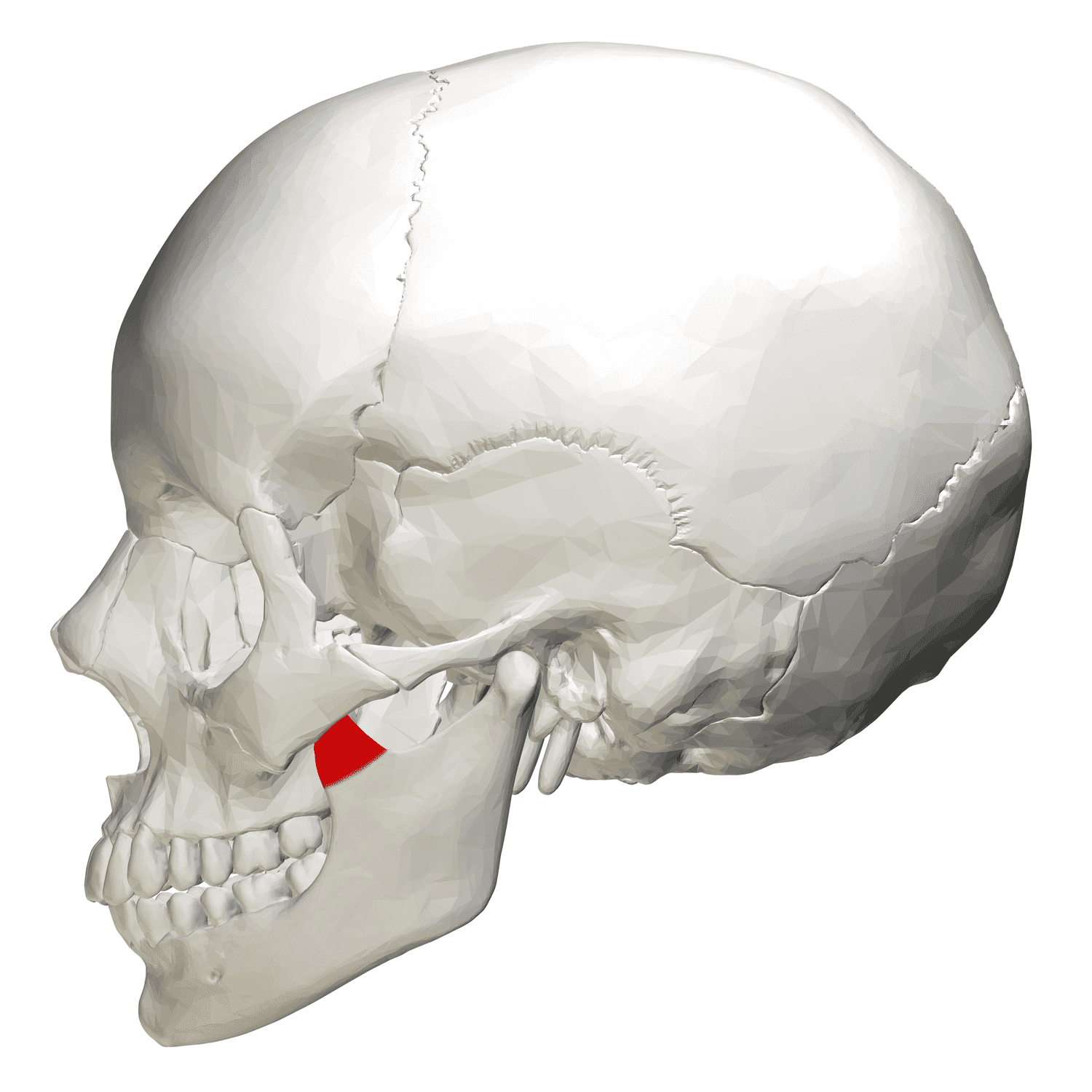
Coronoid process
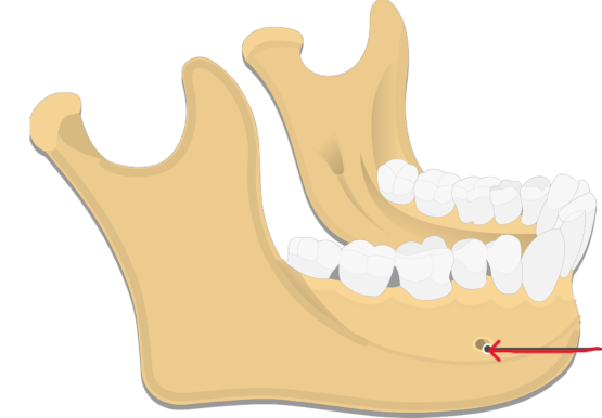
Name and Cranial nerve passing through?
Mental foramen- trigeminal nerve (V)
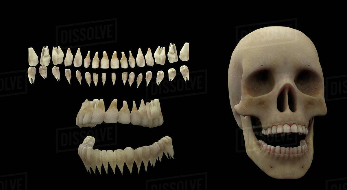
Teeth
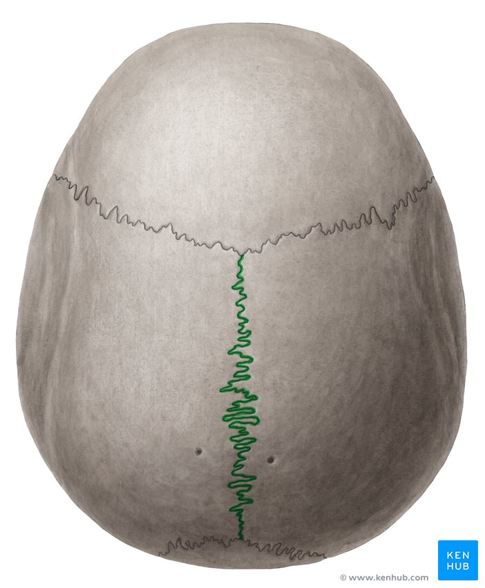
Outlined in green
Sagatittal suture
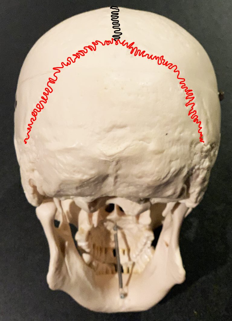
Outlined in red
Lambdoid suture