Lecture 9: Common Disorders of the Equine Forelimb
 Call Kai
 Learn
Learn Practice Test
 Spaced Repetition
 Match
Match Flashcards
 Knowt Play
1/55

There's no tags or description
Looks like no tags are added yet.
Name | Mastery | Learn | Test | Matching | Spaced | Call with Kai | Chat |
|---|
No analytics yet
Send a link to your students to track their progress
56 Terms
foot > below carpus > carpus (most common in racehorses) > above carpus
What is the prevalence of structures of the forelimb lameness in the horse
- comminuted
- chip
- palmar process
What are the types of fractures that can occur at the second phalanx (short pastern)?
- trauma
- twist injuries (comminuted)
What is the etiology of fractures at the second phalanx (short pastern)?
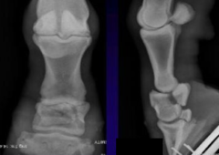
- fracture lame --> non-weight bearing or minimal (4 or 5 lameness)
- radiographs
How do you diagnose a fracture of the equine second phalanx (short pastern)?
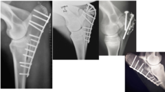
- internal fixation + pastern arthrodesis
- coaptation: half limb cast or transfixation pin cast
How do you treatment for fractures of the equine second phalanx (short pastern)?
- fair to good depending on success of repair, since can fuse the joint
- decreased prognosis with involvement of the coffin joint
What is the prognosis for fractures of the second phalanx (short pastern)?
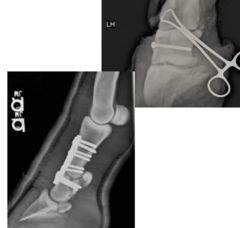
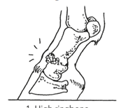
- proximal interphalangeal (pastern) joint OA
What is a high ring bone medically known as?
- conformation
- trauma (since low motion joint)
- OC cysts
What is the etiology of proximal interphalangeal (pastern) joint OA?
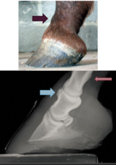
- moderate lameness
- characteristic bony swelling over the dorsal aspect of the pastern due to joint trying to stabilize itself
- positive to lower limb flexion
- blocks to abaxial
- radiographs to assess joint space
How do you diagnose proximal interphalangeal (pastern) joint OA?
Mild to moderate = Corticosteroid injections (high motion) , other OA management
Moderate to Severe: arthrodesis (advanced) or ethyl alcohol facilitated ankylosis (if sx not an option)
How do you treat a horse for proximal interphalangeal (pastern) joint OA?
- fair to good
- 2nd best behind distal hock OA
What is the prognosis for proximal interphalangeal (pastern) joint OA?
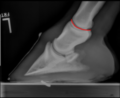
- athletic use
- conformation issues
- trauma
What are the etiologies for metacarpophalangeal (fetlock) joint OA in horses?
- lame
- positive flexion test
- decreased ROM
- effusion
- block to fetlock joint or low 4 point
- radiographs = definitive dx
How do you diagnosis metacarpophalangeal (fetlock) joint OA?
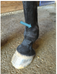
- fluid on the dorsal aspect of the suspensory branch
What does effusion of the fetlock joint look like?
- fluid on the palmar or plantar aspect of the suspensory branch
What does effusion of the tendon sheath look like?
- depends on severity
- fetlock is a high motion joint so cannot fuse and remain sound
- OA causes a lot of pain
What is the prognosis for metacarpophalangeal (fetlock) joint OA?
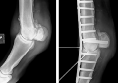
- normally dorsal in the forelimbs
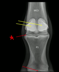
- OCD seen in proximal P1 and mid sagittal ridge of MC3
Where are the common locations for metacarpophalangeal joint osteochondral fragments and OCD in horses?
- trauma --> jumping and racing horses
- OCD lesion
What is the etiology of metacarpophalangeal joint osteochondral fragments in horses?
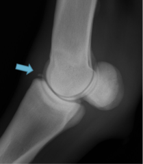
- mild to moderate lameness
- fetlock effusion
- positive to fetlock flexion test
- radiographs for definitive diagnosis
How do you diagnose metacarpophalangeal joint osteochondral fragments in horses?
- arthroscopic removal
- good to excellent
What is the treatment and prognosis for metacarpophalangeal joint osteochondral fragments in a horse?
- trauma --> most commonly the medial splint bone (MC2)
- conformation --> bench knee or toed out
What is the etiology for 2nd and 4th metacarpi (splint bone) exostosis?
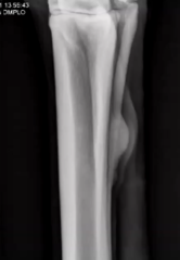
- characteristic swelling = firm over splint bone
- palpation
- +/- lame; usually not lame unless interference of soft tissue structures
- radiographs for definitive diagnosis
- can interfere with suspensory ligament = lame = CT or U/S
How do you diagnose 2nd and 4th metacarpi (splint bone) exostosis?
- if not causing an issue can be monitored
- rest
- NSAIDs
- interfering with suspensory ligament = remove splint bone
2nd and 4th metacarpi (splint bone) exostosis in horses has an excellent prognosis with treatment, what is the treatment?
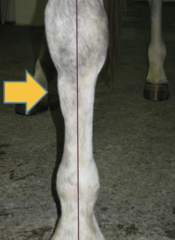
- common in racehorses early in training
- remodeling of MC3 in response to training
What is the etiology for dorsal 3rd metacarpal periostitis (bucked shins)?
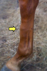
- acutely lame with short stride, especially after hard work
- +/- dorsal swelling
- pain on palpation
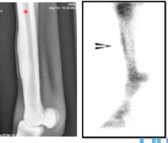
- radiographs
- nuclear scintigraphy
How do you diagnose dorsal 3rd metacarpal periostitis?
- NSAIDs
- rest (length based on severity)
- alter training program (trainers resist this)
- shockwave therapy
- osteoclast inhibitors
treatment for bucked shins
good to excellent with medical management
What is the prognosis for dorsal 3rd metacarpal periostitis?
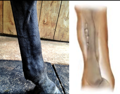
- foals as a pasture accident
- race horses due cumulative stress
What is the etiology for 3rd metacarpal (cannon) bone fractures?
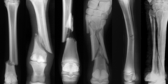
- bone is only covered by skin and some SQ, so will end up with an open fracture if not
Why do 3rd metacarpal (cannon) bone fractures require immediate and correct coaptation?
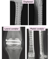
- diaphyseal MC3 fracture
- foals and adults
What is this type 3rd metacarpal (cannon) bone fracture? Who commonly gets it?
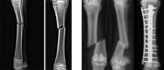
- distal condylar MC3 fracture
- thoroughbreds
What is this type 3rd metacarpal (cannon) bone fracture? Who commonly gets it?
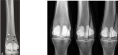
- medial: spirals
- lateral: does not spiral
What is the difference between condylar type fractures of the 3rd metacarpal (cannon) bone?
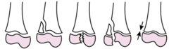
type 1: straight across growth plate
type 2: above growth plate
type 3: below growth plate
type 4: through growth plate
type 5: compression of growth plate
What are the salter harris classifications?
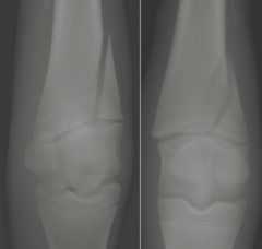
- Salter Harris Type II MC3 fracture
- foals
What is this type 3rd metacarpal (cannon) bone fracture? Who commonly gets it?
- standardbreds
What type of horse get palmar proximal 3rd metacarpal fractures most commonly?
internal fixation
- 2 plates for diaphyseal
- screws for lateral condylar and proximal palmar
- screws and dorsolateral plate for medial condylar
- must take dorsal plate off before returning to work
transfixation pin cats
What is the treatment for 3rd metacarpal fractures?
- open fx: grave
- foals: good
- adults: diaphyseal fair to poor and condylar / proximal palmar good for life /fair for return to race
What is the prognosis for 3rd metacarpal fractures?
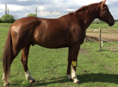
- positive response to flexion
- reduced range of motion = stiff-legged gait
- external swellings = bony or effusion
- acquired "over at the knee" - fails to keep leg straight
- acquired "bow legged" - trying to offload painful structures
What are the general clinical signs associated with disorders of the carpal joint in horses?
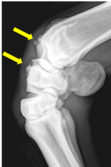
race horses = most common fracture early in their career
- due to hyperextension of limb at speed
What is the etiology for carpal chip fractures?
- has mild to moderate lameness
- carpal effusion**
- +/- nerve blocks --> may be skipped
- radiographs: flexed lateral, flexed skylines
You suspect a young racehorse of having a carpal chip fractures. How do you diagnose it?
- fractious
- obvious effusion associated with a common injury
- severe lameness and don't want to make it worse
Why might nerve blocks be skipped in a lame racing horse?
- arthroscopy
How do you treat carpal chip fractures?
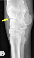
- weight bearing cartilage
- intercarpal ligaments
- good if no other damage
- poor if extensive damage
- range in between
What concurrend damage can impact the prognosis of a carpal chip fracture?
- good if acute
- poor if chronic
How can chronicity impact the prognosis for a carpal chip fracture?
- etiology same as chips --> racing horse due to hyperextension of limb at speed
- C3 frontal plane most common
What is the etiology of carpal slab fractures and where are they most commonly seen?
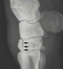
- severe lameness
- severe carpal effusion
- do NOT block because don't want to displace anymore
- radiographs
How do you diagnose carpal slab fractures?
- humeral fracture
- radial nerve injury
- olecranon fracture
What are differential diagnosis for failure of the stay apparatus?
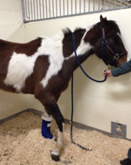
- dropped elbow
- unable to "fix" carpus
- inability to extend limp
What are common clinical signs of failure of the stay apparatus?
- trauma
- avulsion of brachial plexus
- humeral fracture
- prolonged lateral recumbency w/o padding
What is the etiology of radial nerve injuries in horses?
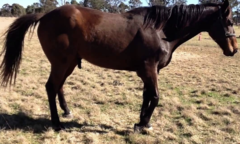
- typical stance = unable to extend limb
- drags limb
- not consistent loss of cutaneous sensation
- muscle atrophy after 2-4 weeks
- EMG?
How do you diagnose radial nerve injuries in horses?
- splint to fix limb
- anti inflammatories like NSAIDs or corticosteroids
How do you treat a radial nerve injuries in horses?
- good if recovers function in 6-8 weeks
- poor otherwise (risk of contralateral limb laminitis)
What is the prognosis of an equine radial nerve injury?
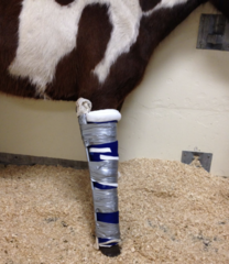
- most common foal fracture
- result of kick = also has a bad wound
What is the common age and etiology for olecranon fractures in horses?
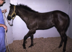
- failure of stay apparatus
- swelling, crepitus
- radiographs
How do you diagnosis an olecranon fracture in a horse?
- incomplete and non-displaced = stall rest
- complete or displaced = surgery (tension band)
How do you treat a foal with an olecranon fracture?
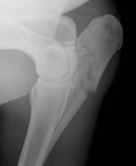
- good in foals and adults if not in joint and surgery
- fair if articular surface involved
- guarded if open fracture
What is the prognosis of olecranon fractures in horses?