DAT Bootcamp Biology 11.6 Skeletal System:
1/87
There's no tags or description
Looks like no tags are added yet.
Name | Mastery | Learn | Test | Matching | Spaced | Call with Kai | Chat |
|---|
No analytics yet
Send a link to your students to track their progress
88 Terms
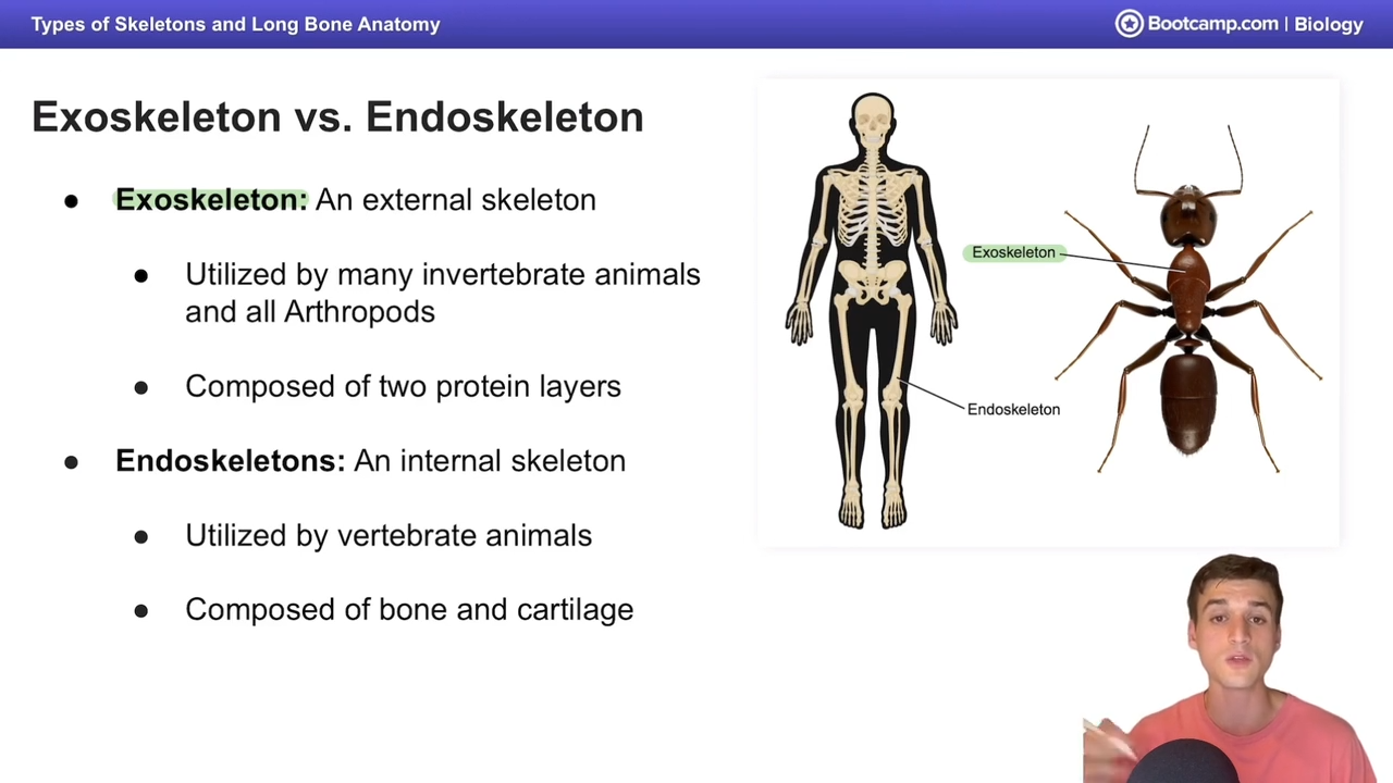
An exoskeleton is an...
external skeleton
Many
invertebrates and all arthropods possess
exoskeletons.
Vertebrates contain an endoskeleton on the
inside.
An endoskeleton can be divided into the
axial
skeleton (central bones: skull, spinal column, and
ribcage) and the appendicular skeleton
(appendages).
Types of bones in the endoskeleton:
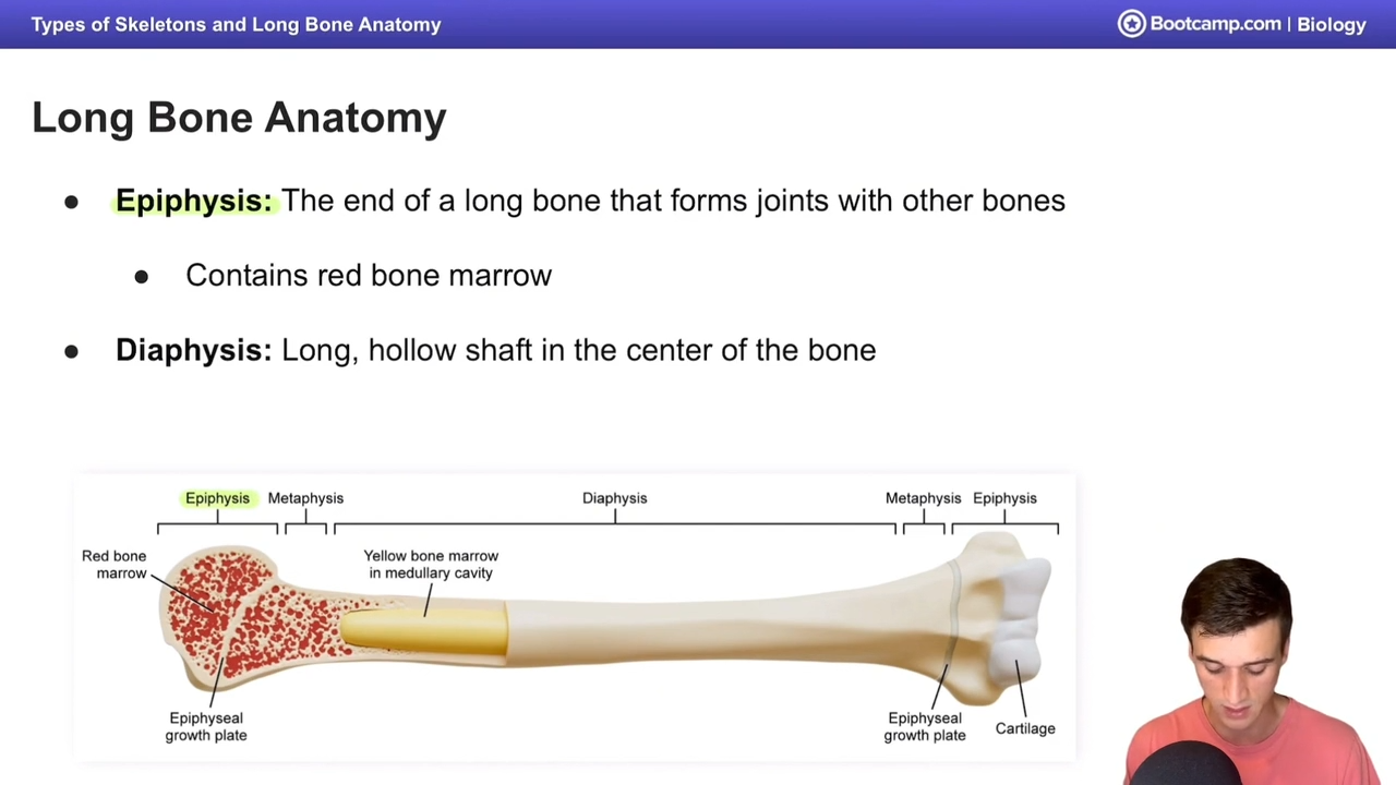
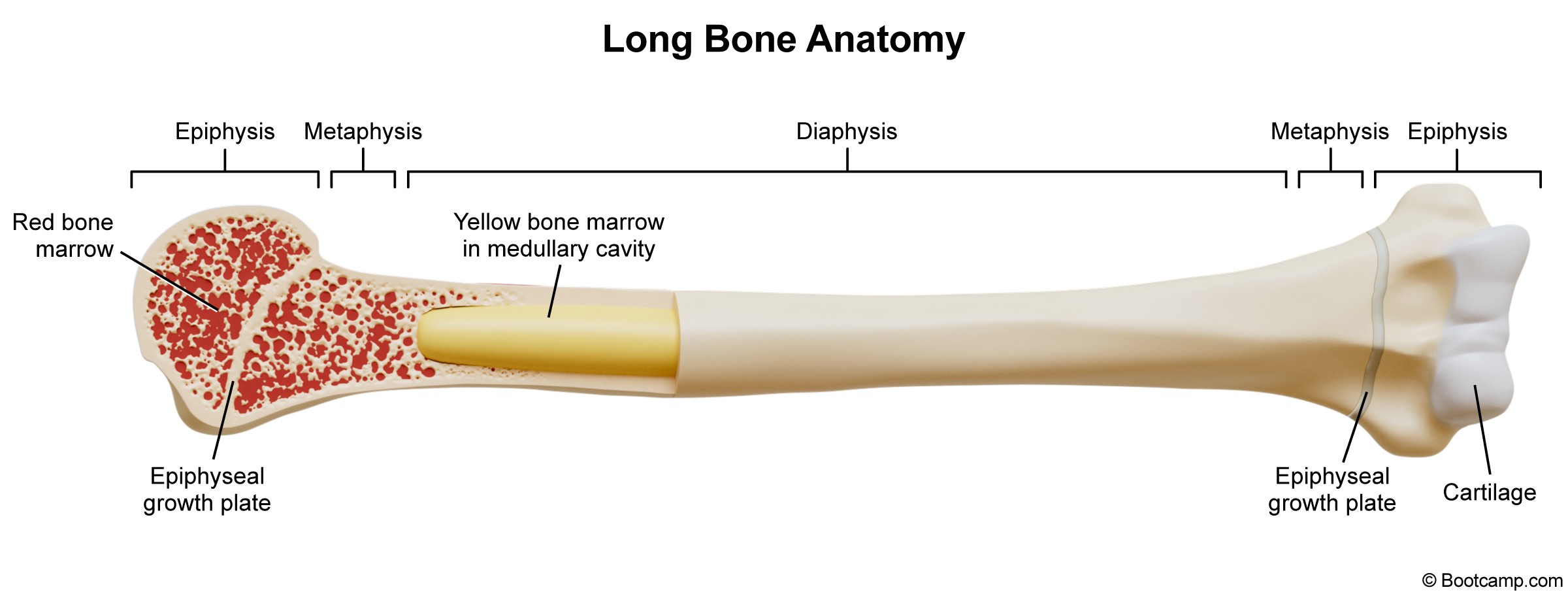
1.
Long bones - made of cortical bone (compact)
and pockets of cancellous bone (spongy).
Important features include the epiphysis,
diaphysis, medullary cavity, metaphysis, and
epiphyseal plate.
Epiphysis -
end of a long bone that forms
joints with other bones and contains red bone
marrow for hematopoiesis (blood cell
synthesis).
Diaphysis -
long hollow shaft in center of
bone.
Medullary cavity -
located within the
diaphysis and contains red and yellow bone
marrow (area of fat storage).
Metaphysis -
similar to epiphyses and found
between the medullary cavity and epiphyseal
plates.
Epiphyseal plate - "growth plate" located
between epiphysis and metaphysis. Made out
of hyaline cartilage and works to lengthen
the diaphysis through growth and ossification.
Short bones -
as wide as they are long and
mainly provide support (eg. parts of the wrist).
3. Flat bones -
mainly provide protection (eg. skull).
4. Sesamoid bones -
found within tendons to help
muscles pull (eg. kneecap).
5. Irregular bones -
irregularly shaped (eg. pelvis).
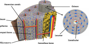
Cortical bone is the dense outer layer of bone that
supports the
weight of our bodies. It is composed of
many microstructures:
Osteons -
cortical bone's functional unit,
composed of tiny multi-layered cylinders. Also
known as haversian systems because they
contain a haversian canal in their center.
Haversian canals -
'tubes' that contain blood
vessels for nutrient supply.
Lamellae -
layers of the osteon.
Lacunae -
small spaces between lamellae that
hold bone cells and interconnect through
canaliculi.
Canaliculi -
small channels that connect lacunae
and the haversian canal.
Volkmann's canals -
connect Haversian canals to
the periosteum, which provides nutrients.
Cancellous bone is the
spongy inner layer of bone
that soaks up red bone marrow via a web of
trabeculae (connective tissue that supports
cancellous bone).
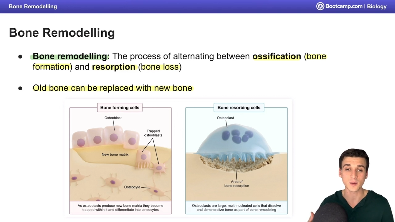
Bone remodeling is the process of going back and
forth between
the processes of ossification (bone
formation) and resorption (bone loss).
Types of cells involved in bone remodeling:
Osteoprogenitors - immature precursor cells that
differentiate into osteoblasts.
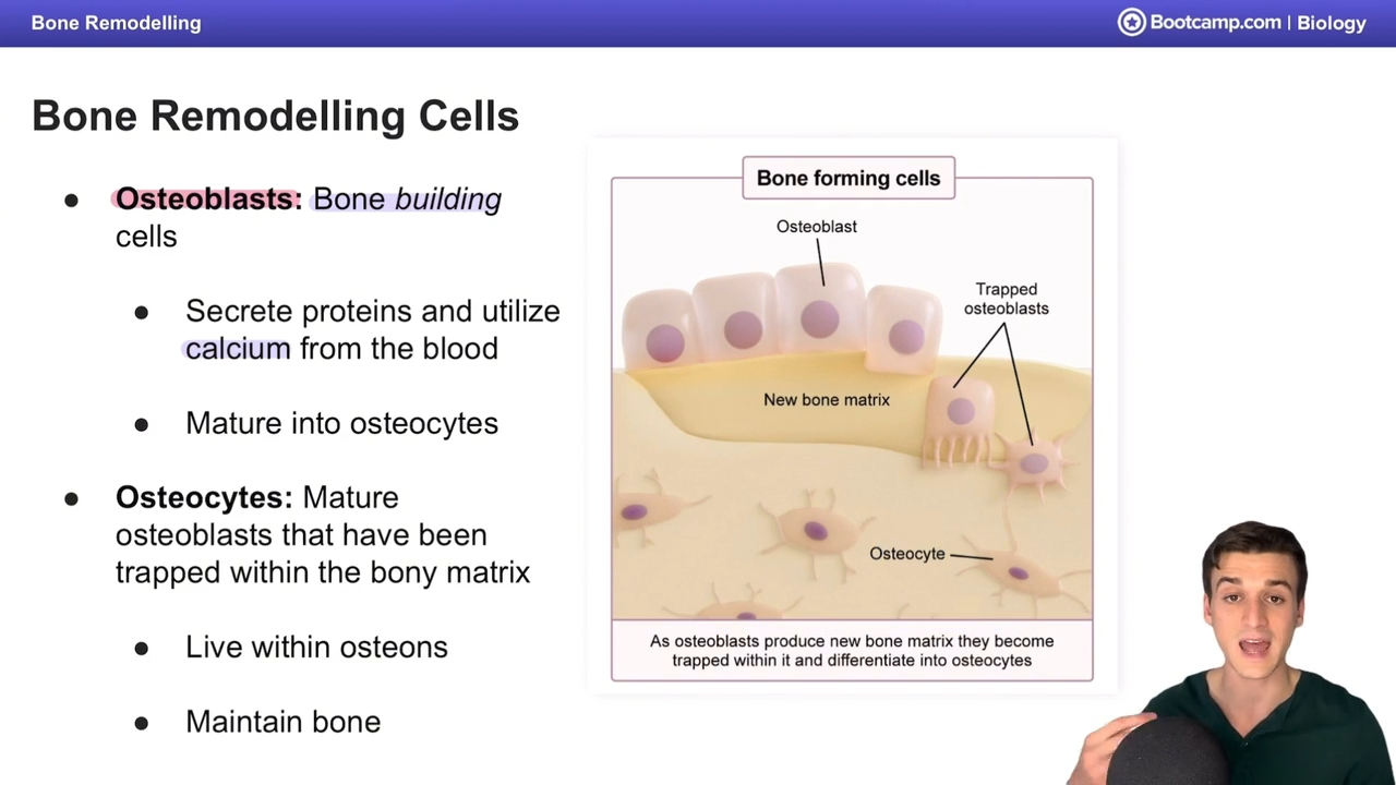
Osteoblasts - build bone by secreting proteins
and utilizing blood calcium. They mature into
osteocytes after getting trapped inside the bone
matrix they create.
Osteocytes - live in lacunae in osteons to maintain
bone.
Osteoclasts - eat and resorb bone, releasing
calcium and phosphate back into the blood.
Derived from monocytes.
Osteoprogenitors -
immature precursor cells that
differentiate into osteoblasts.
Osteoblasts -
build bone by secreting proteins
and utilizing blood calcium. They mature into
osteocytes after getting trapped inside the bone
matrix they create.
Osteocytes -
live in lacunae in osteons to maintain
bone.
Osteoclasts -
eat and resorb bone, releasing
calcium and phosphate back into the blood.
Derived from monocytes.
Mechanisms involved in bone remodeling:
Parathyroid hormone - increases blood calcium
levels by stimulating osteoclasts and depressing
osteoblasts. Secreted by the parathyroid gland.
Vitamin D - increases blood calcium levels by
raising intestinal calcium absorption. Activated by
parathyroid hormone, but provides negative
feedback on PTH production.
Calcitonin - decreases blood calcium levels by
depressing osteoclasts, allowing osteoblasts to
build bone without competition. Secreted by
parafollicular cells (C cells) of the thyroid gland.
Parathyroid hormone -
increases blood calcium
levels by stimulating osteoclasts and depressing
osteoblasts. Secreted by the parathyroid gland.
Vitamin D -
increases blood calcium levels by
raising intestinal calcium absorption. Activated by
parathyroid hormone, but provides negative
feedback on PTH production.
Calcitonin -
decreases blood calcium levels by
depressing osteoclasts, allowing osteoblasts to
build bone without competition. Secreted by
parafollicular cells (C cells) of the thyroid gland.
Mnemonic: CalciTONin =
"Tone down calcium"
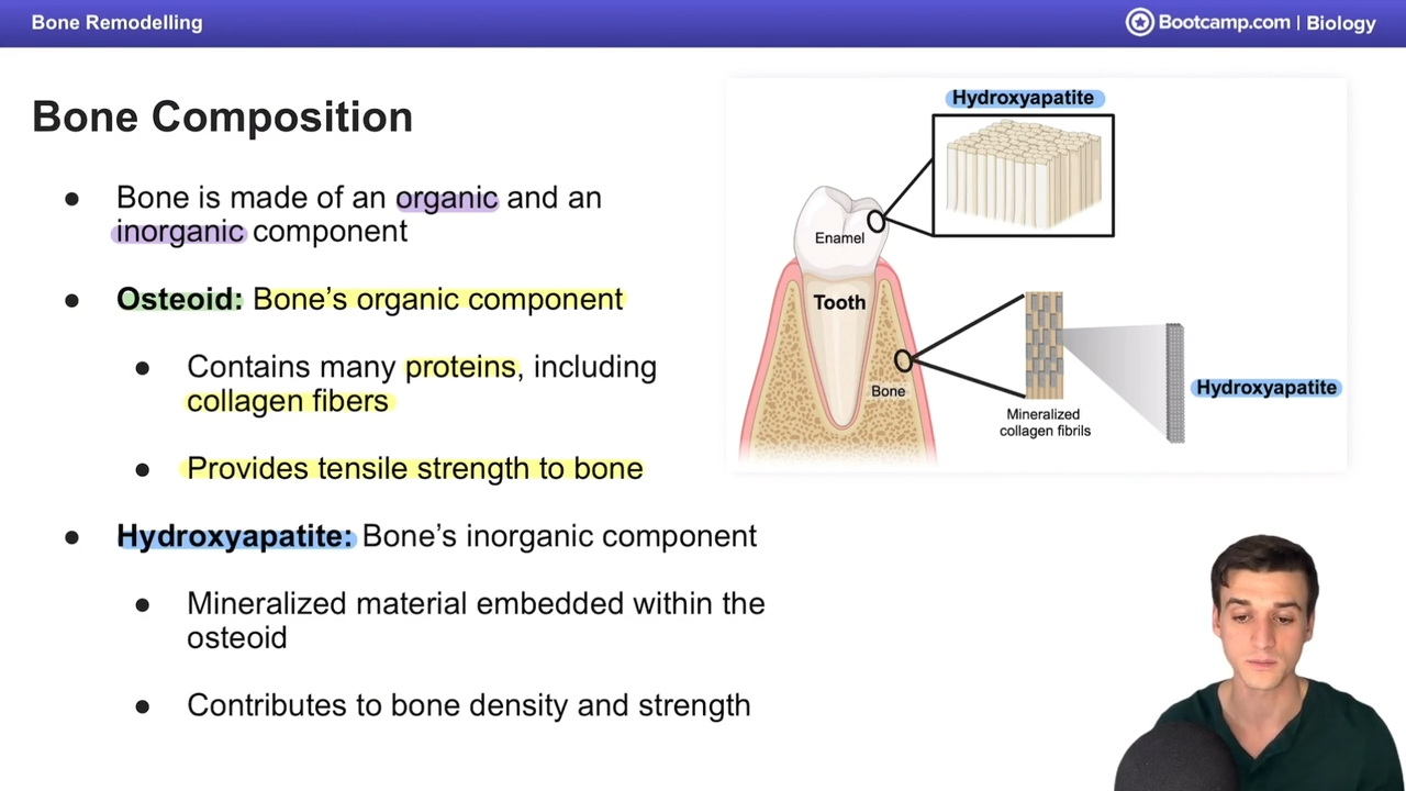
Osteoid is the
organic component of bone containing
many proteins such as collagen (gives bone tensile
strength).
Hydroxyapatite is the inorganic mineral component
of bone that gives the bone density and strength.
Two types of embryonic ossification:
1.
2. Endochondral ossification - bone is created
indirectly through a cartilage model, mainly for
long bones. The cartilage model calcifies during
fetal development, creating ossification centers
that help form the features of long bones.
Intramembranous ossification - bone is created
directly within fibrous membranes, mainly for flat
bones. Osteoblasts start by secreting osteoid,
which hardens and houses osteocytes. Eventually,
cortical bone is created.
Two types of embryonic ossification:
1.
2.
Endochondral ossification - bone is created
indirectly through a cartilage model, mainly for
long bones. The cartilage model calcifies during
fetal development, creating ossification centers
that help form the features of long bones.
Types of connective tissue:
1.
2. Cartilage is avascular (lacks blood vessels) and is
not innervated (as opposed to bone which is
highly vascular and innervated).
Fibrous connective tissue has a matrix made up
of fibers. It is maintained and repaired by
fibroblasts.
● Tendons - connect muscle to bone.
● Ligaments - connect bone to bone.
● Periosteum - membrane that covers cortical
bone with an outer fibrous layer
(vascularized) and an inner/cambium layer
(collagen for attachment to cortical bone)
● Endosteum - membrane located between cortical and cancellous bone.
Types of connective tissue:
1.
2.
Cartilage is avascular (lacks blood vessels) and is
not innervated (as opposed to bone which is
highly vascular and innervated).
Chondroblasts build cartilage by secreting
collagen and elastin.
Hyaline cartilage -
slightly flexible and
important in providing support and stability
to joints.
Fibrous cartilage -
high rigidity and resists
tension, found in intervertebral discs and knee
meniscus.
Elastic cartilage -
highly flexible and found in
ears and epiglottis.
3. Joints are
vascularized and innervated. They are
found between bones. Below are types of joints.
Synarthroses -
dense, fibrous joints that do
not move.
Amphiarthroses -
cartilaginous joints that
partially move.
Diarthroses -
synovial joints that fully move.
Typically contain hyaline cartilage.

What uses an exoskeleton?
invertebrate animals and arthropods

What are exoskeletons composed of?
two protein layers

What are long bones made of?
cortical bone and cancellous bone
In what bones does hematopoiesis occur in?
long bones
What is hematopoiesis?
blood cell production

Describe the anatomy of long bones
epiphysis - end of a long bone that forms joints with other bones, has red bone marrow for hematopoesis
epiphyseal plate - region of cartilage between epiphysis and metapysis that can lengthen diaphysis through ossification
metaphysis - between medullary cavity and epiphyseal palte
diaphysis - long hollow shaft in center of bone, filled with yellow bone marrow in medullary cavity


What is the medullary cavity?
inside of diaphysis, has red and yellow bone marrow
What do red and yellow bone marrow do?
red: produces stem cells capable of generating red and white blood cells
yellow - produces stem cells that can generate fat, bone, cartilage, and muscle. can also store fat.
Where can red bone marrow be found?
diaphysis and epiphysis
What is the metaphysis?
area between the medullary cavity and epiphyseal plate; one on each end of bone
What is the epiphyseal plate?
region of cartilage between epiphysis and metaphysis that can lengthen diaphysis during ossification
What is cortical bone and what is it made of?
dense, outer layer of bone that supports body weight (compact)
made of osteons with cylindrical osteon layers called lamellae
has haversian canals at the center of osteons with blood vessels for nutrient supply - run vertically
has Volkmann's canals with blood vessels connecting Haversian systems to periosteum - run horizontaly

What are the haversian canals?
tubes at the center of osteons with blood vessels for nutrient supply
in cortical bone
run vertically
What are Volkmann’s canals?
channels with blood vessels connecting Haversian systems to periosteum and bone cells outside of cetner of osteons
in cortical bone osteons
run horizontally
What are the layers around the cortical bone?
periosteum - external sheath surronding cortical bone that provides nutrients for growth/healing
endosteum - internal sheath linin the internal surface of cortical bone
What provides nutrients to bone cells outside the center of osteons and periosteum?
volkmann’s canals
What lines the internal surface of cortical bone?
endosteum
What surronds cortical bone and provides nutrients? What brings it nutrients?
periosteum
nutrinets brought by volkmann’s canals
What is cancellous bone?
spongy, inner layer of bones
soaks up red bone marrow via trabeculae

What is trabeculae?
branching structures made of connective tissue that support the cancellous bone
What is bone remodeling?
process of alternating between ossification (bone formation) and resorption (bone loss)
old bone replaced with new bone

What are the cells involved in bone remodeling?
osteoblasts - bone building
osteocytes - bone maintenance
osteoclasts - bone breakdown


What are osteoblasts?
bone building cells
secrete proteins and utilize calcium from blood
mature into osteocytes


What are osteocytes?
mature osteoblasts trapped within bony matrix of osteons
maintain bone

What are osteoclasts?
bone degrading cells
eat and reabsorb bone by releasing enzymes and decreasing pH
release Ca and phosphate back into blood
What are the organic and inorganic components of bones?
organic - osteoid
inorganic - hydroxyapatite

What is osteoid?
bone’s organic component
proteins, collagen fibers
gives strength and flexibility


What is hydroxyapatite?
bone’s inorganic component
imbedded with osteoid
gives hardness and density
When blood calcium is low, what happens to bones?
broken down (bone resorption) to release calcium
What hormone increases calcium in the blood?
parathyroid hormone
What does parathyroid hormone do?
increase Ca in the bloodsteam
stimulates osteoclasts to break down bone, increasing bone resorption and releasing ca
increases reabsorption of calcium in the kidney, decreasing Ca excretion in urine
How does PTH affect the kidney?
increase reabsorption of Ca at the kidney, decreasing Ca exertion in urine
What does calcitonin do?
decreases ca in bloodstream
inhibits osteoclast activity, decreasing bone resorption to allow ca to remain in blood
decreases reabsorption of ca in the kidneys, increasing Ca exretion in the urine
“tones down” calcium in blood
What gland causes secretion of calcitonin?
thyroid gland
What are the models of embryonic ossificaton?
intramembranous and endochondral
Describe intramembranous ossification
direct bone formation from sheets of embryonic connective tissue without first forming cartilage
osteoblasts develop osteoid, which calcifies and forms cortical bone
occurs mostly in flat bones, like skull
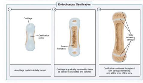
Describe endochondral ossification
when bone forms by replacing hyaline cartilage
cartilage forms, then osteoid is deposited at ossification centers, gradually replacing cartilage at the diaphysis then epiphysis
osteoid calcifies and forms cortical bone
most bones use this, including long bones and ribs

Long bones are formed through what type of ossification
endochrondral
What is found in the epiphysis of a long bone?
spongy bone with red bone marrow
