pathology endocrine practical
 Call Kai
 Learn
Learn Practice Test
 Spaced Repetition
 Match
Match Flashcards
 Knowt Play
1/43

There's no tags or description
Looks like no tags are added yet.
Name | Mastery | Learn | Test | Matching | Spaced | Call with Kai | Chat |
|---|
No analytics yet
Send a link to your students to track their progress
44 Terms
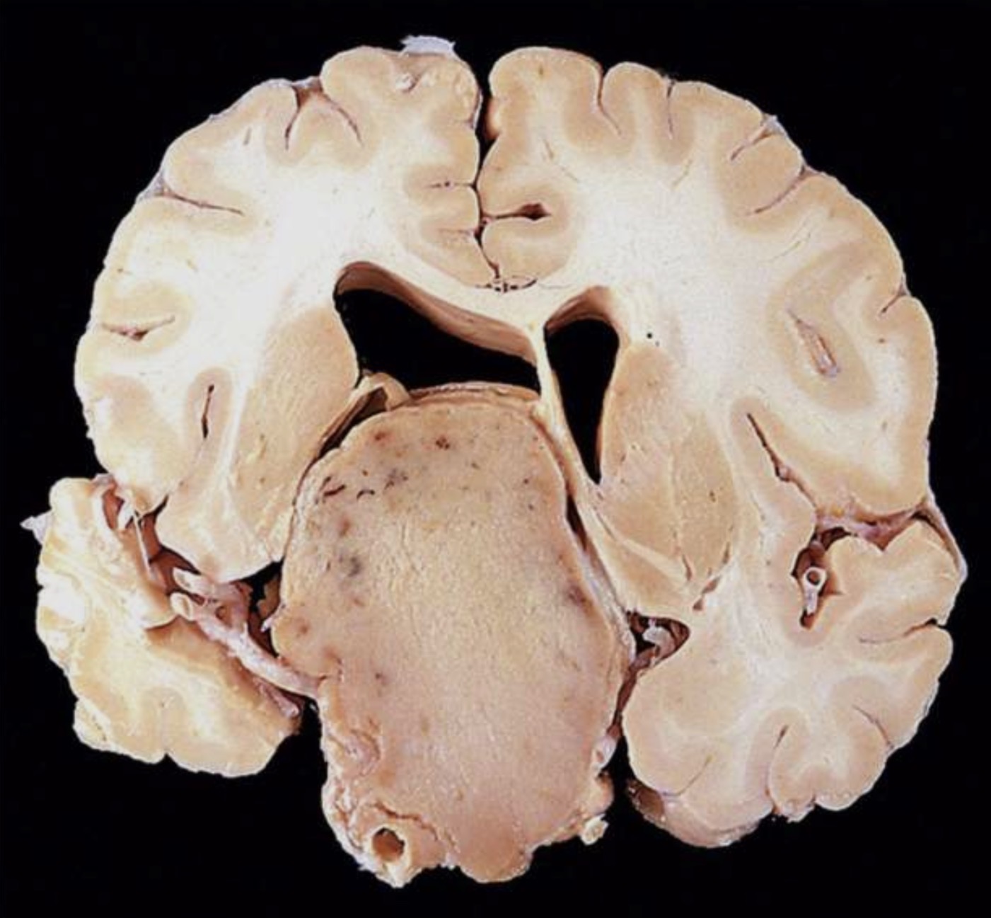
Pituitary adenoma
massive non functioning
Distorted the overlying brain
(Non functioning adenomas are larger at time of diagnosis than those that secret a hormone )
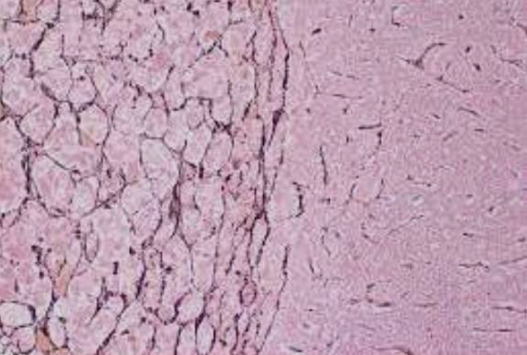
Pituitary adenoma
absence of the reticulin network
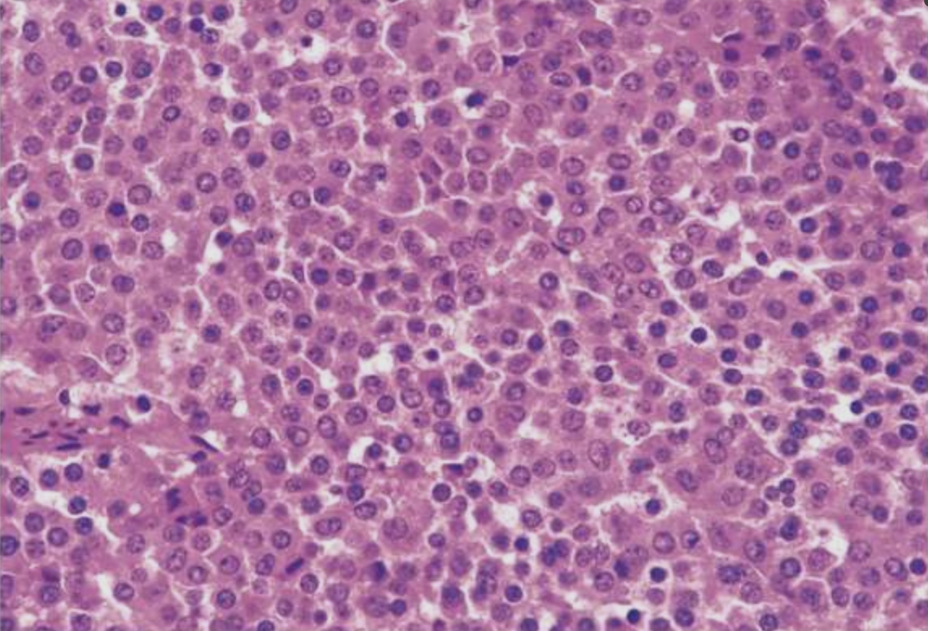
Pituitary adenoma
absence of the reticulin network
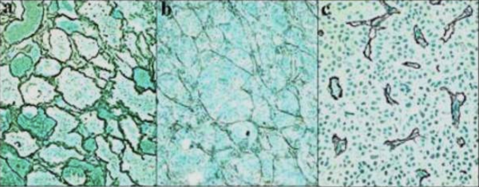
Pituitary adenoma
absence of the reticulin network
1- normal
2- hyperplasia
3- adenoma
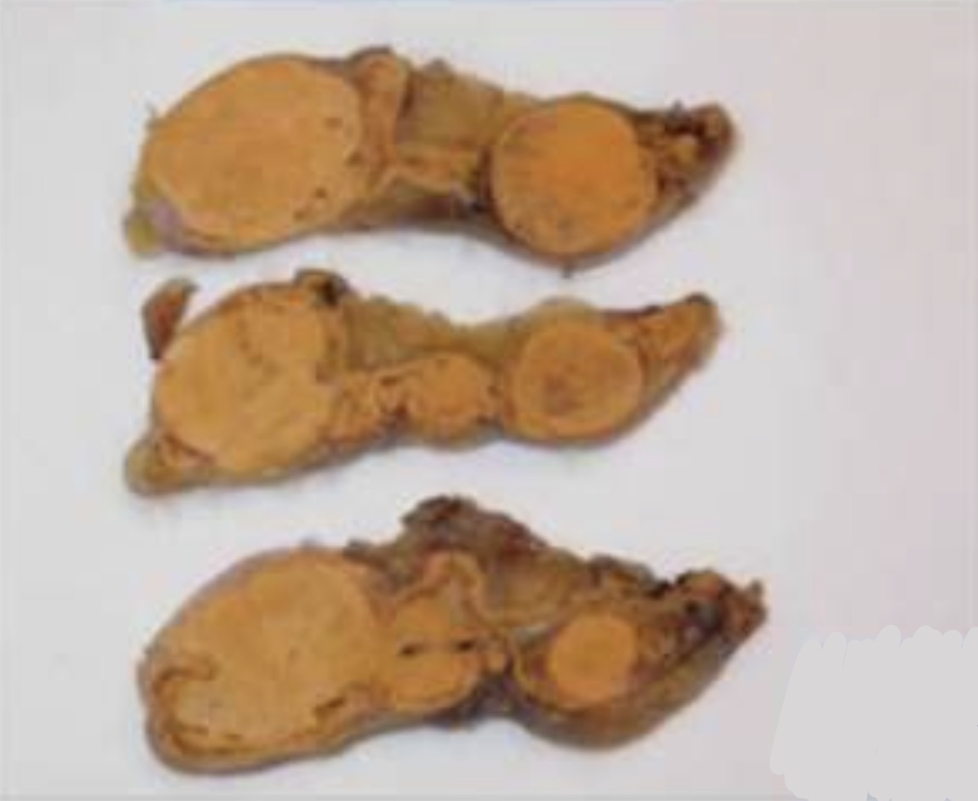
Primary = nodular hyperplasia
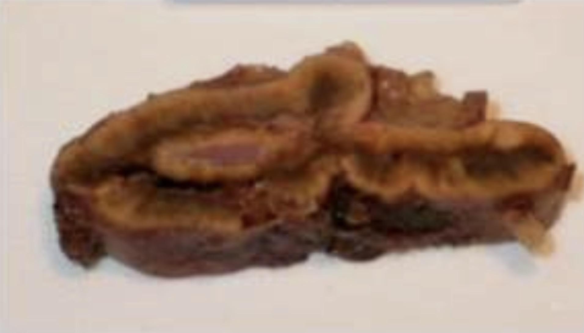
Secondary = diffuse , hyperplasia
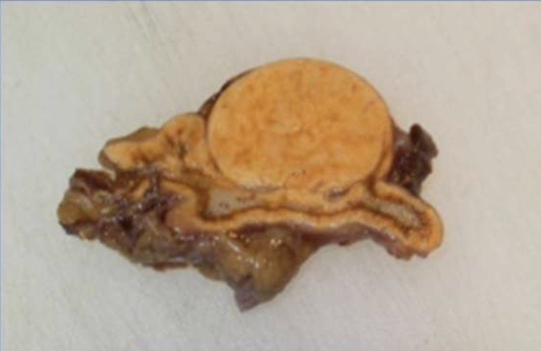
Adrenal Adenoma
-small
-solitary
-spherical
-encapsulated, bright yellow
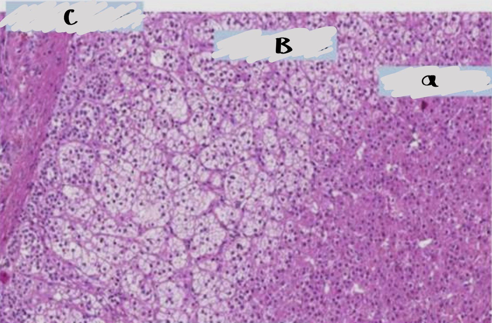
Normal cortex
A- zona reticularis
B- zona fasciculata
C- zona glomerulosa
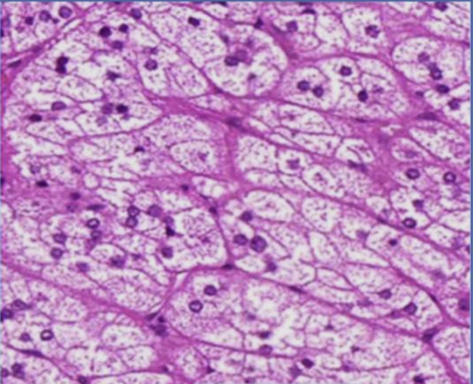
Adrenal adenoma
Resemble cells of zona fasciculata
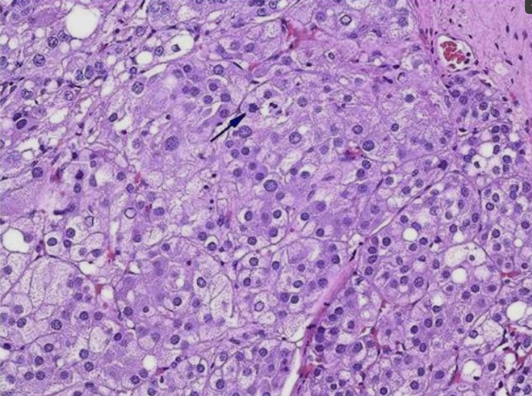
Adrenal cortical carcinoma
Hyperchromatic nuclei
Necrosis
Mitotic figures
Eosinophilic cytoplasm
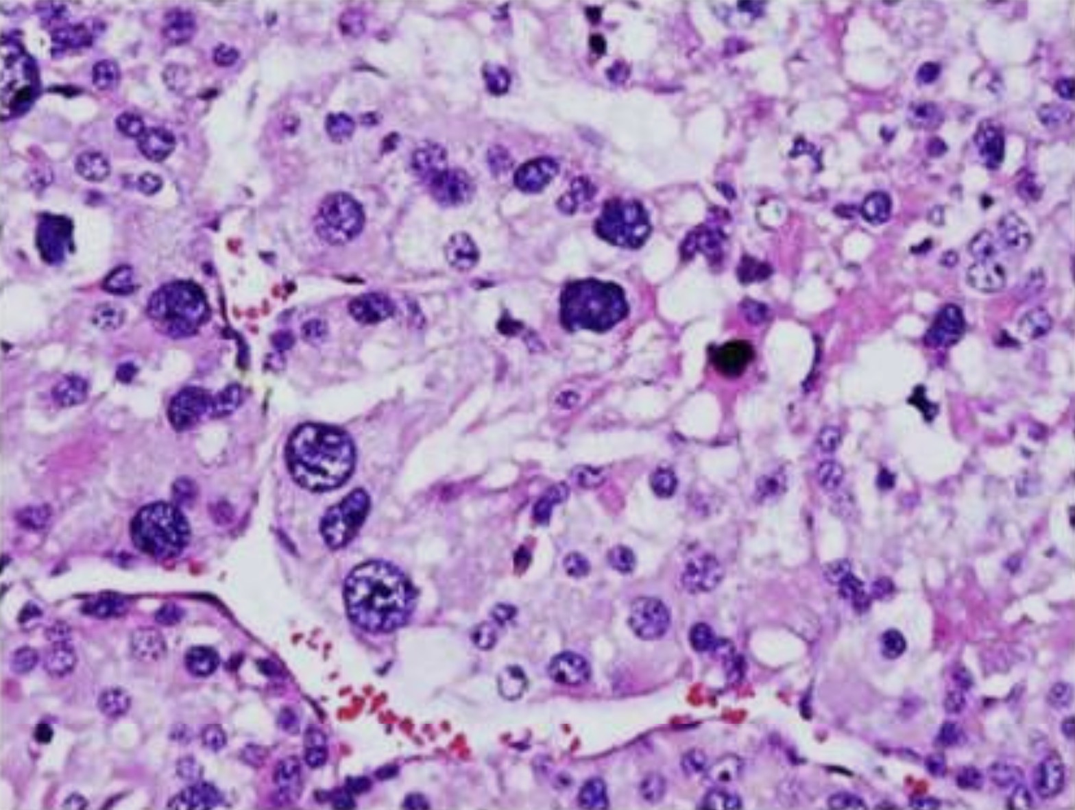
Adrenal cortical carcinoma
Hyperchromatic nuclei
Necrosis
Mitotic figures
Eosinophilic cytoplasm
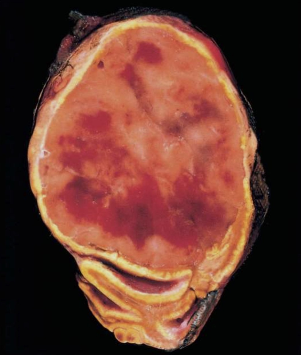
Pheochromocytoma
attenuated cortex and areas of hemorrhage
Comma-shaped residual adrenal gland is seen at the bottom
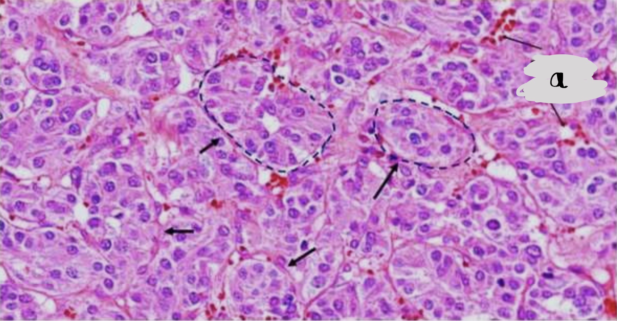
Pheochromocytoma
zellballen pattern separated by abundant fibrovascular stroma
Tumor cells are large , polyhedral and pleomorphic with abundant granular amphophilic or basophilic cytoplasm and vesicular nuclei
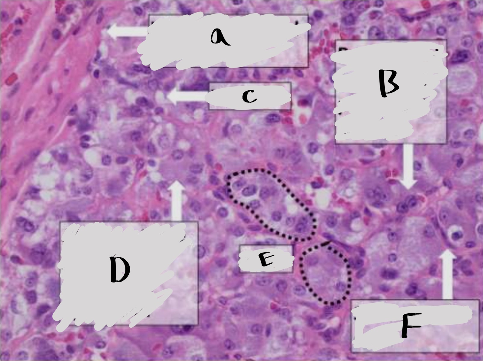
Pheochromocytoma
A- well demarcated border
B- round oval nuclei with prominent nucleoli
C- vacuoles
D- abundant fine granular red purple cytoplasm
E- nests
F- sustentacular cells
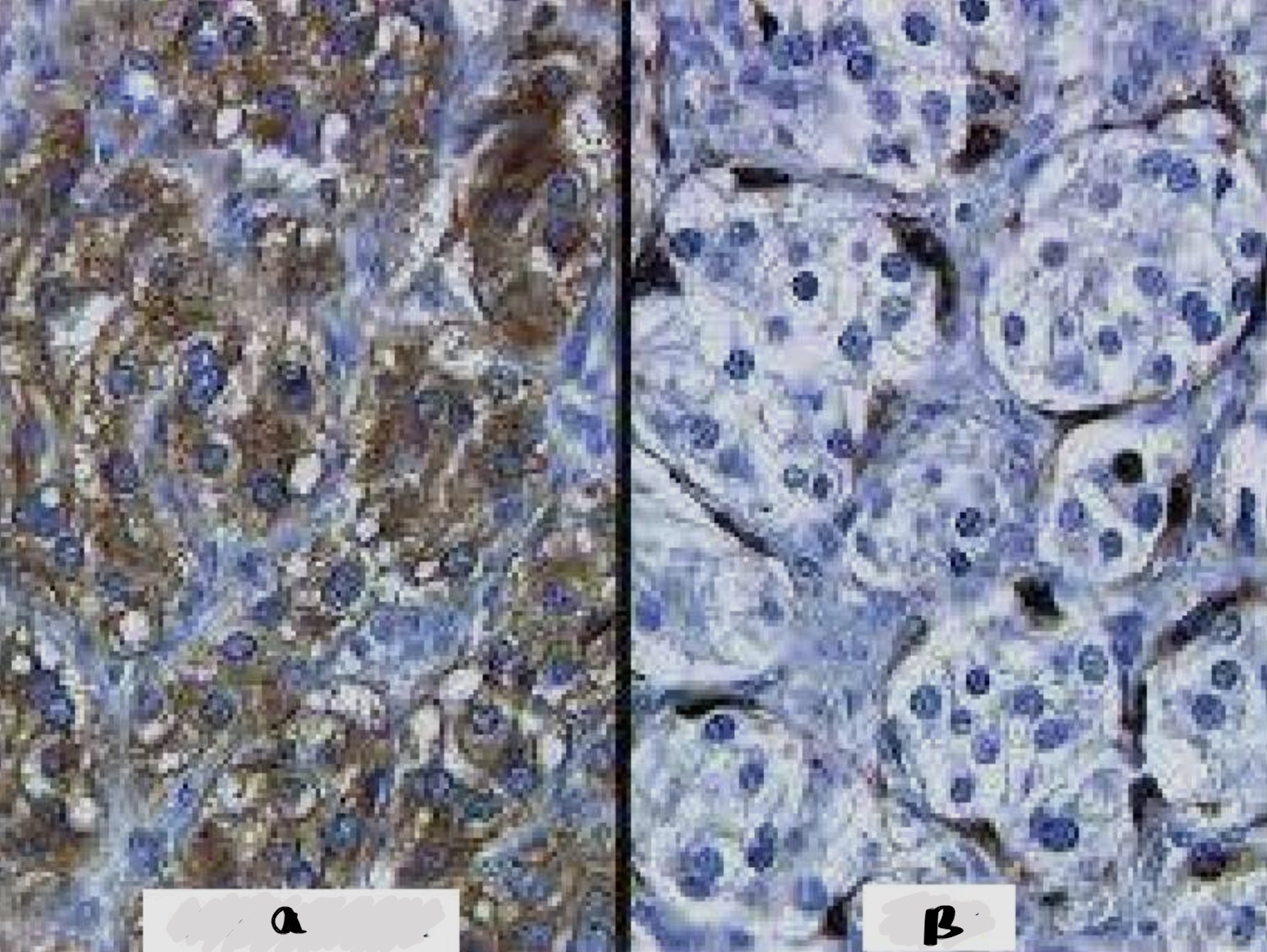
Pheochromocytoma
A- chromogranin in chromaffin cells
B- positive S100 in sustentacular cells
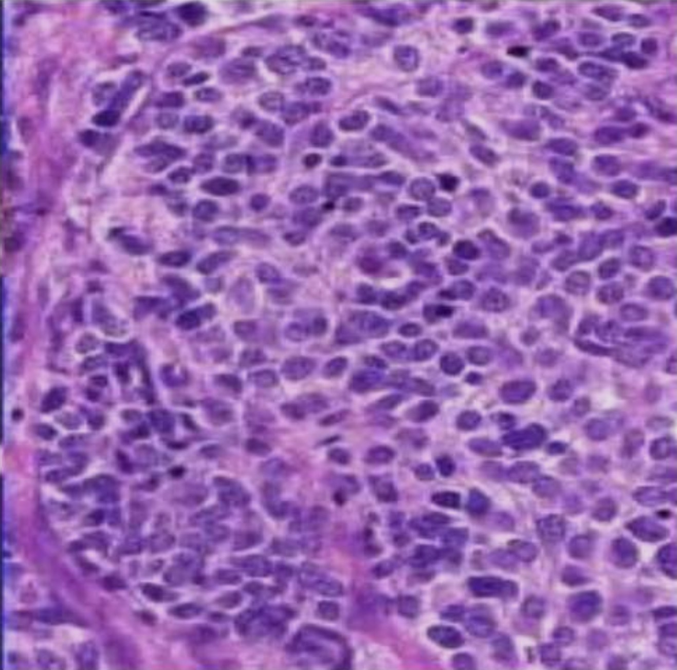
Neuroblastoma (pediatric tumor)
Small round and oval with scanty cytoplasm
Sheets separated by fibrovascular stroma
Homer-wrights rosettes
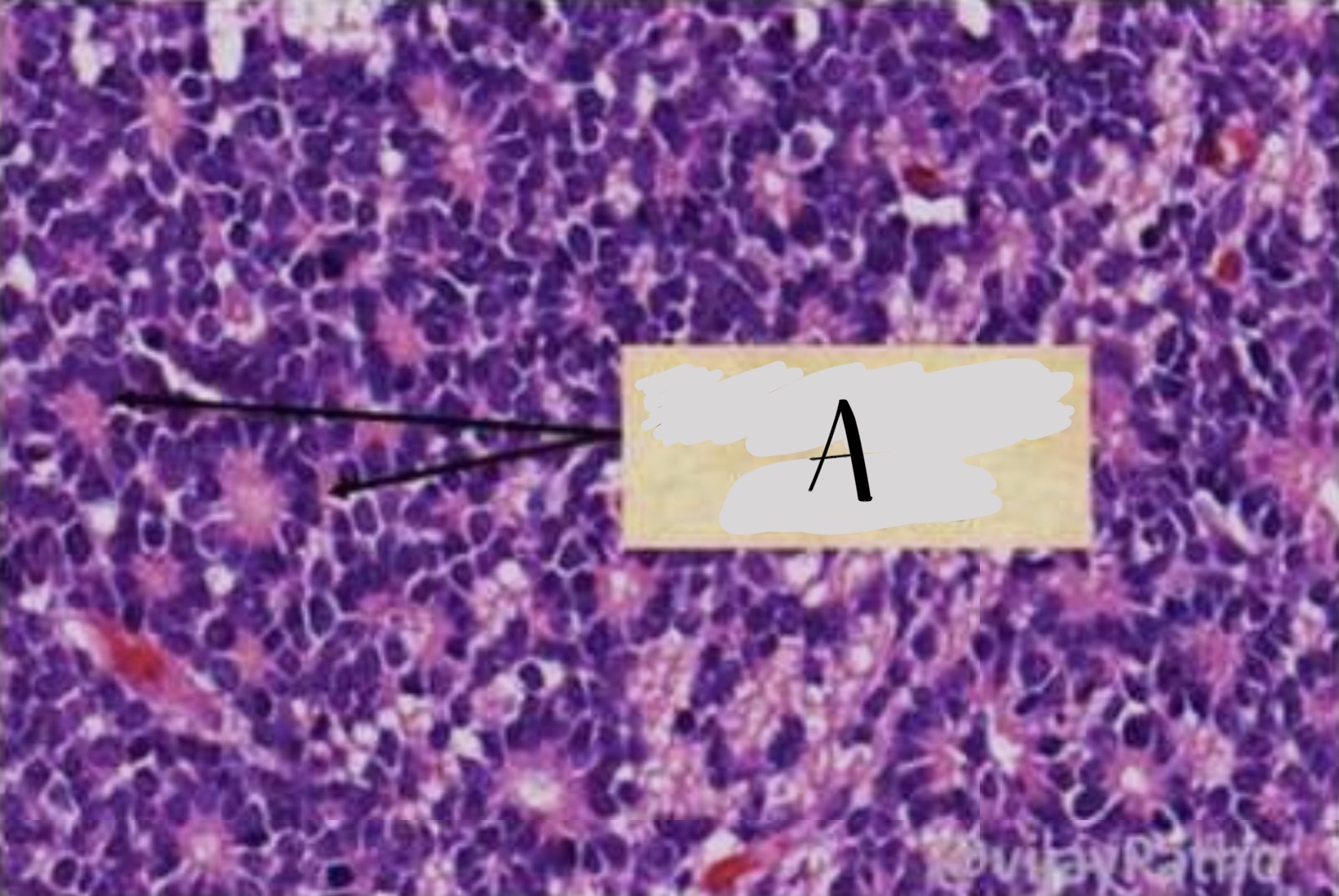
Neuroblastoma (pediatric tumor)
A. Homer wright rosettes
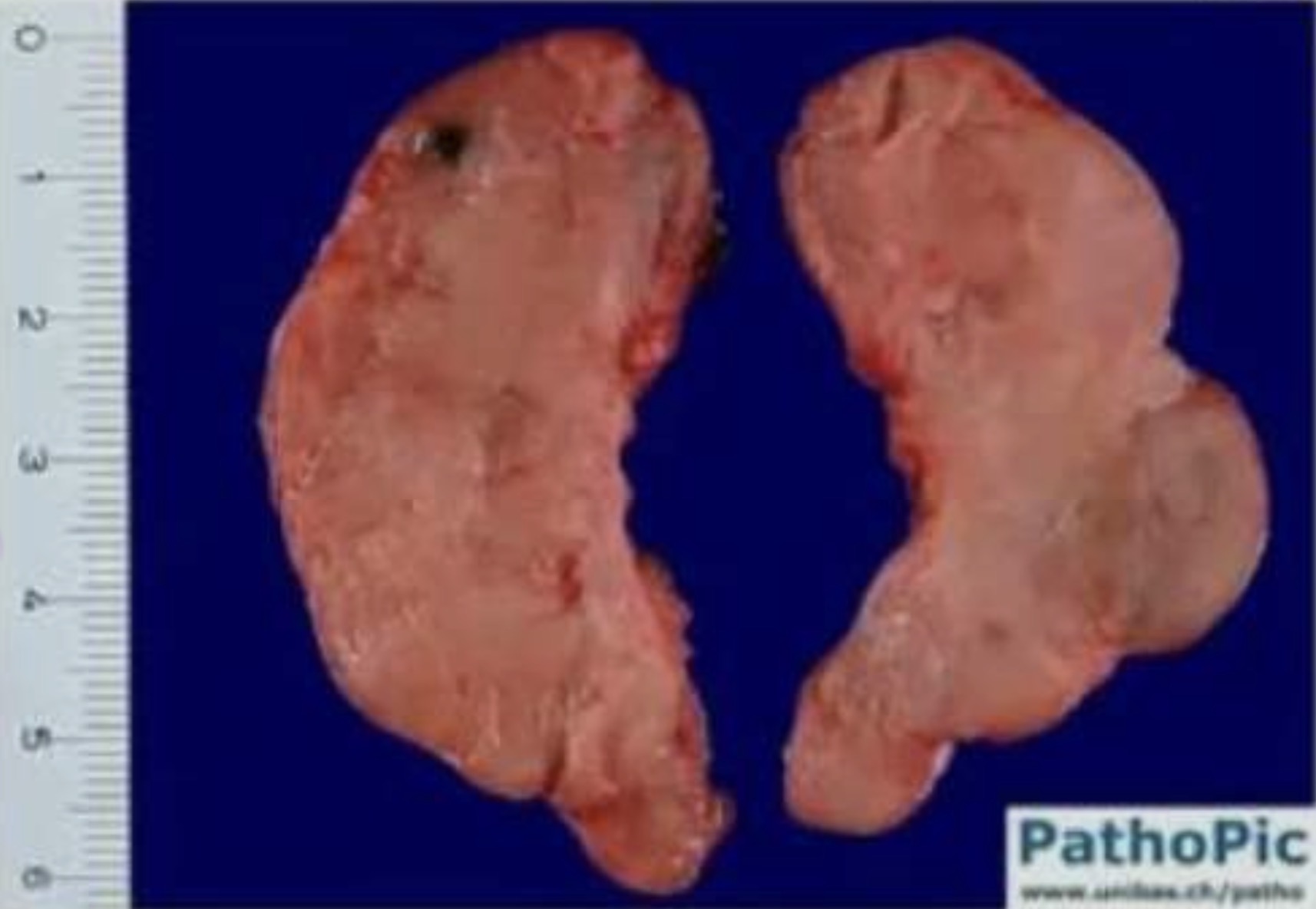
Hashimoto’s Thyroiditis
symmetric , firm and rubbery enlargement of the thyroid
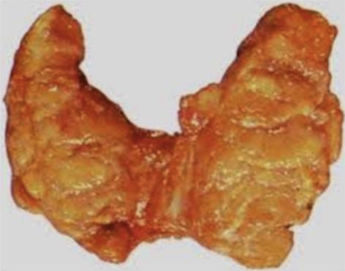
Hashimoto’s Thyroiditis
symmetric , firm and rubbery enlargement of the thyroid
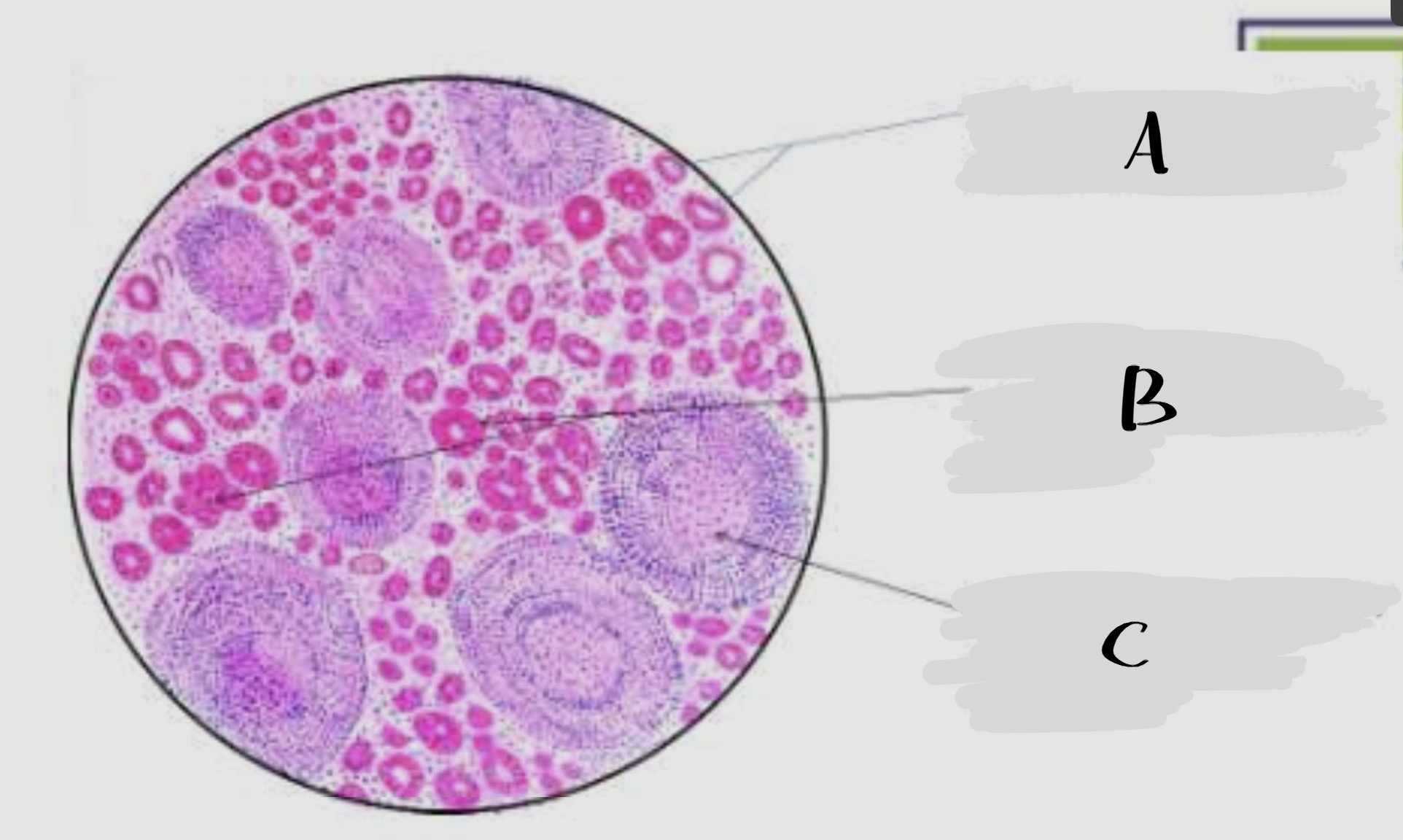
Hashimoto’s Thyroiditis
A. Follicles are atrophied and show no colloid
B. Follicles lined by hurtle cells and also in sheets
C. Lymphoid follicles with germinal center formation
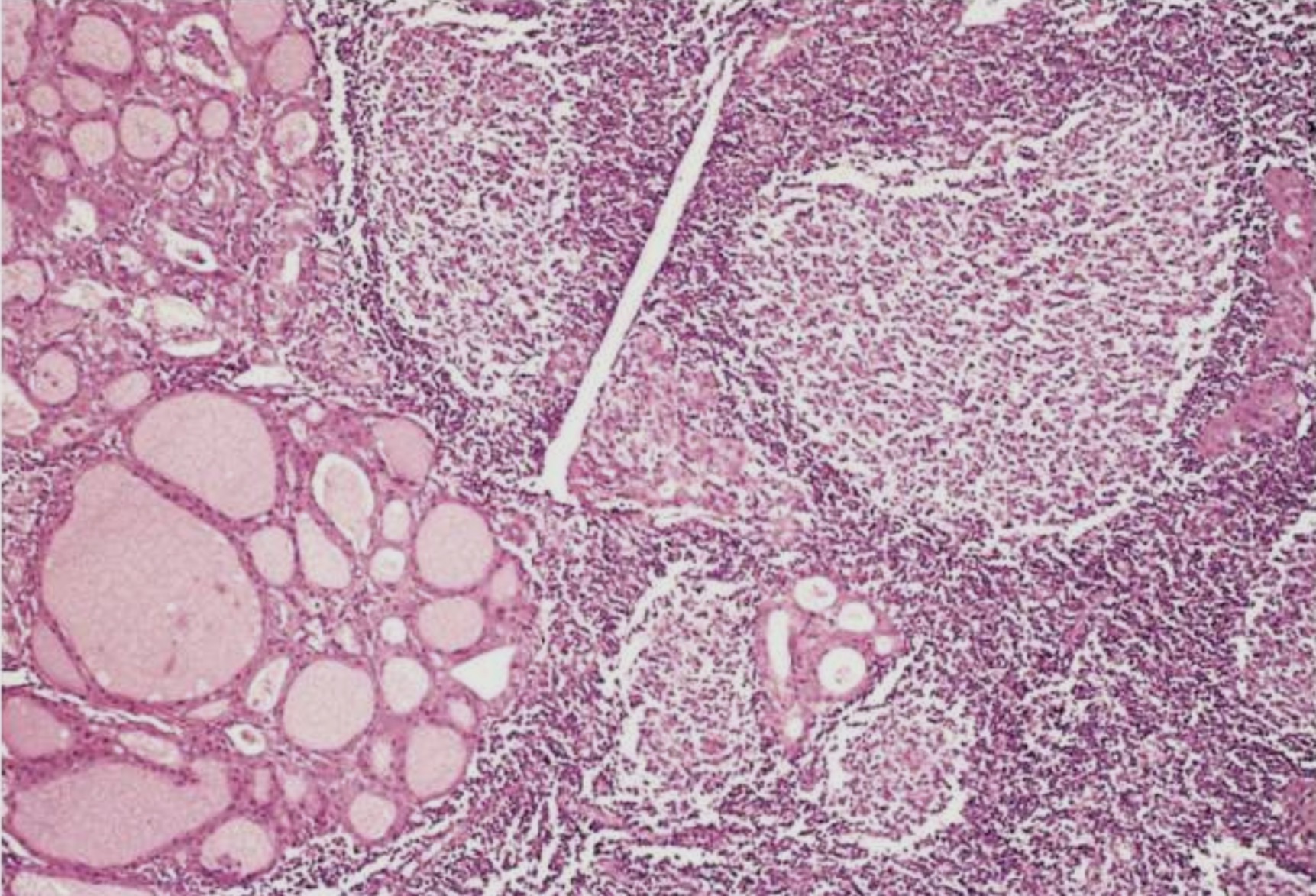
Hashimoto’s Thyroiditis
A. Follicles are atrophied and show no colloid
B. Follicles lined by hurtle cells and also in sheets
C. Lymphoid follicles with germinal center formation
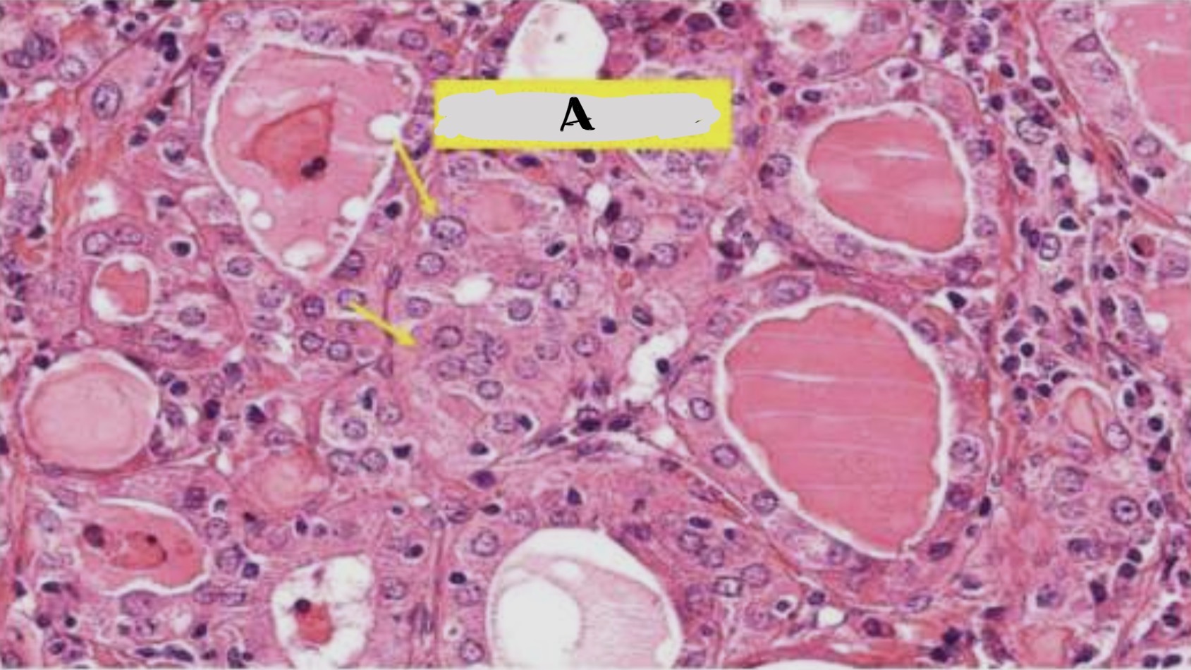
Hashimoto’s Thyroiditis
A. Hurthle cells
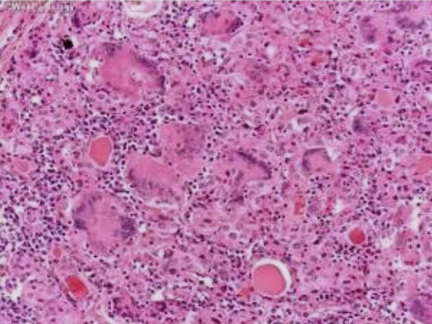
Subacute (De Quervain) granulomatous thyroiditis
Granulomatous inflammations with giant cells
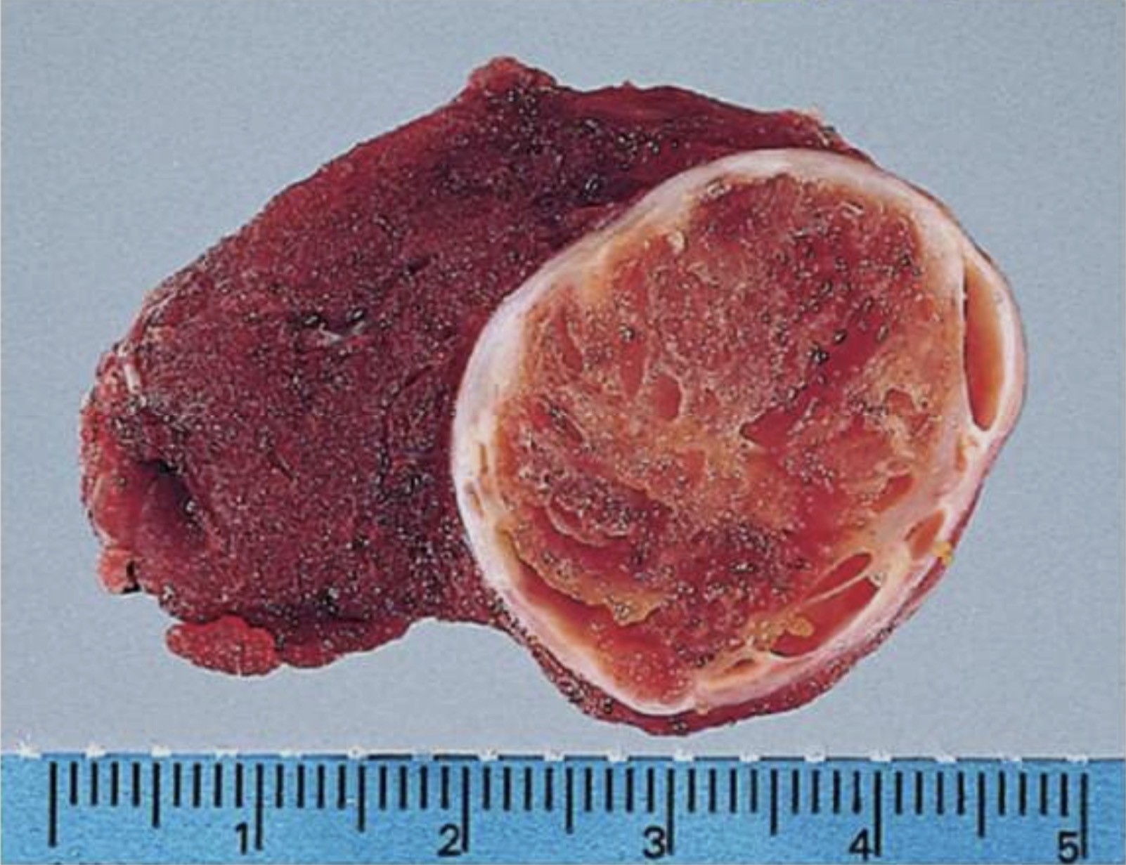
Follicular Adenoma
solitary nodule
Complete encapsulation
Clear distinct architecture inside and outside the capsule
Compressed thyroid parenchyma outside the capsule
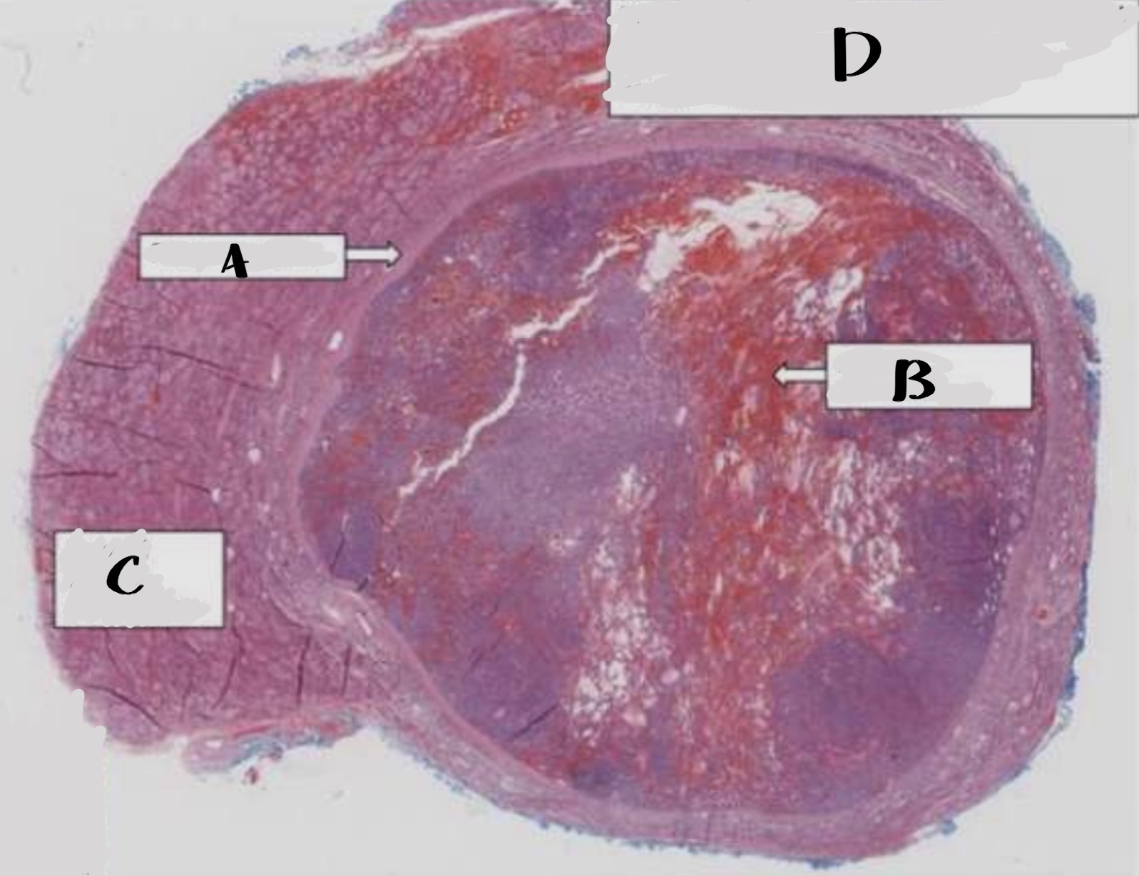
Follicular Adenoma
A. Fibrous capsule
B. Areas of haemorrhage
C. Normal thyroid parenchyma
D. Well circumscribed , encapsulated nodule compressing thyroid parenchyma
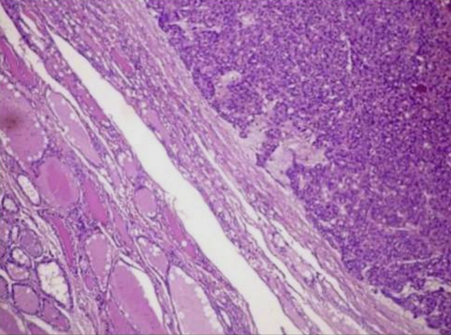
Follicular Adenoma
Encapsulated
Tumor cells are benign follicular epithelial cells
Microfollicular (foetal) pattern , no colloid
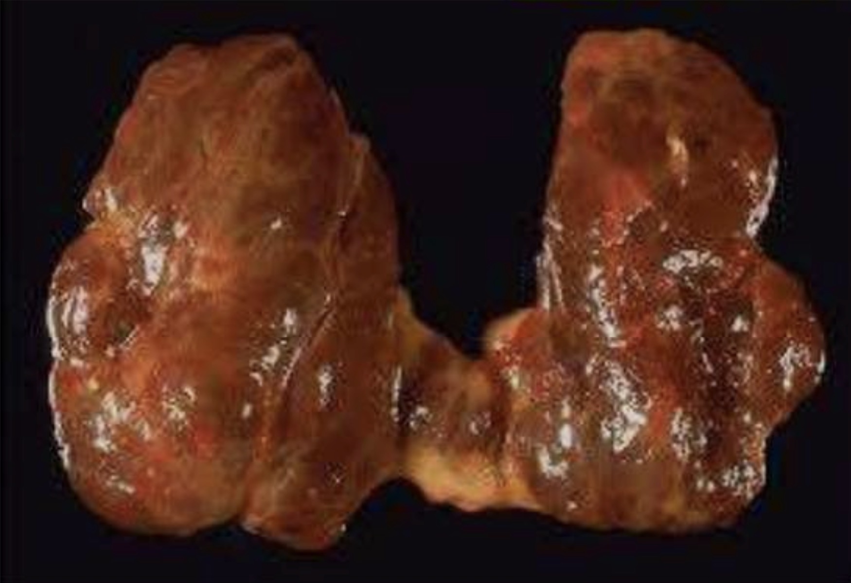
Nodular Goitre
nodularity with poor encapsulation
Fibrous scarring
Haemorrhages
Focal calcification
Cystic degeneration
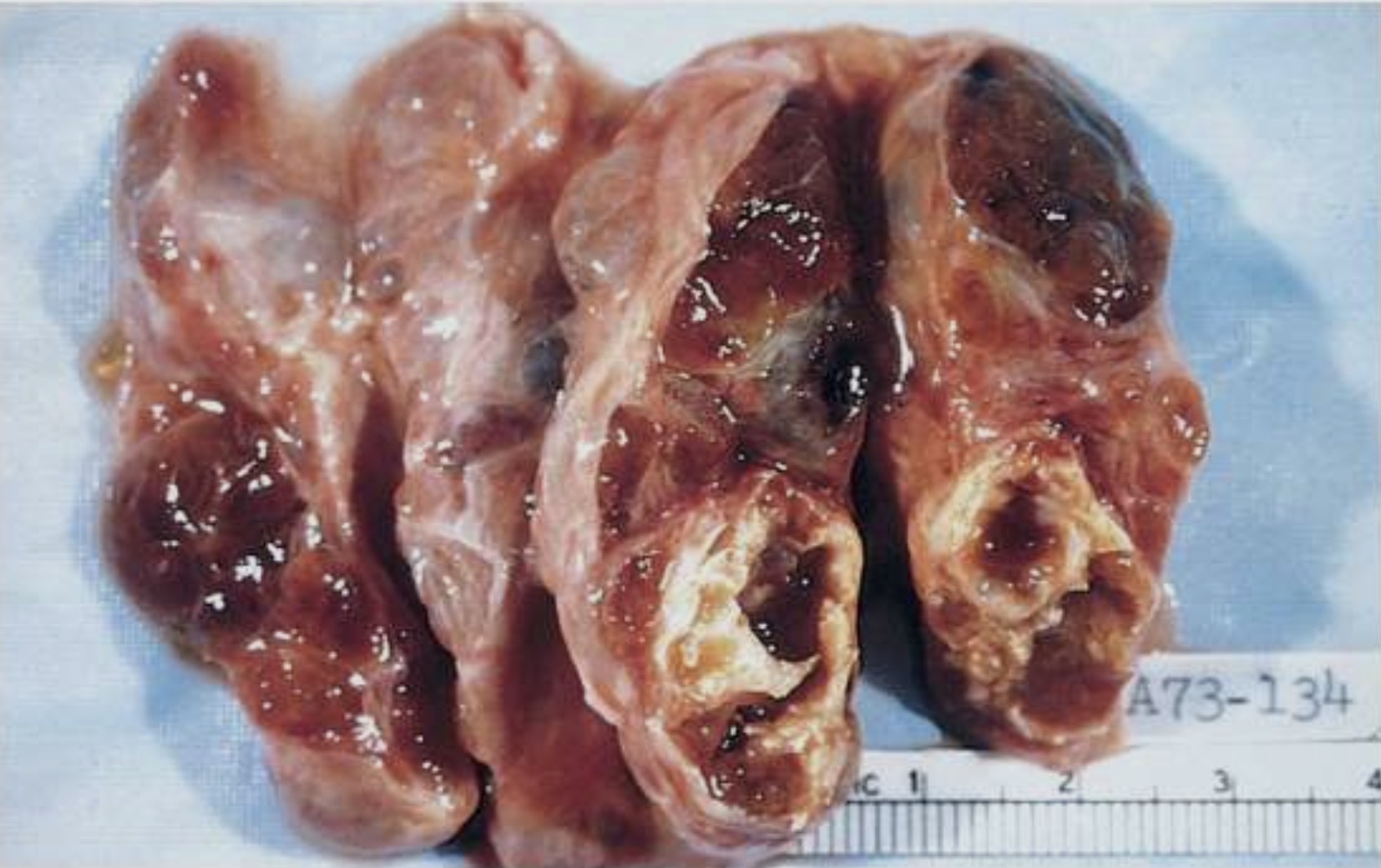
Nodular Goitre
nodularity with poor encapsulation
Fibrous scarring
Haemorrhages
Focal calcification
Cystic degeneration
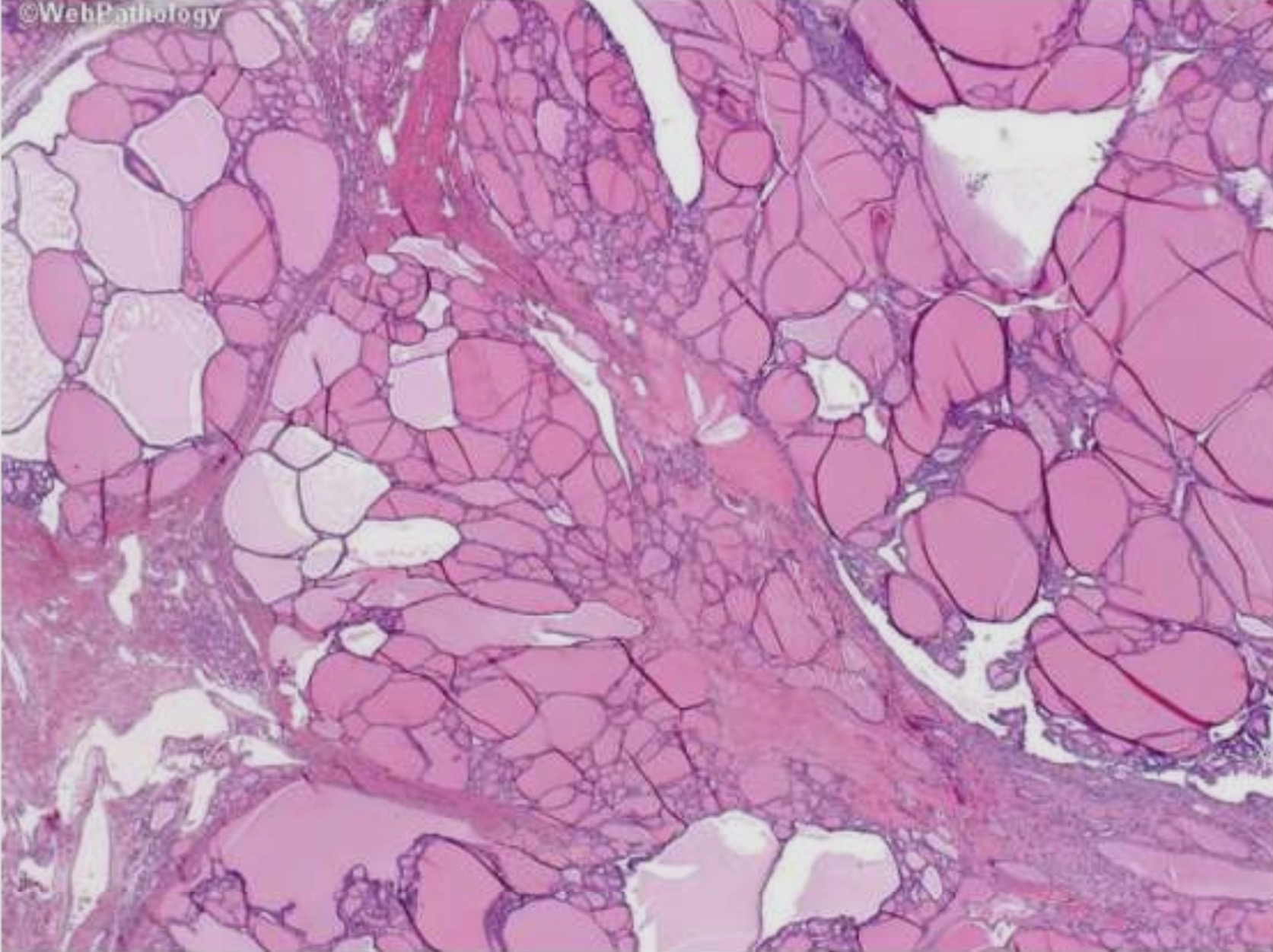
Nodular Goitre
Incomplete or partial encapsulation
Fibrous scarring
Areas of Haemorrhage
calcification
Cystic degeneration
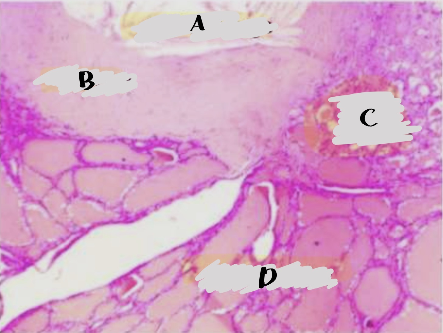
Nodular Goitre
A. Cystic change
B. Fibrosis
C. Small follicles
D. Large follicles
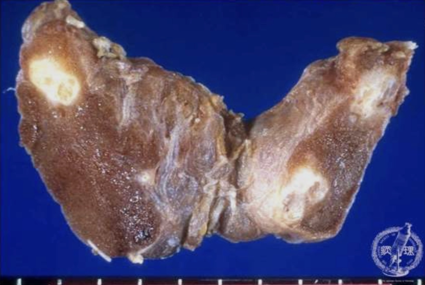
Papillary Carcinoma
Poorly delineated
Cute surface is grayish white
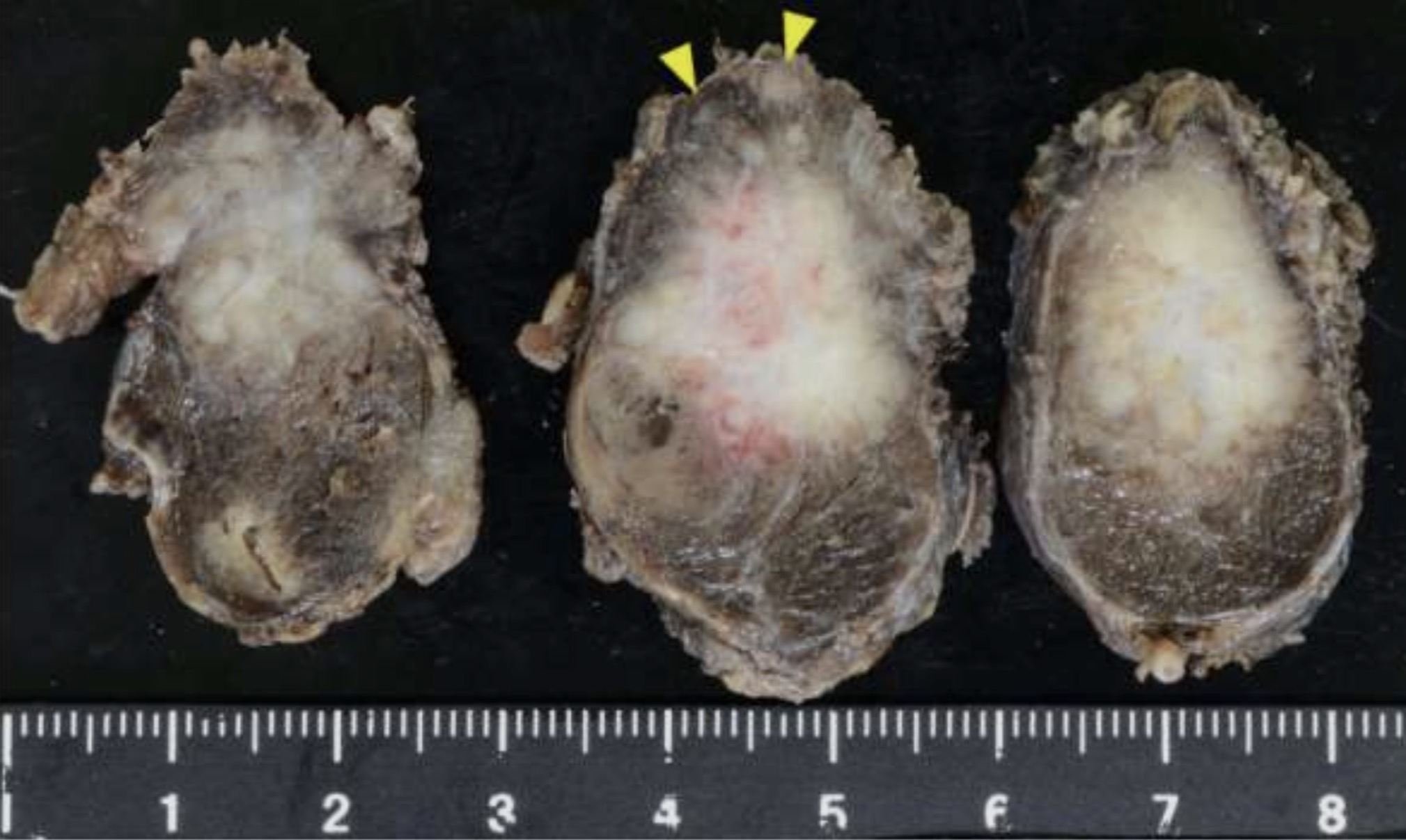
Papillary Carcinoma
Poorly delineated
Cute surface is grayish white
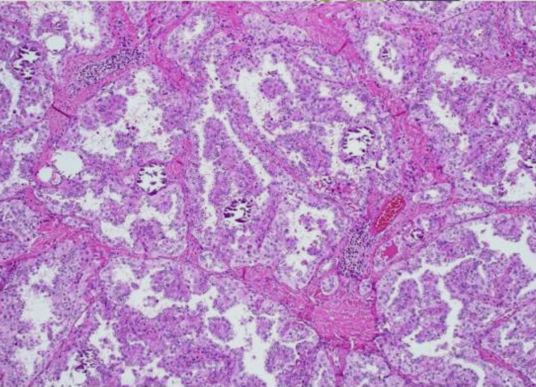
Papillary Carcinoma
Papillary pattern
tumor cells have ground glass appearance and oxyphil cytoplasm
Invasion
Psammoma bodies
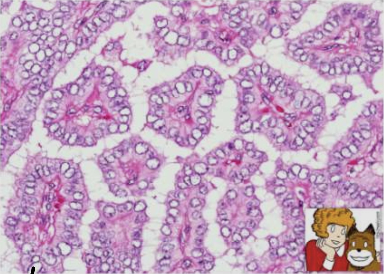
Papillary Carcinoma
Papillary pattern
tumor cells have ground glass appearance and oxyphil cytoplasm
Invasion
Psammoma bodies
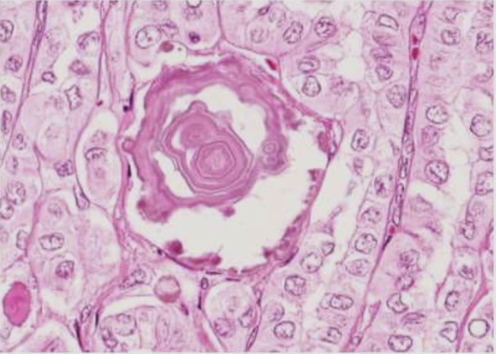
Papillary Carcinoma
Papillary pattern
tumor cells have ground glass appearance and oxyphil cytoplasm
Invasion
Psammoma bodies
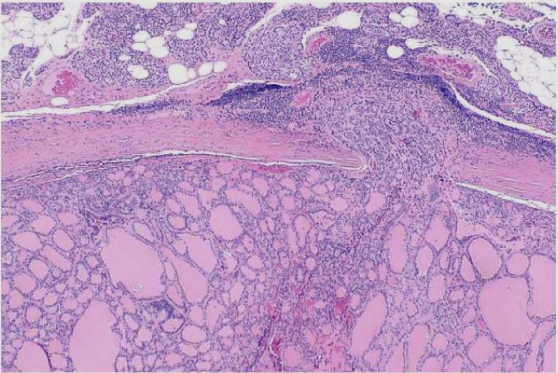
Follicular Thyroid Carcinoma
uniform follicles invading the capsule
Blood vessels ( diagnostic hallmark )
No nuclear features of papillary carcinoma
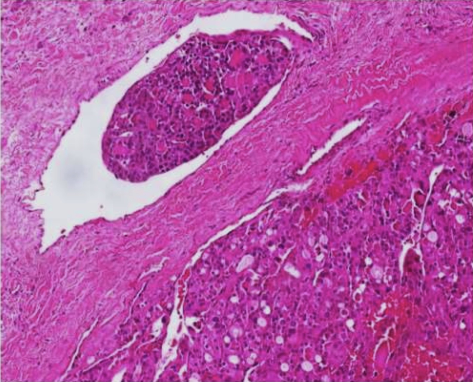
Follicular Thyroid Carcinoma
uniform follicles invading the capsule
Blood vessels ( diagnostic hallmark )
No nuclear features of papillary carcinoma
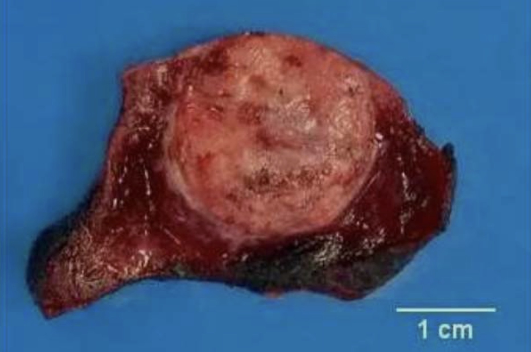
Medullary Thyroid carcinoma
Solid, firm, gray tan mass
Bilateral/ can be multi focal in MEN syndromes
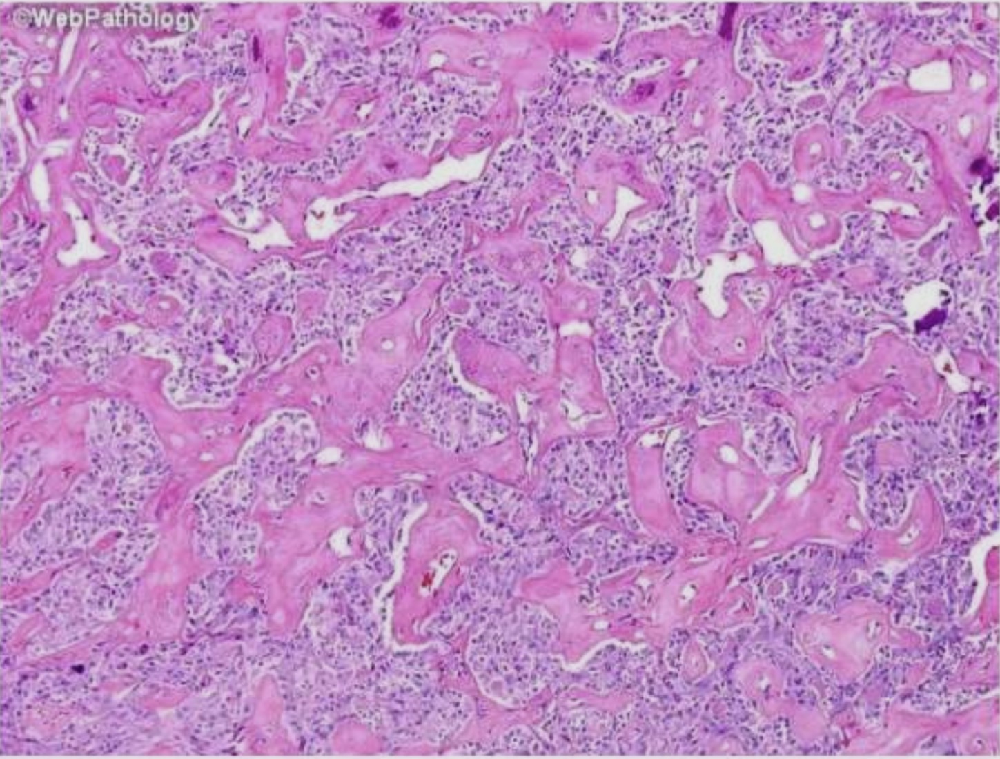
Medullary Thyroid carcinoma
polygonal to spindle-shaped cells in nests , trabeculae or follicles
Amyloid stroma (derived from altered calcitonin) - Congo red positive
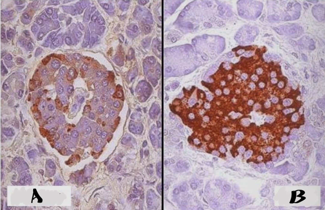
Islet of Langerhans , immunohistochemical staining with antibody to insulin and glucagon
A. Glucagon→ identifies alpha cells
B. Insulin → identifies beta cells
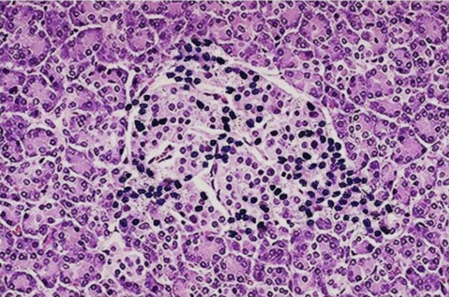
Type 1 diabetes mellitus - insulitis of an islet of langerhans
Lymphocytic infiltrates
Destruction of the islets leads to absolute lack of insulin
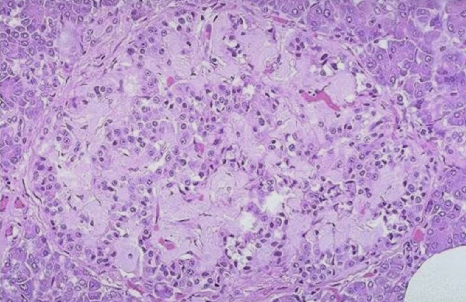
Type 2 diabetes mellitus - Amyloid deposition in islet
Pink hyalinization
deposition of amyloid in many islet cells
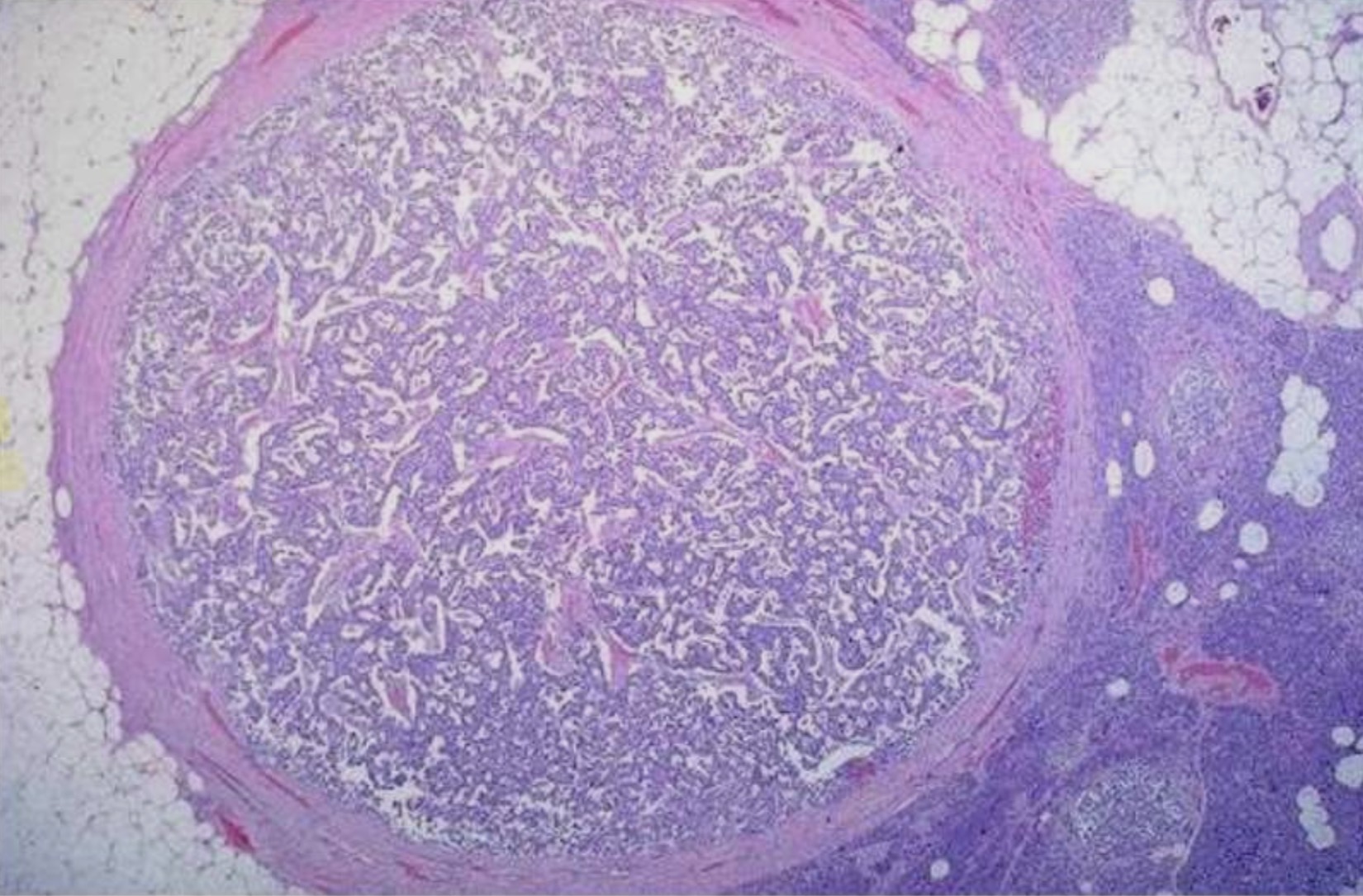
Pancreatic neuroendocrine tumor
Cells resembling those in islets of langerhans separated from pancreas by thin collagenous capsule
Normal islets are on the right
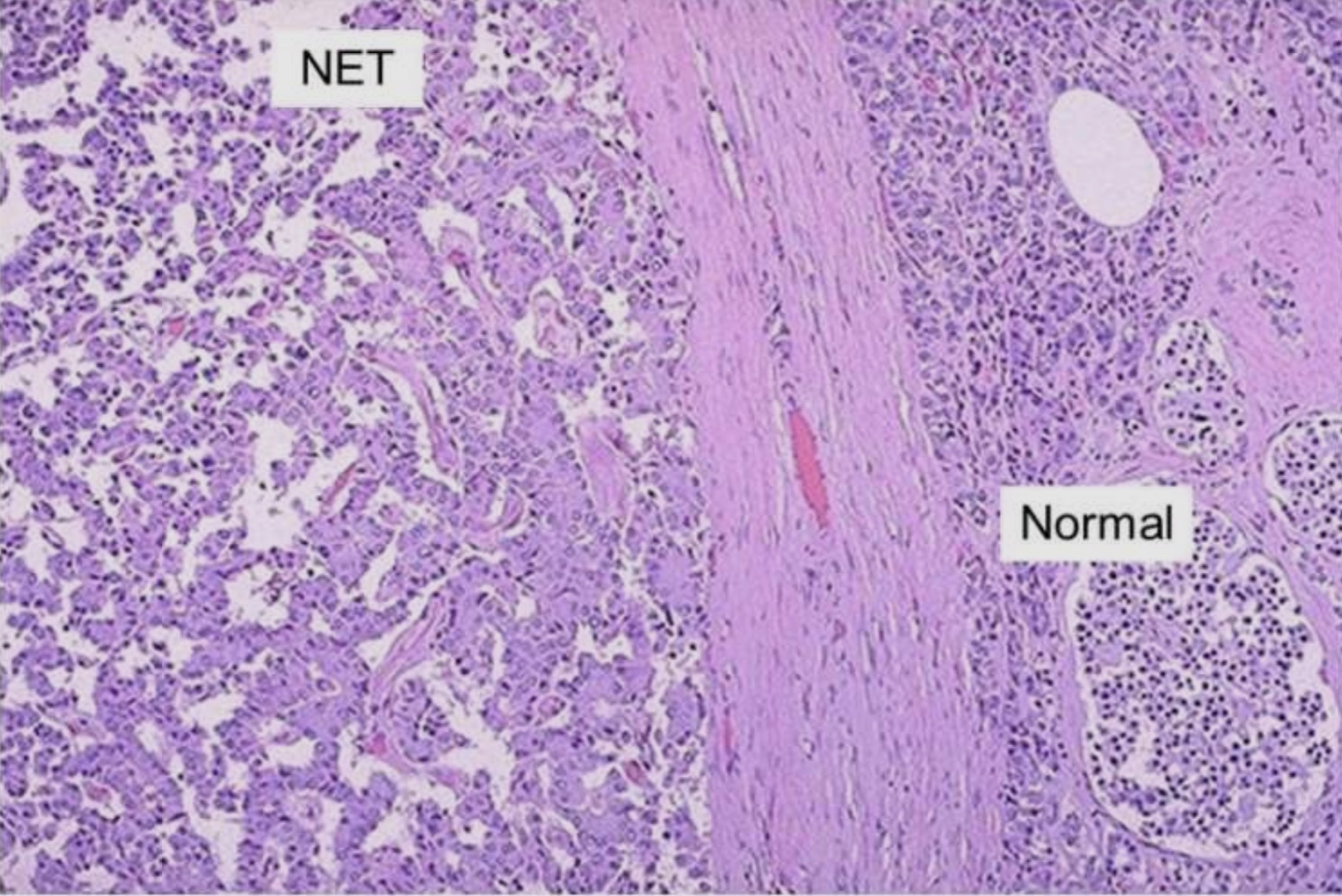
Pancreatic neuroendocrine tumor
Round central nuclei , salt and pepper chromatin , scant to moderate eosinophilic granular cytoplasm
Nucleoli vary depending on grade cells grow in:
solid
Gyriform
Glandular
Diffuse patterns