Human Skull
 Call Kai
 Learn
Learn Practice Test
 Spaced Repetition
 Match
Match Flashcards
 Knowt Play
1/18

There's no tags or description
Looks like no tags are added yet.
Name | Mastery | Learn | Test | Matching | Spaced | Call with Kai | Chat |
|---|
No analytics yet
Send a link to your students to track their progress
19 Terms

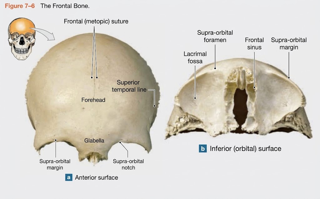
Name the bone (2)
Frontal Bone, Frontal Sinus, Supraoptic. foramen/notch
Frontal sinus: Hollow cavity inside the frontal bone above the eyes. (Often seen on a split skull).
Supraorbital foramen/notch: The hole or groove right above the eye socket (orbit) where nerves/blood vessels pass.

Which part is the Parietal Bone?
Parietal Bone
Large, paired bones forming the bulk of the superior and lateral skull


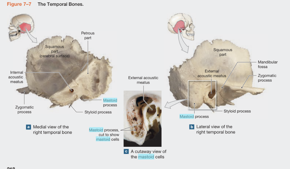
Name the Bones (5) missing carotid canal, Jugular Foramen, Zygomatic Arch
Temporal bone:
Mastoid process: Rough, conical projection behind the ear. (⚠ Look-alike: Styloid process is thin and needle-like).
· External auditory (acoustic) meatus: The ear canal opening.
· Zygomatic process: The bridge of bone extending forward to meet the cheekbone.
· Mandibular fossa: The smooth depression where the lower jaw (mandible) articulates.
· Styloid process: Sharp, needle-like projection pointing downward from the temporal bone.
· Carotid canal: A circular opening on the inferior surface of the temporal bone (where the carotid artery enters).
· Jugular foramen: A large opening at the base of the skull between the temporal and occipital bones.
Zygomatic arch: The complete cheekbone bridge
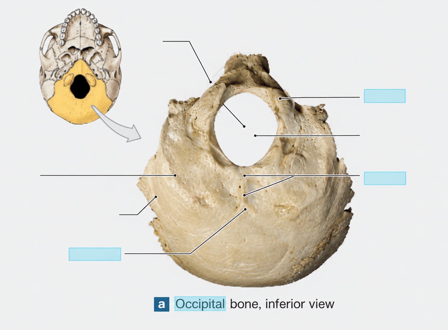
Name the Bones (3)
Occipital Bone
· Foramen magnum: The large hole at the base of the skull where the spinal cord exits.
· Occipital condyles: Two rounded projections on either side of the foramen magnum that articulate with the atlas (C1).
· Occiput (external occipital protuberance): The prominent bump on the back of the head.
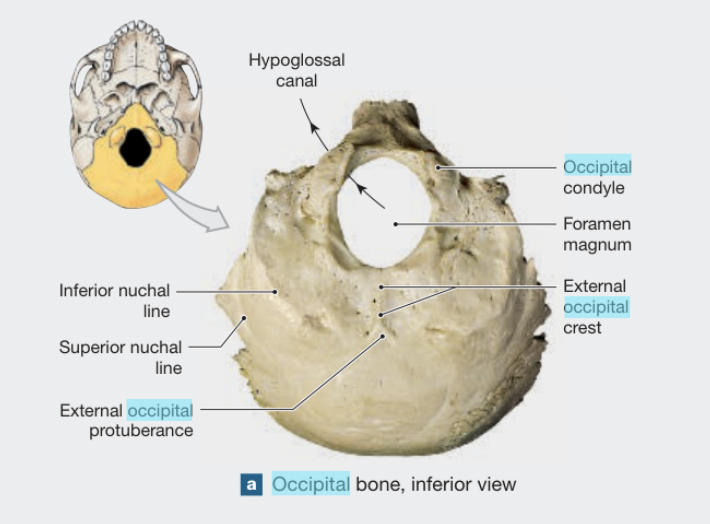
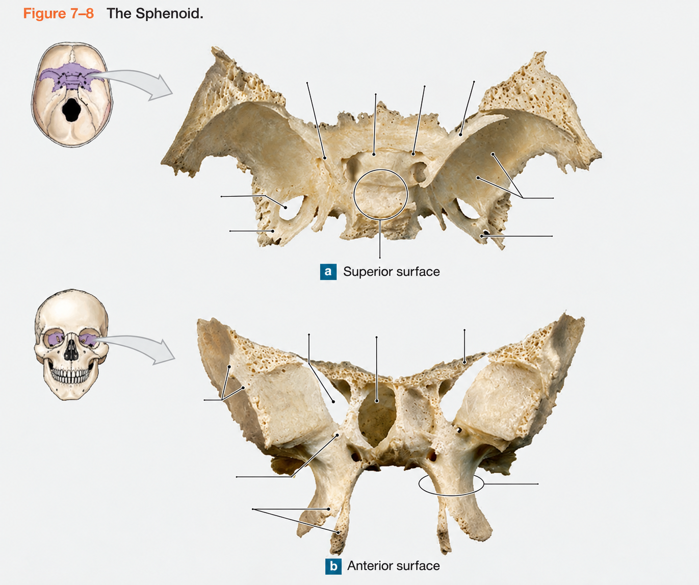
Name the Bones (10)
Sphenoid Bone
· Sella turcica: The saddle-shaped depression in the middle of the skull that houses the pituitary gland. (⚠ Look-alike: This is a depression, not a hole. Look for the "Turk's saddle" shape.)
· Optic foramen (canal): The hole in the back of the orbit where the optic nerve passes.
· Sphenoid sinus: Air cavity inside the sphenoid bone.
· Foramen ovale: A large oval hole in the greater wing of the sphenoid.
· Foramen rotundum: A round hole in the greater wing of the sphenoid.
· Foramen lacerum: A jagged opening between the sphenoid and temporal bones.
· Greater & lesser wings: The wing-like projections of the sphenoid bone.
· Orbital surface: The smooth part of the sphenoid that forms part of the eye socket.
· Pterygoid process (lateral & medial plates): Two downward projections on the inferior sphenoid.
· Foramen spinosum: A small hole near the foramen ovale.


Name the bone (6)
Ethmoid Bone
· Cribriform plate: The horizontal plate with tiny holes (olfactory foramina) that forms the roof of the nasal cavity.
· Concha: The curled, scroll-like bones in the nasal cavity.
· Crista galli: The "rooster's comb" projection pointing up from the cribriform plate.
· Ethmoid sinus (air cells): The honeycomb-like air spaces within the ethmoid.
· Perpendicular plate: The vertical plate that forms the upper part of the nasal septum.
· Olfactory foramina: The tiny holes in the cribriform plate for smell nerves.

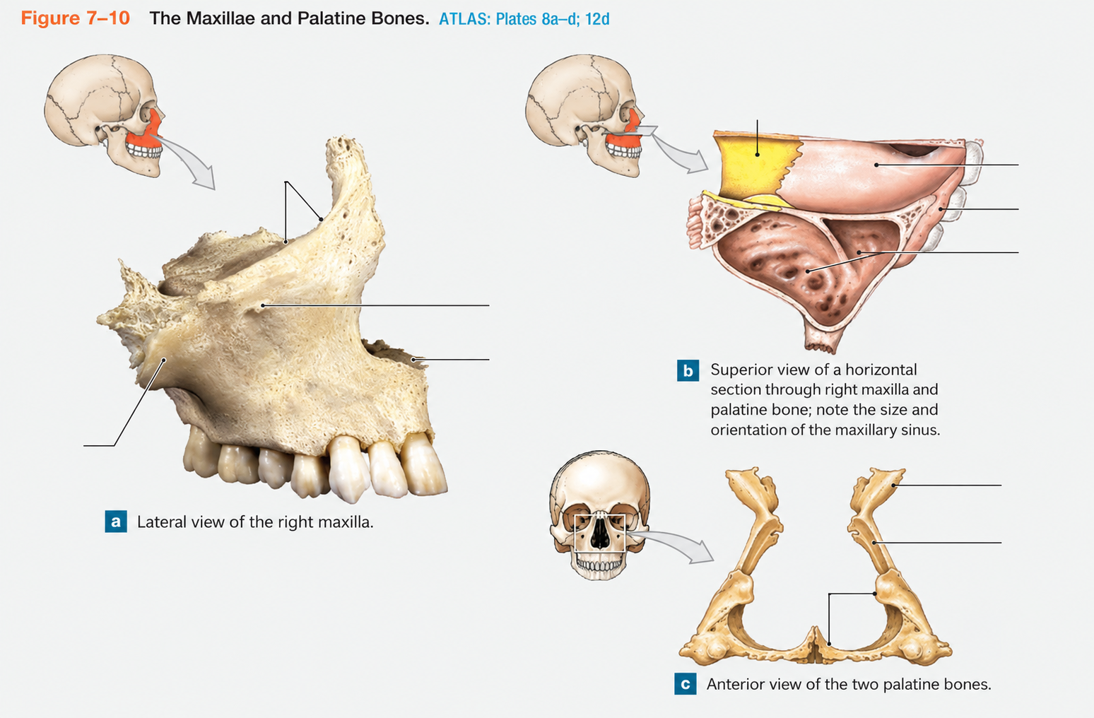
Name the bone (3)
Maxillary Bone (Maxilla)
· Alveolar process: The ridge containing the upper teeth sockets.
· Maxillary sinus: The large air cavity inside the cheek area of the maxilla.
· Infraorbital foramen: The hole below the eye socket.


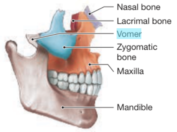
Which is the Zygomatic Bone and Name the bones (2)
Zygomatic Bone
· Temporal process: The extension that meets the temporal bone to form the zygomatic arch.
· Zygomatic arch: The complete cheekbone bridge (formed by the zygomatic and temporal bones).


Which is the Lacrimal + where is the sulcus
Lacrimal Bone
· Lacrimal sulcus: The groove that forms part of the tear duct pathway


Where is the palatine bone?
The L-shaped bone forming the posterior part of the hard palate.


Where is the nasal bone?
Nasal Bone
· The small, rectangular bones forming the bridge of the nose.


Where is the vomer bone?
The thin, flat bone forming the lower part of the nasal septum

Name the bones (9)
Mandible Bone
· Body: The horizontal, horseshoe-shaped part of the jaw.
· Condyloid (condylar) process: The rounded posterior projection that articulates with the temporal bone.
· Alveolar process: The ridge containing the lower teeth sockets.
· Coronoid process: The flat, triangular anterior projection. (⚠ Look-alike: Coronoid is flat and anterior; Condyloid is round and posterior.)
· Ramus: The vertical part of the jaw.
· Angle: The corner where the body meets the ramus.
· Mandibular notch: The U-shaped depression between the coronoid and condyloid processes.
· Mental foramen: The hole on the anterior lateral side of the mandible (where nerves pass to the chin).
· Mandibular foramen: The hole on the medial side of the ramus (for the inferior alveolar nerve).

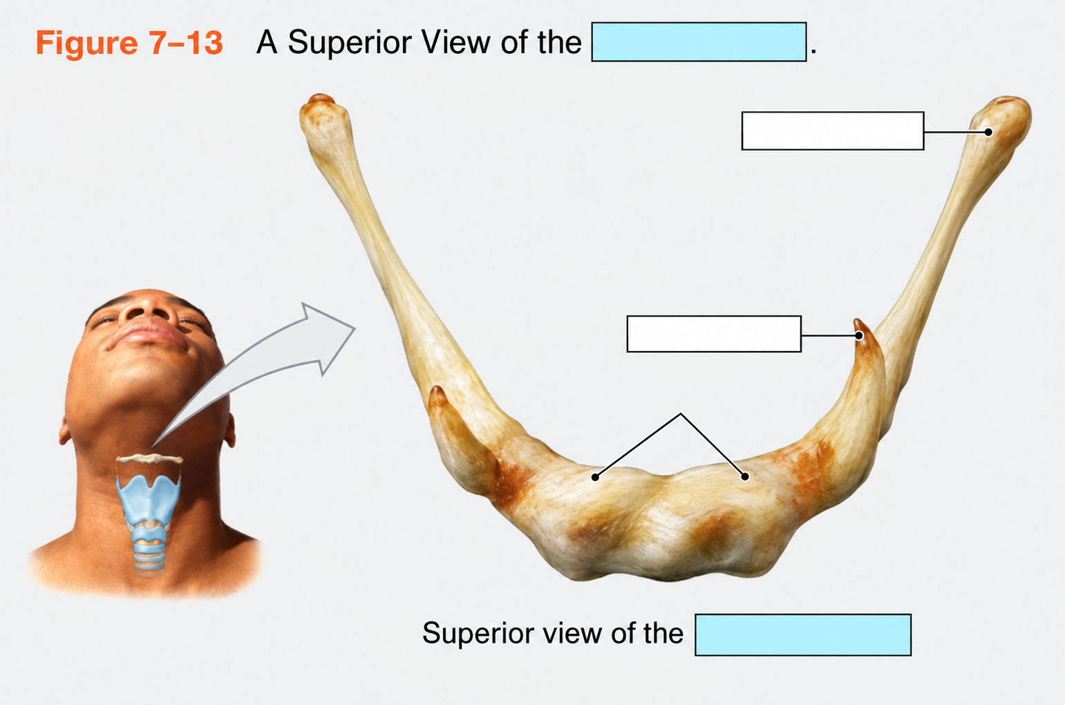
What bone is this?
Hyoid- It is a U-shaped bone in the neck, not attached to any other bone.


Where is the orbits? Which ones are inferior and which are superior?
The eye sockets and the cracks within them

What is the Sagittal suture?
The immovable joint between the two parietal bones (top of head).
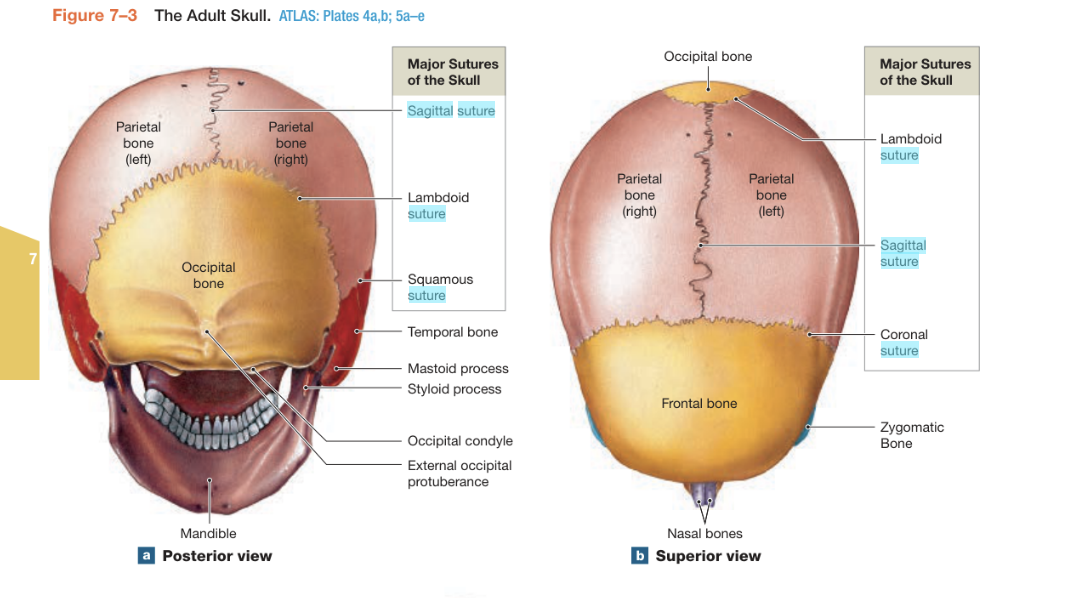
Where and what is the coronal suture?
The joint between the frontal and parietal bones.
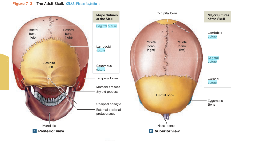
Where and what is the lambdoid suture?
The joint between the occipital and parietal bones (back of head).
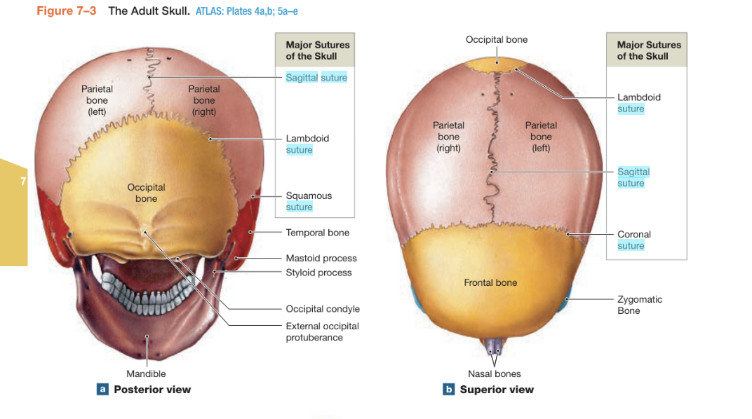
Where and what is the squamosal suture?
The joint between the temporal and parietal bones (side of head)
