DAT Biology: Nervous System
 Call Kai
 Learn
Learn Practice Test
 Spaced Repetition
 Match
Match Flashcards
 Knowt Play
1/102

There's no tags or description
Looks like no tags are added yet.
Name | Mastery | Learn | Test | Matching | Spaced | Call with Kai | Chat |
|---|
No analytics yet
Send a link to your students to track their progress
103 Terms
Neuron
Functional unit of the nervous system
Soma
Neuron cell body, nucleated region, integrating all the incoming electrical signals from the dendrites
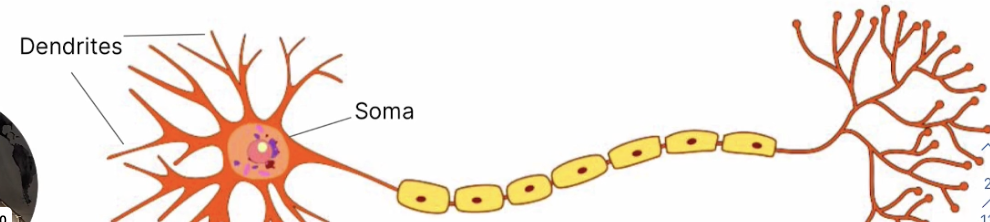
Dendrites
Branch-like structures for receiving signals from neighboring neurons and converting them into electrical impulses
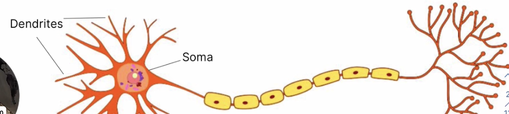
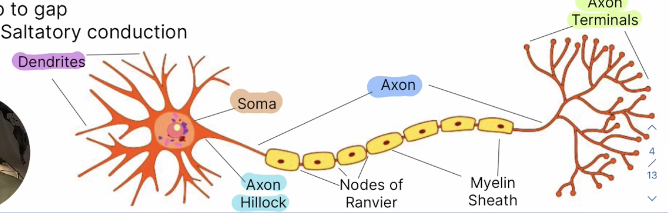
Myelin Sheath
Fatty insulation of the axon, accelerating action potential propogation
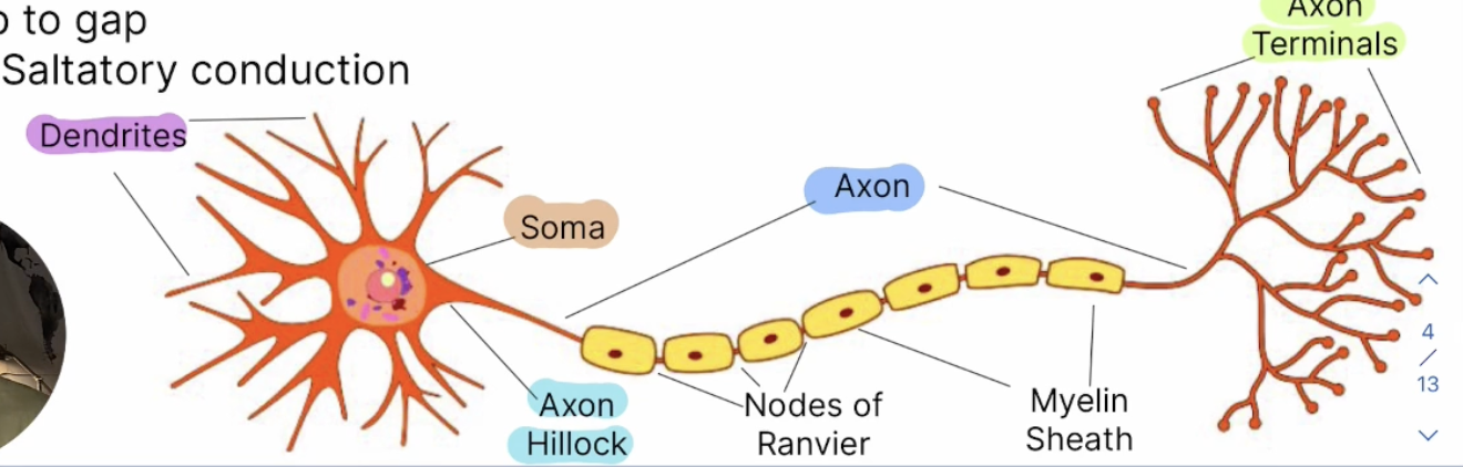
Axon
Process for sending signals to neighboring neurons, long-cable-like projection conducting electrical impulses (action potentials) away from the cell body toward other neurons/target cells
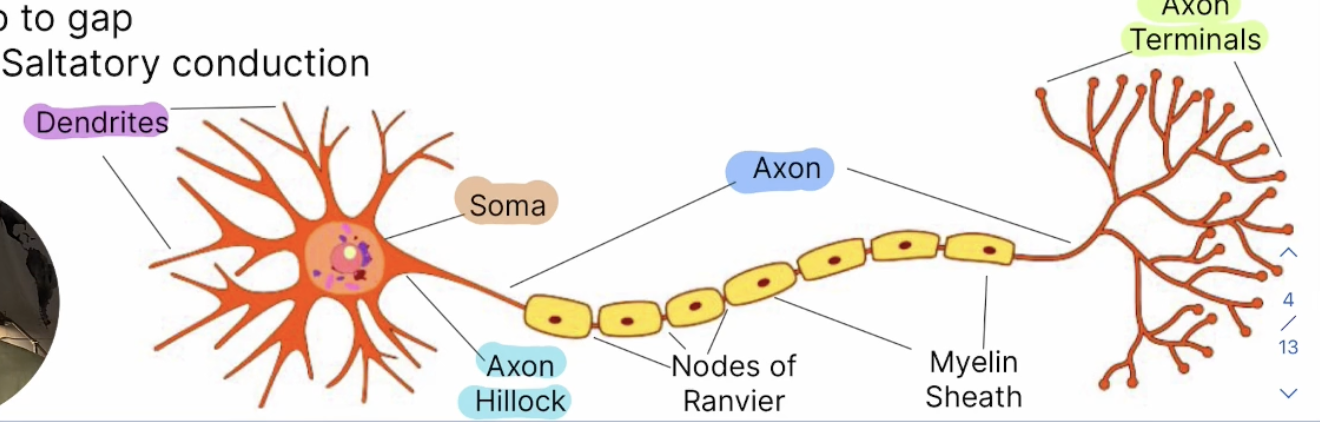
Axon Terminals
Site of synaptic signal transmission, branched endpoints of the axon releasing neurotransmitter molecules into the synaptic cleft to communicate with the next cell
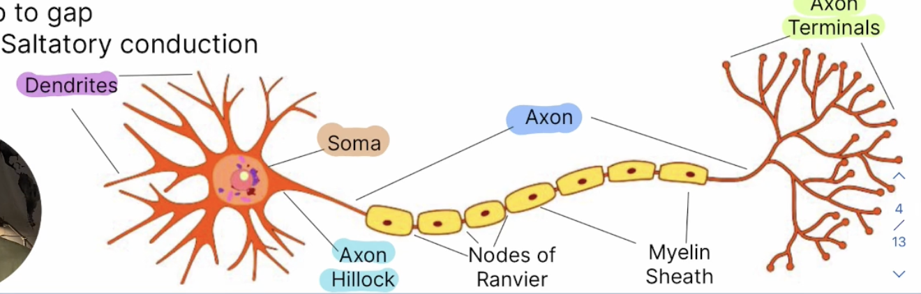
Axon Hillock
Site of connection between the soma and the axon, cone-shaped, trigger zone, determines whether the summed signals are storng enough to generate an action potential
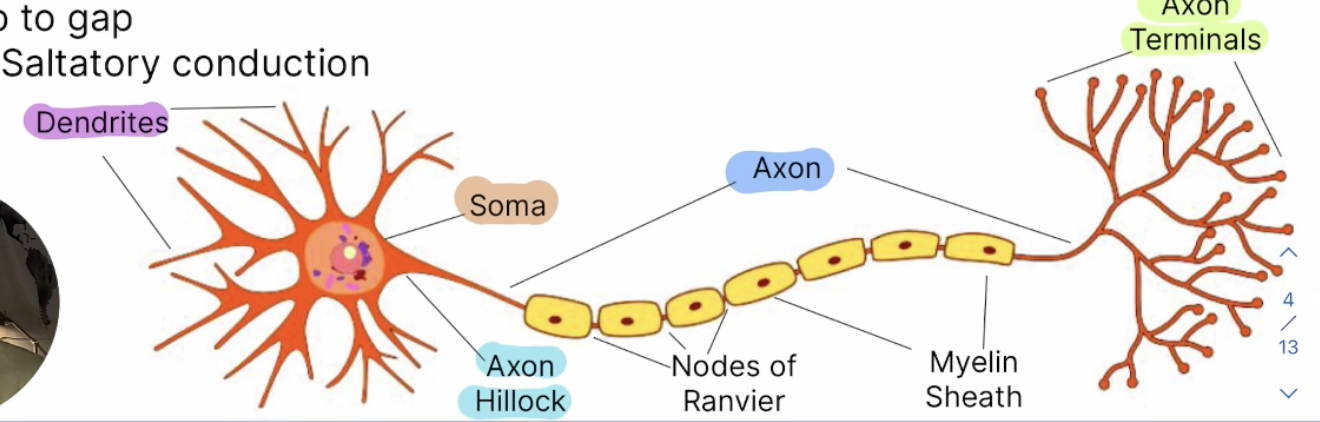
Nodes of Ranvier
Gaps in the myelin sheath, action potential can jump from gap to gap, where axon membrane is exposed and packed with ion channels allowing the electrical signal to be regenerated
Site of ion exchange in saltatory conductoin
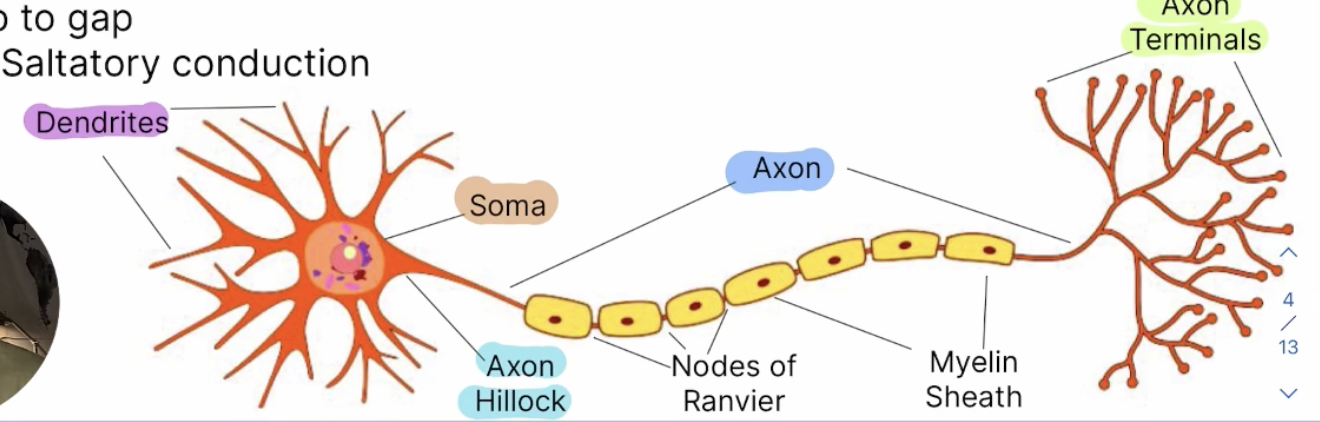
Saltatory Conduction
The process where an action potential rapidly "jumps" from one node of Ranvier to the next, rather than traveling continuously down the axon, which speeds up transmission and conserves metabolic energy.
Myelin Sheath
Fatty, insulating layer wrapping around axon to electrically insulate it and prevent ion leakage, increasing speed of nerve signal transmission
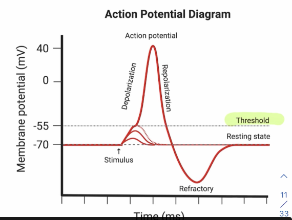
Action Potential
Rapid change in the membrane potential that exists across a membrane, dictated by relative ion ratios in the intracellular/extracellular spaces, finishing at the axon terminal of the presynaptic neuron
Na+/K+, all or nothing (either has one or doesn’t)
Resting State
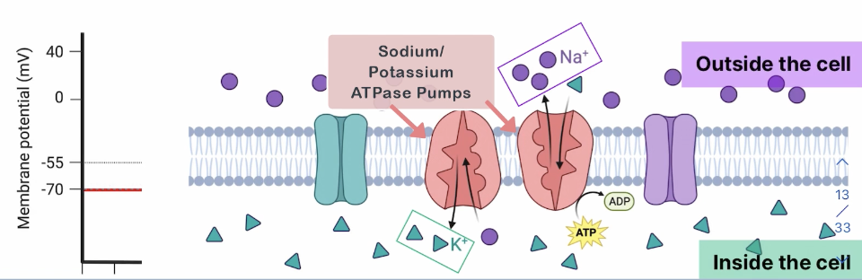
Prior to firing of the action potential, maintained around -70 mV
Voltage more negative inside the cell than outside
Pump 3 Na+ OUT of the cell for every 2 K+ pumped INTO the cell
Depolarization
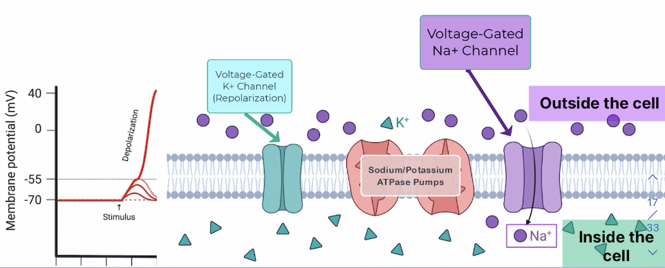
Stimulated neurons depolarize IF the threshold potential is reached (-55 mV), causing some Na+ to flow back into the cell as voltage-gated Na+ channels open
Voltage-Gated Calcium Channels open (Ca2+ enters the cell), and large influx in Ca2+ triggers exocytosis of NTs; synaptic vesicles house NTs bind to the plasma membrane to initiate exocytosis
Repolarization
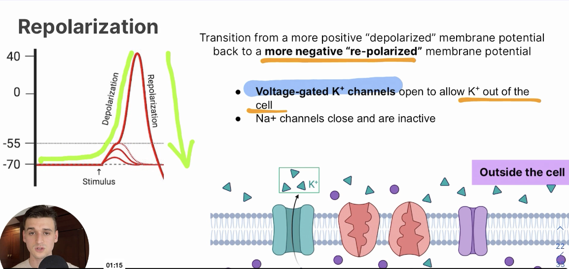
Transition from a more positive “depolarized” membrane potential back to a more negative “repolarized” membrane potential
Voltage-gated K+ channels open to allow K+ out of the cell, Na+ channels close and are inactive
Hyperpolarization
A membrane potential even more negative than the resting potential
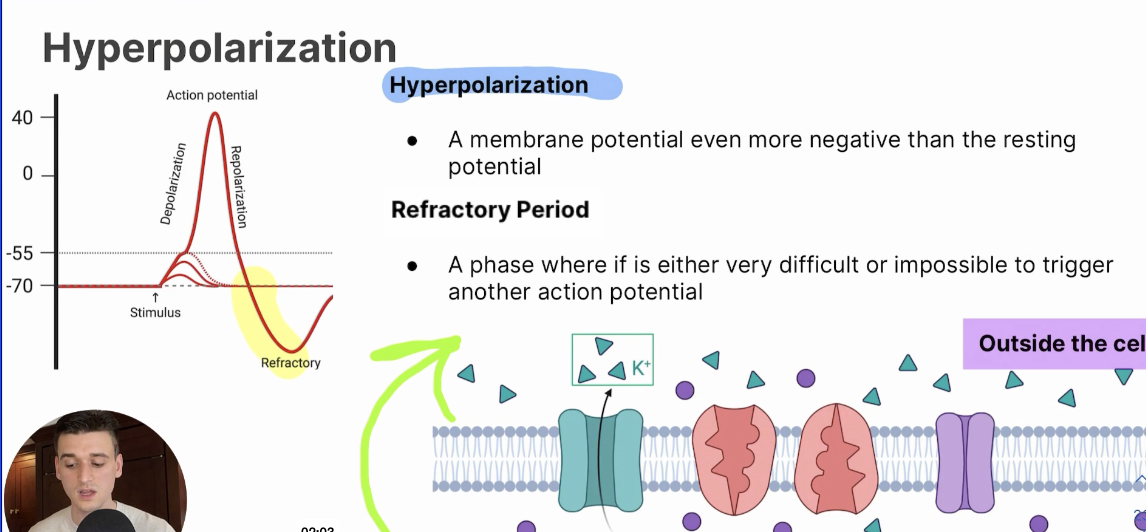
Refractory Period
Phase where its either very difficult or impossible to trigger another action potential
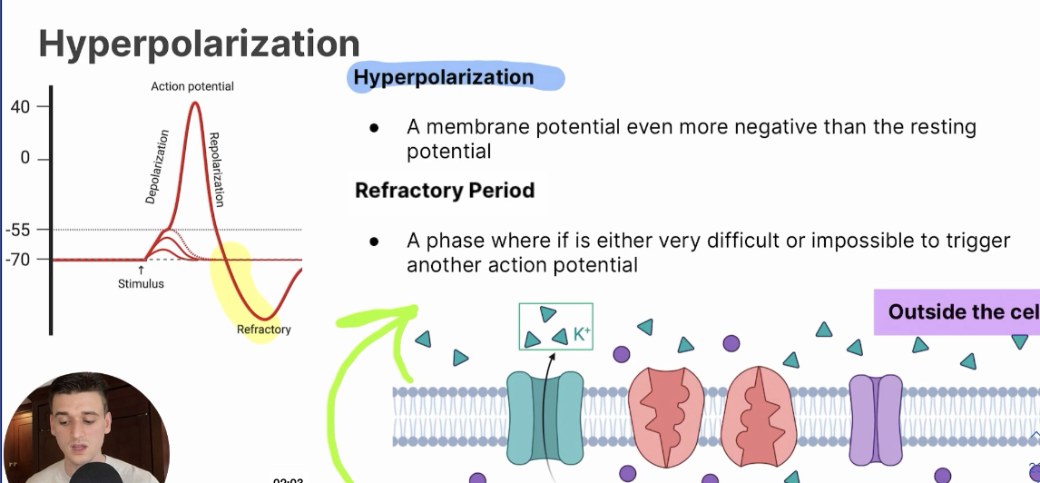
Absolute Refractory Period
Impossible to trigger a second action potential
Na+ channels are inactivated as K+ channels continue to repolarize the membrane back to resting state from + charge
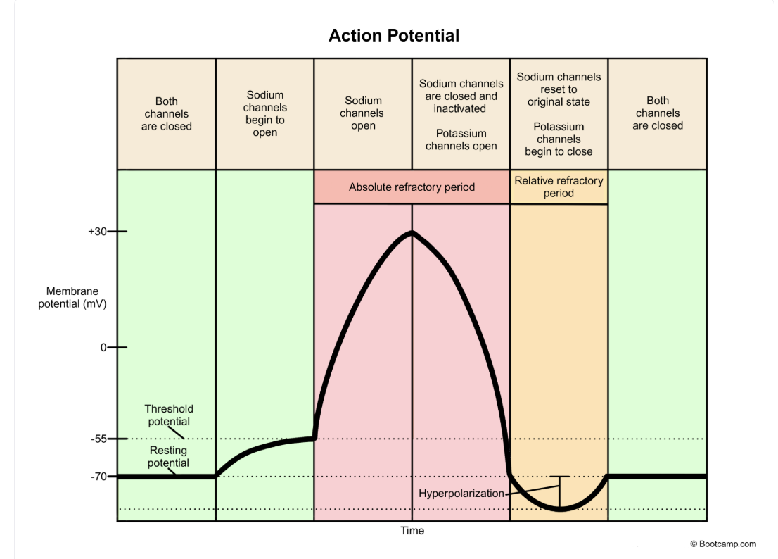
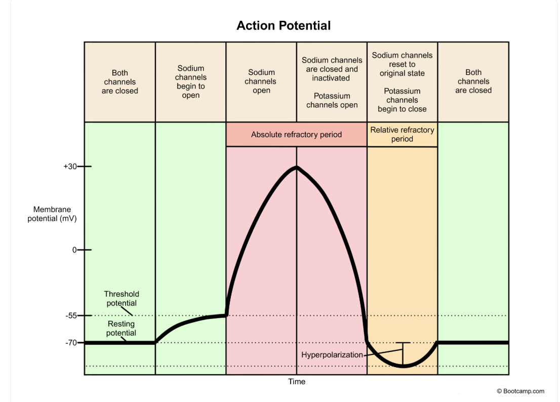
Relative Refractory Period
Very difficult to trigger a second action potential
Membrane potential drops below -40 mv and Na+ channels are closed but not inactivated
Synaptic Transmission
Transmission of chhemical signals from one neuron to another across an interneuron space called a synapse
Neurotransmitters (NTs)
Chemical signals generated in the presynaptic neuron and received at the postsynaptic neuron
Synapse
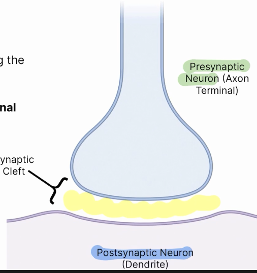
Space between two neurons, including the presynaptic and postsynaptic neurons
Presynaptic Neuron
Responsible for sending the signal and releasing neurotransmitters into the synaptic cleft via the axon terminal
Synaptic Cleft
Zone between two neurons, not including either neuron
Synaptic Vesicles
Responsible for the transport of neurotransmitters from the axon to the synaptic cleft
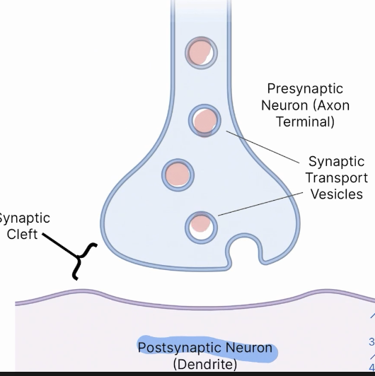
Postsynaptic Neuron
Responsible for signal reception and interacting with neurotransmitters
Neurotransmitters (NTs)
Messenger molecules released by presynaptic neuron and received by the postsynaptic neuron
Postsynaptic Ligand-Gated Ion Channels
Directly responsible for binding of free NTs at the postsynaptic neuron, where released NTs are received
Graded Potential
Local, short-lived change in membrane potential (depolarization or hyperpolarization) varying in strength and intensity and diminishing as it travels passively along the dendrites/soma toward the axon hillock
Excitatory Graded Potential
Graded depolarization (more positive) caused by Na+ influx (opens Na+ channels), pushing the membrane closer to the threshold making firing an action potential more likely
Inhibitory Graded Potential
Graded hyperpolarization (becomes more negative) caused by Cl- influx or K+ efflux (opens K+ channels), pushing membrane farther from threshold making firing an action potential unlikely,
Excitatory Neurotransmitters (ENTs)
Neurotransmitters invoking an excitatory response at the postsynaptic membrane, causing depolarization at synaptic membrane and pass on the action potential signal
Glutamate
Main ENT of the central nervous system
Dopamine
ENT involved in reward-motivated behavior (catecholamine)
Catecholamine
Class of neurotransmitters and hormones derived from tyrosine, consisting of dopamine, norepinephrine, and epinephrine (adrenaline)
Epinephrine and Norepinephrine
Involved in fight/flight response
Postsynaptic ENTs of the sympathetic nervous system, reroute blood towards most critical parts of body
Catecholamine
Acetylcholine
Most common neurotransmitter in muscular system, signaling muscle fibers by binding to ligand-gated sodium channels
Inhibitory Neurotransmitters (INTs)
Neurotransmitters invoking an “inhibitory” response at the postsynaptic membrane
Hyperpolarizes a cell at its synaptic membrane refrains from passing down the action potential
Gamma-Aminobutyric Acid (GABA)
Main INT of the CNS
Glycine
Another INT of the CNS
Seratonin
INT of the brain
Voltage-Gated Ca2+ Channels
This channel’s opening is responsible for the exocytosis of neurotransmitters into the synaptic cleft
Action potential works its way down axon to axon terminal, electric signal stimulates voltage gated calcium channels to open causing an influx of calcium into the cell, causing synaptic vesicles to undergo exocytosis to release neurotransmitters into synaptic cleft
Central Nervous System (CNS)
Brain and spinal chord
Peripheral Nervous System (PNS)
Nerves branching off of the CNS
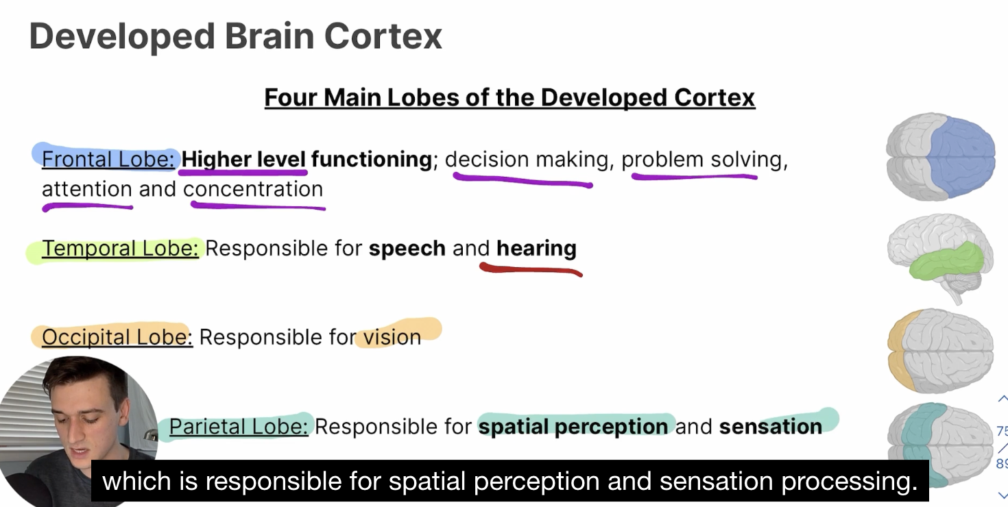
Cerebral Cortex
Largest, outermost, deeply grooved portion of the brain (what you picture typically), made up of 4 regions—frontal, temporal, occipital, and parietal lobe
Frontal Lobe
Higher-level functioning, decision making, problem solving, attention and concentration
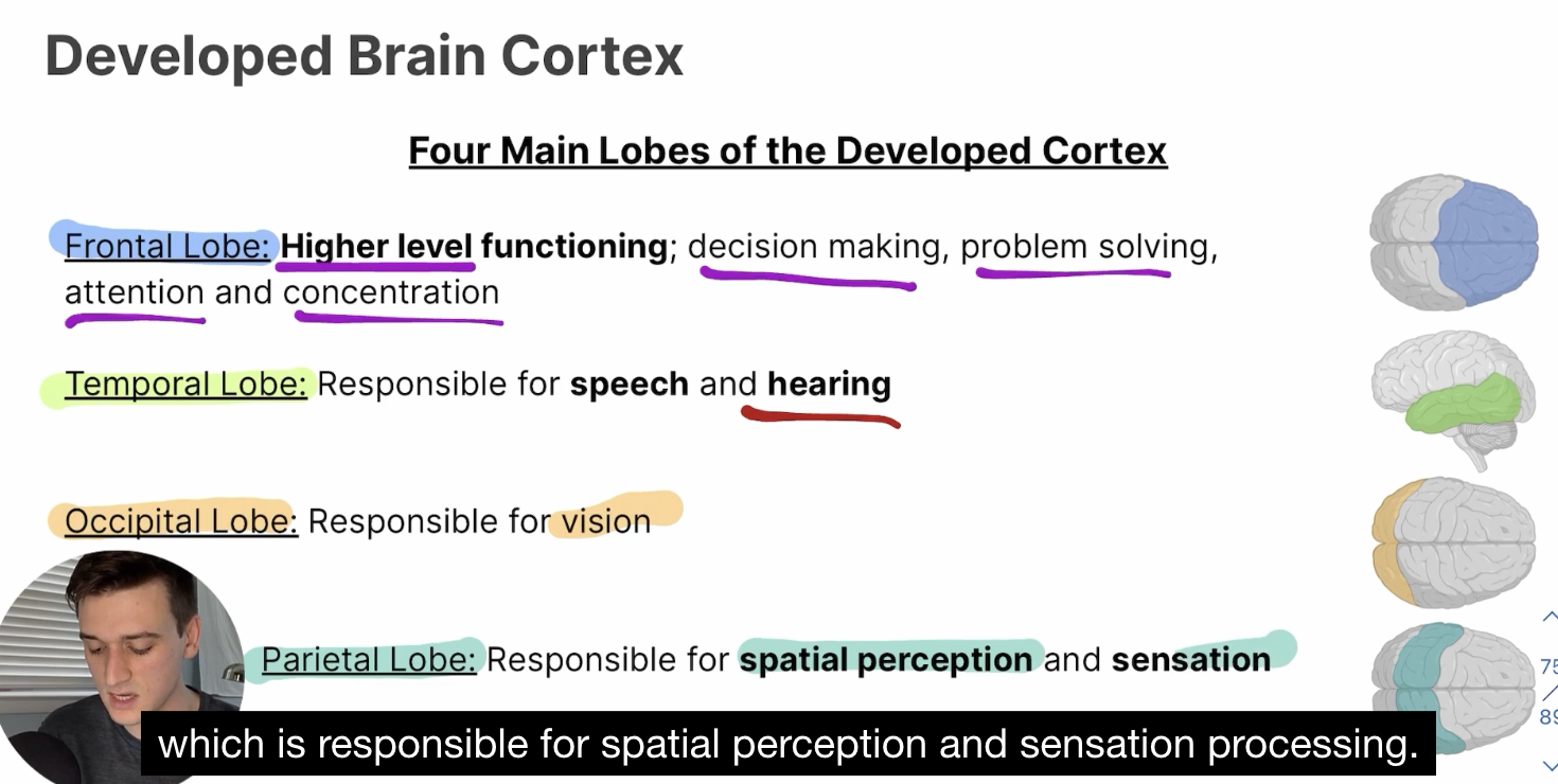
Temporal Lobe
Responsible for speech and hearing
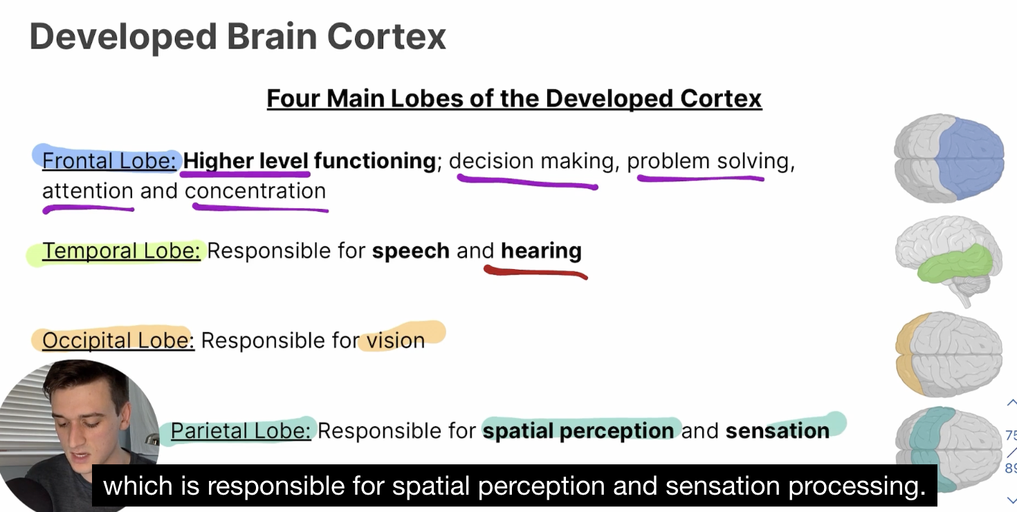
Occipital Lobe
REsponsible for vision
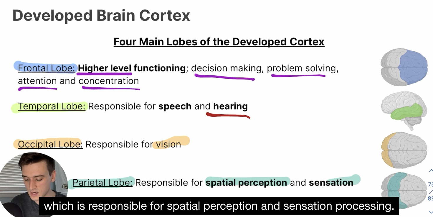
Parietal Lobe
Responsible for spatial perception and sensation
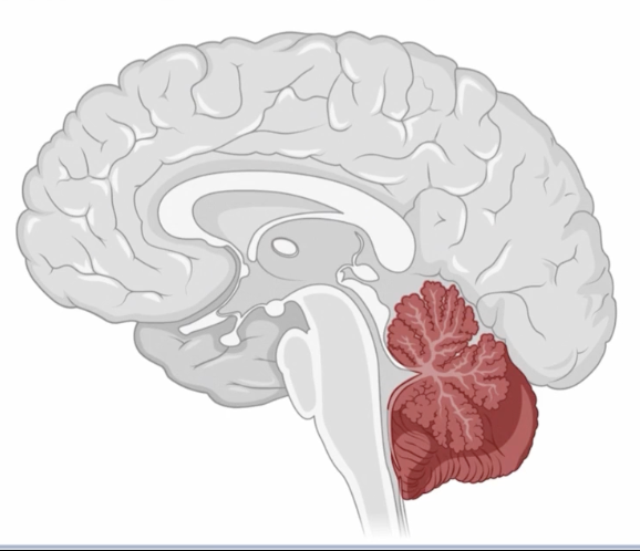
Cerebellum
Responsible for the coordination of movement, independent of the cerebral cortex, between the occipital lobe and the brain stem
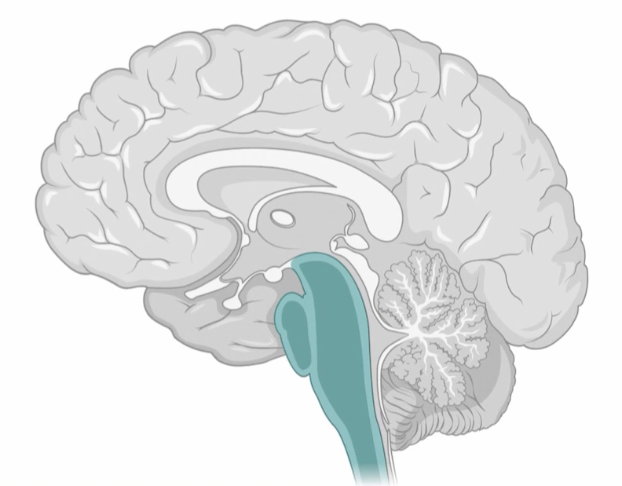
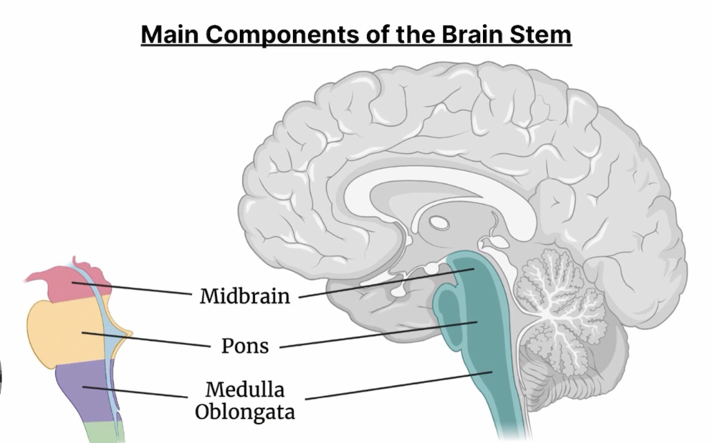
Brain Stem
Involved in the control of automatic functions (heart rate and signal relay), between cerebral cortex and spinal cord
Main Components: midbrain, pons, medulla oblongata, reticular formations
Midbrain
Responsible for the relay of senses to other parts of the brain
Pons
Responsible for the relay of messages between the forebrain, cerebellum, and medulla
Medulla Oblongata
Multi-functional part of the brain stem, responsible for controlling heart rate, breathing rate, blood pressure, and for sensing toxins
Reticular Formations
Neurons existing throughout the brainstem, responsible for corticol arousal and consciousness
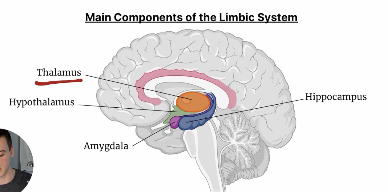
Limbic System
Responsible for emotion, memory, learning, motivation
Main Components: thalamus, hypothalamus, hippocampus, amygdala
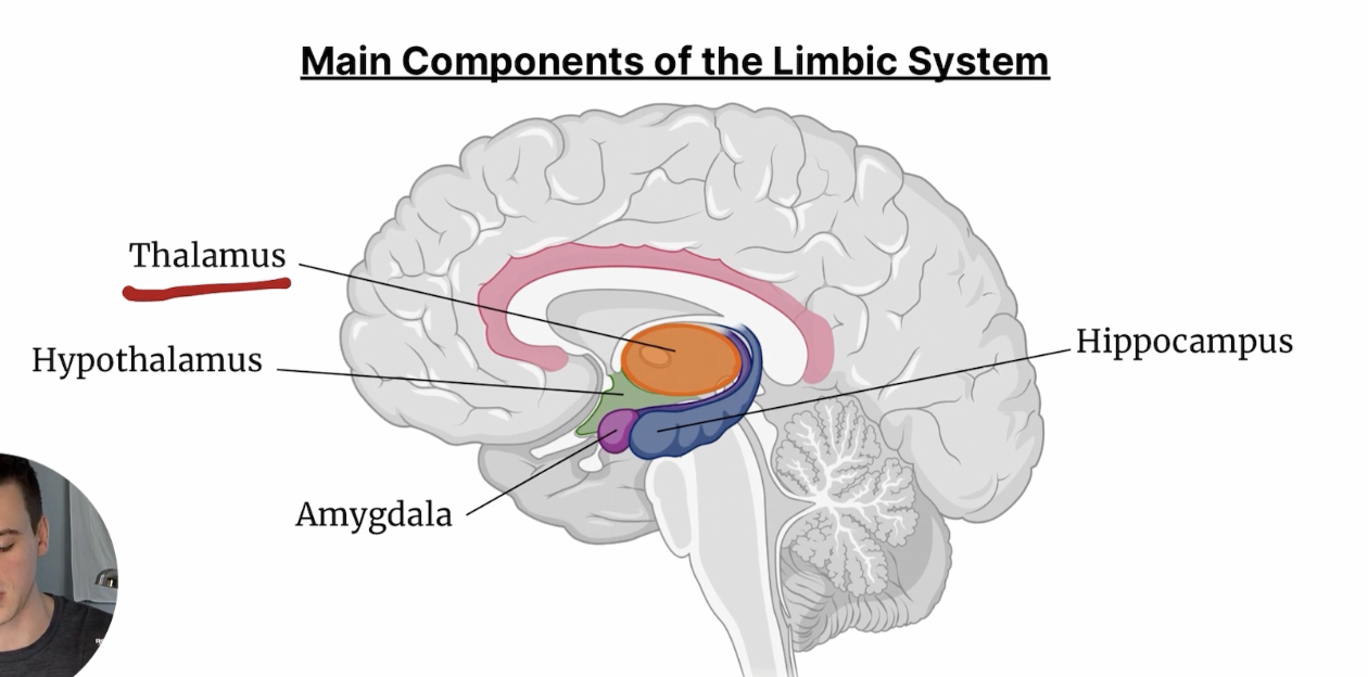
Thalamus
Brain’s relay center, relaying sensory and motor signals from the body to the brain, between the cerebrum and the midbrain
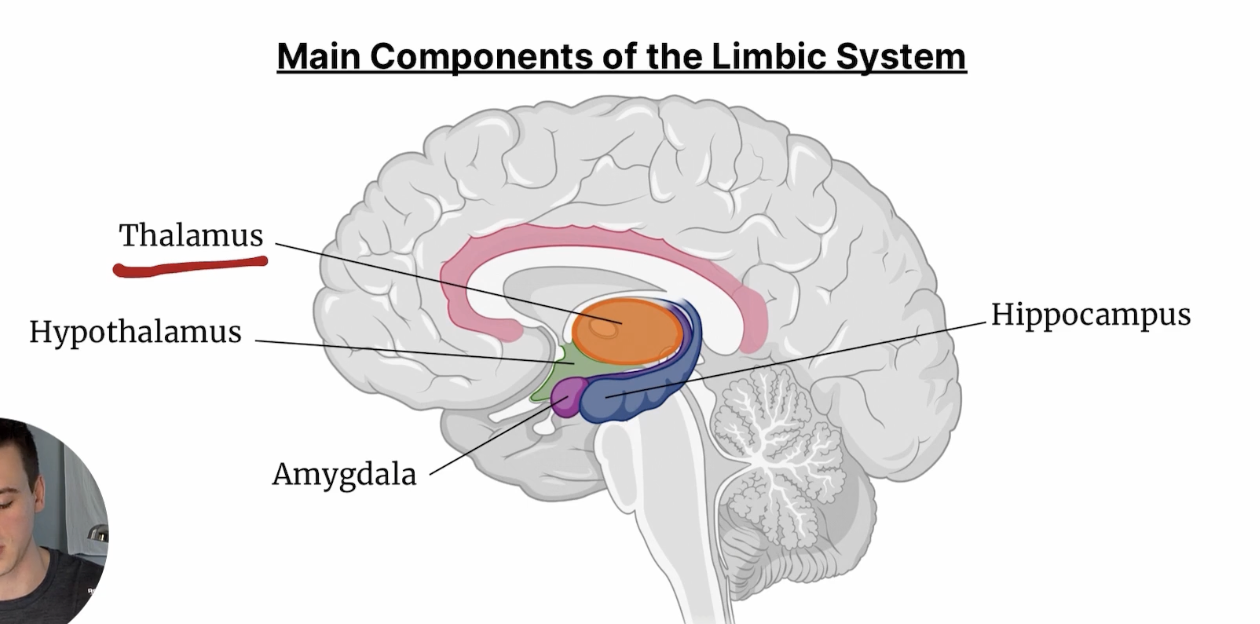
Hypothalamus
Responsible for the regulation of hormone secretion in the body
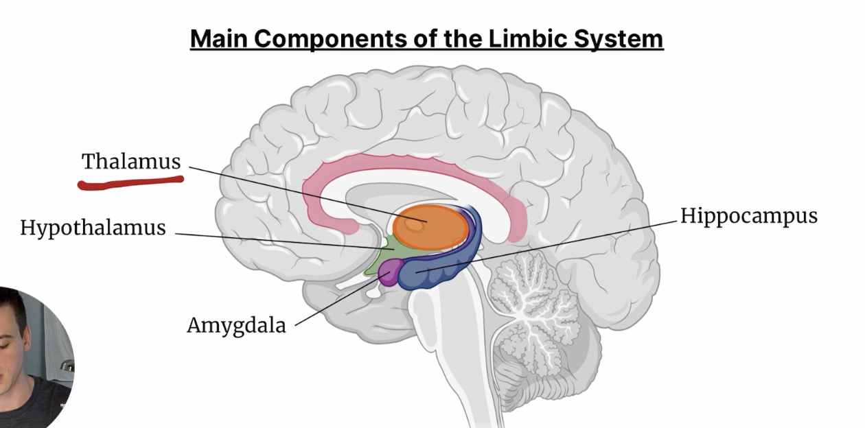
Hippocampus
Responsible for the consolidation of memory
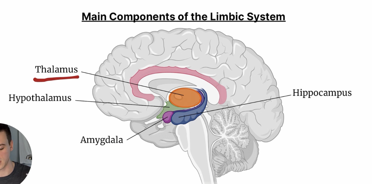
Amygdala
Responsible for the emotional reaction to scents
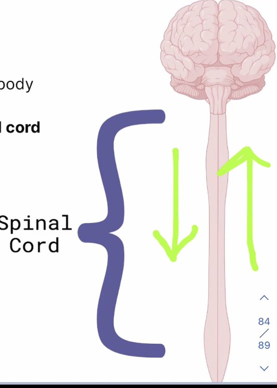
Spinal Cord
Nervous tissue connecting the brain to the body, containing sensory (afferent) neurons and motor (efferent) neurons
Sensory (Afferent) Neurons
Relay signals to the spinal cord and eventually the brain via dorsal roots
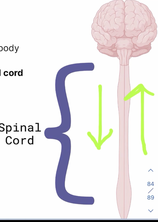
Motor (Efferent) Neurons
Relay signals from the brain to the muscles via ventral roots
PNS Motor (Efferent) Neurons
Somatic Nervous System + Automatic Nervous System
Somatic Nervous System
Involved in voluntary motor action, including skeletal muscles
Automatic Nervous System
Involved in involuntary actions, including cardiac muscle, effector organs, smooth muscles
PNS Sensory (Afferent) Neurons
Mechanoreceptors, nociceptors, thermoreceptors, chemoreceptors, electroreceptors
Mechanoreceptors
PNS Sensory (Afferent Neuron) Responsible for reception of mechanical stimuli
Nociceptors
PNS Sensory (Afferent Neuron) Responsible for reception of pain stimuli
Thermoreceptors
PNS Sensory (Afferent Neuron) Responsible for reception of stimuli related to changes in temperature
Chemoreceptors
PNS Sensory (Afferent Neuron) Responsible for reception of chemical stimuli
Electroreceptors
PNS Sensory (Afferent Neuron) responsible for reception of light, electrical, and magnetic stimuli
Sympathetic Nervous System
Responsible for aiding the fight/flight response
Parasympathetic Nervous System
REsponsible for relaxing the body out fo a sympathetic state (rest and digest), effects occurring via the vagus nerve
Ganglion
Cluster of nerve cell bodies in the peripheral nervous system outside of the brain and spinal cord, site of synapsing between the preganglionic and postganglionic neurons
Preganglionic Neuron
Neuron coming from the CNS that enters the ganglia
Postganglionic Neuron
Neuron coming from the ganglia and exiting to the effector organs
Sympathetic Nervous System (Toward Effector Organ)
Contains Short preganglionic neuron and long postganglionic neuron, where acetylcholine is the neurotransmitter between them, and norepinephrine/epinephrine as NT from postganglionic neuron to effector organ
Parasympathetic Nervous System
Long preganglionic neuron, short postganglionic neuron, with acetylcholine as the sole neurotransmitter
Sympathetic Nervous System (Toward Blood Stream)
Stimulates adrenal medulla to release norepinephrine or epinephrine into the bloodstream
Acetylcholinesterase
Enzyme responsible for the rapid hydrolysis of acetylcholine to stop the propagation of a signal
Outer Ear
Responsible for taking in sound waves
Tympanic Membrane
Responsible for transferring sounds from the outer ear to the middle ear
Middle Ear
Responsible for carrying sound waves from the outer to inner ear
3 Bony Ossicles
Malleus, incus, stapes
Transfer and amplifies vibrations through the middle ear
Stapes
Responsible for transferring sound from the middle ear to the inner ear via the oval window
Inner Ear
Begin the transformation of sound waves into electrical impulses
Cochlea
Use fluid and hairs to convert mechanical signals into neural signals, known as transduction
Round Window
Membrane covered opening between the middle ear and inner ear, helping the cochlear expand and vibrate
Semicircular Canal
Uses fluid and hairs to convey information about a person’s movement
Cornea
Responsible for focusing light and protecting the eye, transparent
Iris
Controls the size of the pupil
Pupil
Controls the amount of light that enters the rest of the eye
Lens
Focuses the relevant image onto the retina
Retina
Located at the back of the eye and contains photoreceptors, and retinal photoreceptors can either be rods/cones
Rods
Responsible for vision in low light (greyscale)
Cones
Responsible for color perception in high light environments
Fovea
Retinal region with the highest concentration of photoreceptors, responsible for hgih acuity vision
Amacrine and Bipolar Cells
Transmits the info from the retinal rods/cones to ganglion cells of the optic nerve fibers
Optic Nerve
Bundel of axons transmitting visual info to the brain
Optic Disk
Eye’s blind spot, site of passage for the optic nerve heading towards the brain, no photoreceptors present