Surgical Principles
 Call Kai
 Learn
Learn Practice Test
 Spaced Repetition
 Match
Match Flashcards
 Knowt Play
1/244

There's no tags or description
Looks like no tags are added yet.
Name | Mastery | Learn | Test | Matching | Spaced | Call with Kai | Chat |
|---|
No analytics yet
Send a link to your students to track their progress
245 Terms
Compare use of a scalpel vs. scissors
Scalpel
Best for dense tissue like skin or fascia
Minimal collateral damage
Scissors
Best for loose tissue like adventitia or fat
More crushing damage
Compare mechanism of cautery vs. electrosurgery
Cautery = Direct heat produced AT THE TIP by using low-voltage current (current does NOT pass through the patient)
Electrosurgery = Heat produced WITHIN the tissue via resistance of flow as current passes through the patient
Compare uses of cautery vs. electrosurgery
Cautery = Disbudding
Electrosurgery = Cutting OR coagulation depending on the electrical waveform selected
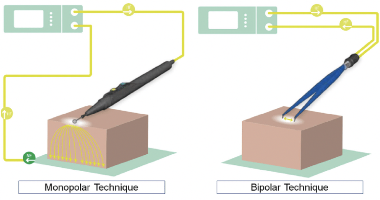
Compare the 2 types of electrosurgery (mechanism and advantages/disadvantages)
Monopolar Electrosurgery
Mechanism: Active electrode at surgical site and return electrode (dispersive pad) elsewhere on patient
Current passes through patient and resistance generated produces heat
Resistance inversely proportional to SA (resistance decreases as current spread through body)
Disadvantage: Cannot work in wet field (eg. blood) as current will disperse throughout fluid and target tissue will not heat effectively
Bipolar Electrosurgery
Mechanism: Active AND return electrode at surgical site
Current passes through tissue grasped by forceps
Advantage: Works in ALL types of environments (eg. fluid)
Laser Surgery
Mechanism
2 Uses
Mechanism: Light amplification by stimulated emission of radiation (monochromatic, highly coherent in infra-red, visible or near Uv)
Uses:
Tol to ablate/vapourise/weld tissue
Photodynamic cancer therapy = Photosensitive drug taken up by tumour and laser applied to kill tumour
Fluoroscopy (eg. C-arm)
Imaging modality to produce real-time radiographs to assist with orthopaedic repair
6 Reasons to perform surgery
Therapeutic
Diagnostic (eg. ex-lap, biopsy)
Increase suitability of use (eg. desexing, dehorning)
Palliative
Biomedical research
Cosmesis
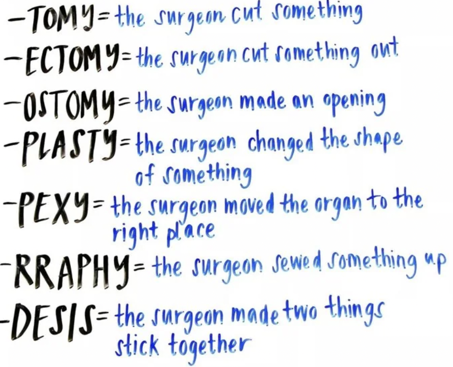
Definitions
-ostomy
-oplasty
-rrhaphy
-pexy
-desis
-ostomy = Permanent/semi-permanent stoma (opening) eg. colostomy bag or gastrostomy tube
-oplasty = Reconstructive/reparative surgery to restore function or appearance eg. rhinoplasty
-rrhaphy = Suturing a structure to close a defect/rupture eg. herniorraphy/tenorraphy
-pexy = Fixation eg. gastropexy
-desis = Attach together eg. arthordesis
6 Advantages and 4 disadvantages of minimally invasive procedures (eg. endoscopy)
Advantages
Reduces duration of hospital stay (less $)
Faster surgery time (minimal closure)
Less exercise restriction (minimal closure)
Superior cosmesis (smaller scar)
Reduced pain (do not need to stretch abdominal wall with retractors)
Magnified field and excellent lighting
Disadvantages
Surgeon’s hands remove from tissue (no manipulation/palpation)
Haemorrhage = Visibility issues and more difficult to stop
Requires specialised/expensive equipment and new skill
Arthroscopy impossible for canine hip
Triangulation in endoscopy
Concept required for skin incision for endoscopic techniques
Apex = Lesion on interest
Point in base = Scope viewing point
Other point on base = Instrument
Separate incisions for scope and instrument
Halsted’s Principles (7/8)
Handle tissue gently
Reduce necrosis and post-op infection
Reduce pain and dysfunction
Speed up healing
Meticulous haemostasis
Better surgical field visualisation
Reduce haemorrhagic shock
Reduce post-op infection
Preserve blood supply
Strict aseptic technique
Reduce post-op infection
Avoid tension on wound edges
Reduce tissue strangulation (necrosis) and dehiscence
Eliminate dead space between tissue planes
Dead space accumulates blood/serum which delays healing is a growth medium for bacteria
Meticulous approximation of all wound layers
Promotes rapid healing
Increases wound strength
Reduce post-op infection
Minimise foreign material in wound (eg. suture material and glove powder)
5 Questions to ask when deciding to operate
Has a definitive diagnosis been made?
Is surgery the ONLY/BEST treatment? What are the alternatives? Minimally invasive techniques?
Logistics: When? How? Who? Where?
Harm to the patient during/after surgery
Owner considerations: Coping with aftercare, expectations, cost
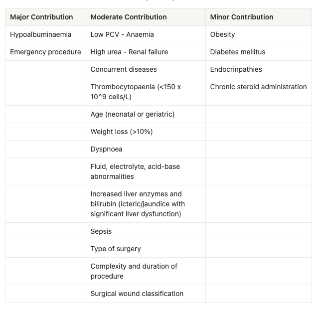
Determinants of surgical risk
Table
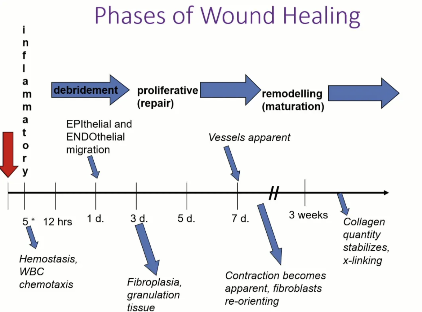
List the 4 phases of wound healing (+ duration)
Acute inflammatory (lag) phase = Day 0 - 5
Debridement phase
Proliferative (repair) phase = Day 3 - 14
Remodelling (maturation) phase = Week 2 - year 1
Phases overlap and problems arise if one phase is absent or prolonged
Acute Inflammatory (Lag) Phase
3 Functions
6 steps (+ descriptions)
Functions:
Haemostasis
Prevent infection
Lay groundwork for subsequent steps of healing
6 Steps:
Skin retraction = Defects becomes larger due to normal skin elasticity and external tension from muscle pull
Haemorrhage = Haemorrhage due to injured blood vessel to clean wound surface and inoculate wound with cells for healing
Vessel reaction/vasoconstriction = Traumatised epithelium → Phospholipid membrane injury and arachidonic acid release
Arachidonic acid cascade induces peripheral vasoconstriction (5 - 10 minutes post-injury)
→ Hypoxia and decreased pH within wound and tissue
Vasodilation and increased permeability of venules
In response to cytokines (eg. histamine, serotonin, bradykinin, prostaglandins, leukotrienes) secreted from mast cells in damaged tissue
Cellular response (<30 minutes) = Recruitment and activation of platelets from peripheral bloodstream → Degranulation results in mitogen and chemoattractant release (important for directing and initiating wound healing)
Clot formation = Plasma proteins (eg. fibrin and clotting factors) to form haemostatic plug, reduce dead space and form framework for repair
2 Functions of a scab
Scab = Clot exposed to air causing the surface to dry and contract
Function:
Protect wound from external contamination
Maintain internal homeostasis to allow epithelial cells to migrate beneath the scab
Debridement Phase (Inflammatory)
Timing
5 Steps
Timing: ~6hr after injury
Steps:
Fibrin released (by activation of intrinsic and extrinsic haemostasis mechanisms) to stabilise provisional wound matrix (clot)
“Provisional” = Lag
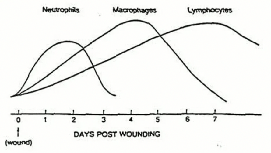
Neutrophil recruitment (peaks at day 2) stimulated by exposed collagen → Phagocytosis of debris and bacteria AND release of chemoattractants to augment inflammatory response
Neutrophils die or become phagocytosed
Monocyte migration = Phagocytosis, Ag presenting cells and release of growth factors which induces the proliferative phase
Lymphocytes
Proliferative (Repair) Phase
4 features (+ descriptions)
Fibroplasia = Migration of fibroblasts for collagen synthesis and granulation tissue formation
Fibroblasts predominate the wound bed by day 4 (arrive from day 2) → Necessary for ECM and type I collagen synthesis
Peak at day 7 - 8 after injury → Release collagen and
ECM rich in fibronectin, hyaluronan and proteoglycan (provide a scaffold for cell migration and tissue remodelling)
Type III collagen deposited → Maturation to type I collagen (increase tensile strength)
High vascularity and beginning of granulation tissue
Fibroblasts → Myofibroblasts which contribute to wound contraction
Epithelialisation = Epithelial cells proliferate and migrate from wound edges via contact-guidance and -inhibition
Contact guidance = Migrating epidermal cells roll/slide over each other across the basal lamina or along fibrin deposits
Relies on keratinocyte stem cells from adjacent tissue (basement membrane, hair follicles, sweat and sebaceous glands) → Recruitment and proliferation
Hence, deep wounds with complete disruption of the basement membrane are slower to heal (lack of keratinocyte stem cells for epithelialisation)
Requires healthy granulation tissue
Migrate from residual epithelium (periphery)
New epidermis at wound margins by day 4 - 5
Rapid re-epithelialisation when partial thickness abrasions (basal membrane and keratinocyte population still present)
Angiogenesis = New blood vessel formation
Occurs with decreased O2 tension, high lactate and low pH within wound
Initiated by vasculogenesis (NEW blood vessel formation) → Dense capillary network
Initiated from bone marrow progenitor cells
Driven by angiogenic growth factors:
VEGF (Vascular Endothelial Growth Factor)
FGF-2 (Fibroblast Growth Factor-2)
PDGF (Platelet-Derived Growth Factor)
Endothelial cell migration = Involves migration and proliferation to create new capillary structures
Reversal of hypoxia → Increased blood flow and reversal of hypoxia
Wound contraction = Wound borders are drawn together to reduce SA for epithelialisation
Occurs when fibroblasts predominate
Reduces wound surface area by 40 - 80% depending on location of wound
Dependent on differentiation of fibroblasts → Myofibroblasts
Collagen reinforces contracted wound
6 Disadvantages of wound contraction/2nd intention as a method of healing
Increases skin tightness and reduces function
Limited movement when formed over flexor surfaces
Stenosis of body opening (eg. anus)
Insufficient contraction = Large areas of thin epithelium which can abrade and split open
Cosmesis
Expensive bandage changes and hospital visits
Prolonged healing
5 Functions of granulation tissue
Provide surface area for epithelial cell migration
Provide fibroblasts for collagen formation
Assists in wound contraction
Provides blood supply
Barrier against systemic infection and local infection
4 Features of maturation (remodelling) phase
Continued epithelialisation and wound contraction
Hyaluronan from provision wound matrix replaced with proteoglycans within ECM
Angiogenesis and wound metabolic activity decreases
Cytokines and growth factors stimulate decline
Collagen content increases with organisation/alignment of collagen into bundles and collagen cross-linking
Remodelling via proteinases
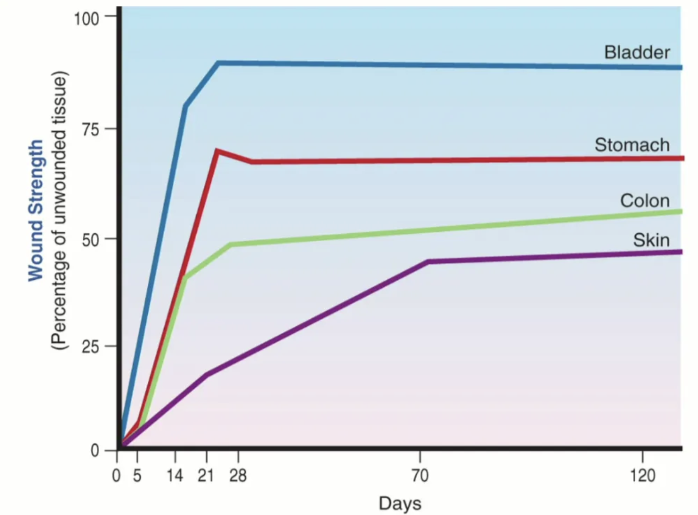
Scar tissue strength over time
Weaker than original tissue with gradual gain over time (never as strong as original tissue)
20% at 3 weeks
50% by 3 months
70 - 80% at end of maturation
Tissue appearance at each stage of wound healing (8)
Swelling (inflammatory phase)
Receding wound edges
Exudative (vasodilation in inflammatory phase bringing in fluid for wound clotting and phagocytosis)
Decreased tissue strength
Pink, cobblestone appearance (granulation)
Thin, whitish edges (epithelialisation)
Puckered appearance around edges
Restored tissue strength
Compare surgical vs. open wound healing
Blood clot
Inflammatory phase
Debridement
Epithelialisation
Fibrin
Granulation tissue/collagen
Surgical Wound
Blood clot forms in incision
Brief inflammatory phase
Minimal debridement
Epithelial cover by 48hr
Vertically oriented fibrin by day 3 - 4
Collagen formed by day 5 and parallel collagen and capillary orientation by day 6
Open Wound
Blood clot fills wound bed
Inflammatory response depends on damage
Prolonged debridement phase due to presence of devitalised tissue, infection and foreign material
Epithelialisation requires granulation bed and may be delayed ≥7 days
Fibrin clot covers wound and granulation tissue forms by day 4 - 5
3 Potential complications of surgical wound closure
Sutures too tight = Skin necrosis, suture abscess and delayed healing, removed prematurely by patient as uncomfortable
Sutures too loose = Gaping wound and open wound healing with scar tissue
Migrating epithelial cells down suture tracts may become keratinised and form suture abscess
4 Potential complications of open wound healing
Excessive granulation tissue (proud flesh) inhibits wound contraction and epithelialisation
Inhibited function associated with wound contraction
Slow epithelialisation and scar tissue formation when contraction stops before wound closure
Large areas of scar tissue are easily abraded by external trauma (eg. licking)
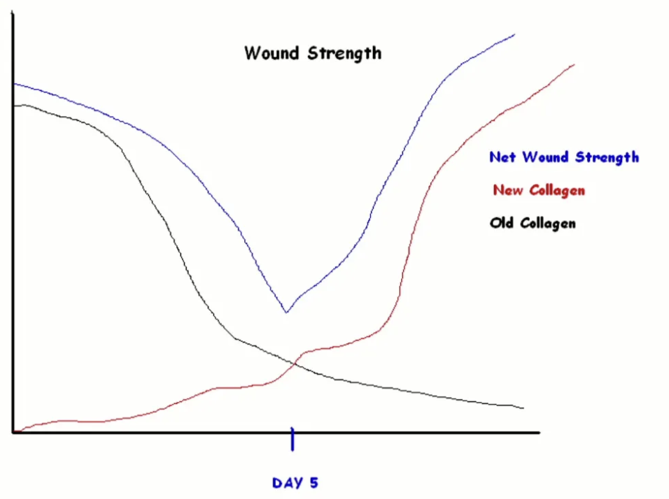
What day is wound strength the lowest? Why?
Day 5 = Old collagen broken down by neutrophils BUT delay of laying down new collagen
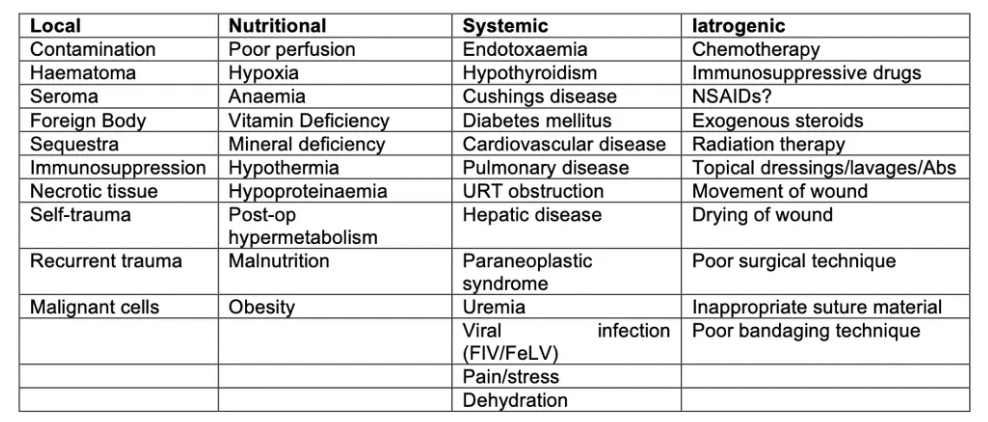
7 Most common causes of delayed wound healing
Infection
Excess motion (horses)
Self-mutilation
Excess tension
Debilitated patient: Systemic disease
Recumbency
Poor vascularity
7 Factors affecting wound infection
Degree of bacterial contamination (>10^5 bacteria/g tissue or mL of fluid required for infection)
Virulence of organism (eg. Klebsiella, Pseudomonas, E. coli vs. commensals)
Type of injury
Crushing = Severe devitalisation and vascular injury
Sharp laceration = Little necrotic tissue
Vascular insufficiency/ischaemia (eg. dehydration, shock, tissue tension, vascular injury, excessive electrocautery)
Foreign material
Harbours bacteria which the inflammatory reaction cannot reach
Creates chronic infection/sinus formation as macrophages cannot remove foreign material
Free fluid accumulation (eg. seroma)
Due to improper closure which creates dead space
Creates pressure that interferes with blood supply to adjacent tissue
Good bacterial growth medium
Immunological incompetence (eg. immunocompromised, secondary to drugs, malnutrition, irradiation)
3 Causes of seroma formation after wound closure
Tissue rich in lymphatic vessels is transected
Excessive movement in wound
Tissue response to suture material
List 7 principles of wound management
History
Important to know timeline of injury as suturing sooner carried better prognosis
Cause of injury: Sharp laceration vs. HBC
Previous wound treatment (eg. blue spray) = Suturing NOT appropriate
FULL physical examination (avoid missing crucial problems)
Prevent further contamination
Lavage
Wound exploration
Debride
Wound therapy = Drain, closure and bandaging
6 Steps of wound management to prevent further contamination
Ensure owner covers wound with sterile dressing
Saline-soaked gauze ideal to prevent desiccation and further contamination
Remove bandage on admission and cover wound with sterile gauze impregnated with K-Y jelly
Prevents further contaminants (eg. hair and dirt) falling into the wound
Clip surrounding hair and use vacuum cleaner to remove
Trim hair at wound edges with scissors dipped in mineral oil (hair sticks to blade and not wound)
Scrub skin with betadine to prepare for aseptic surgery
Drape-off surgical site and remove wound cover
Main indication for systemic broad-spectrum antibiotics for wound management
ONLY indicated with bacterial culture and sensitivity of sterile swab from deep in the wound
Culture AFTER flushing and consider waiting as bugs present on admission are NOT causative agents of infection later
Do NOT use antiseptics, ointments, powders as this leads to further contamination and chemical injury to tissue
Ideal pressure for wound lavage
8 psi (high pressure may open fascial planes leading to further damage and contamination)
5 Ideal solutions for wound lavage
Tap water = Gross contamination before more careful lavage
0.9% NaCl = Any wound to cleanse wound in conjunction with debridement and dilute bacterial load
LRS = Any wound as for sterile saline
Chlorhexidine = Contaminated/infected wounds
Broad-spectrum 24hr activity but some Staphylococcus aureus and G- resistance
Toxic to fibroblasts
Povidone iodine = Contaminated/infected wounds
Broad-spectrum activity but only for 4 - 6hr
Inactivated by debris, pus or blood
2 Indications and caution for antiseptic solutions with wound lavage
Indications:
Severely infected wounds
Immunocompromised patients
Caution: Use at correct concentration to avoid damage to host cells (in addition to bacteria)
3 Lavage solutions to avoid
Hypochlorite/Dakin’s solution
Hydrogen peroxide
Savlon
Debridement
2 Functions
Best method
Function:
Remove all debris, necrotic/devitalised tissue to prepare the wound for suturing or selective debridement
Explore wound to determine limits and affected anatomical structures
Method: Sharp scalpel (NOT scissors) changed and disinfected repeatedly to prevent contamination of clean areas
2 Methods of surgical debridement (+ comparison)
Layered debridement = Tissue cut back to clean/viable tissue
Conserves vital structures
Ideal when closure is difficult to keep as much tissue as possible
En bloc debridement = Complete wide excision
Only when non-vital tissue is involved
Reduce time and cost of wound management
Debridement methods for:
Skin
Fat
Muscle
Nerves
Joints
Tendons/ligaments
Bone
Skin = Trim edges and consider staged debridement (“wait and see”) as viability of tissue may be difficult to determine/misleading due to vascular spasm
Fat = Debride all exposed fatty tissue to a clean plane
Muscle = Debride muscle which is dark, friable or fails to contract
Nerves = Conserve and protect from other damaged tissue
Joints = Lavage thoroughly, repair and immobilise
Tendons/ligaments = Preserve as much as possible
Bone = In open wounds >4hr, discard small avascular fragments and carefully preserve all bone with soft tissue attachments
Debride to prevent overgrowth of granulation tissue from exposed bone
Currettage cortex until tiny bleeding vessels are seen
Types of wound drainage
Surgical drain
Passive drain
Active drain
Pack wound open with saline-soaked gauze
Saline maintains hydration, breaks down thick discharge and absorbs drainage material
Wound classification
2 Functions
Ways to classify
Functions:
Predict likelihood of complications
Select appropriate management (closure type, drainage etc.)
Classification:
Degree of contamination
Duration
Depth
Location
Cause
List the types of wound closure
PRIMARY WOUND HEALING
Primary closure (1st intention)
Delayed primary closure
Secondary closure (3rd intention)
SECONDARY WOUND HEALING (2nd intention)
Primary closure (1st intention wound healing)
Definition
Indications
Examples
Definition: Close wound immediately after infliction
Indications: Clean or clean-contaminated wounds
<4 - 6hr with minimal contamination
Sufficient skin for closure
Examples:
Spay wound
Small contaminated wound with loose skin (resect contaminated area)
Delayed primary closure
Definition and method
Indication
Example
Definition: Closure of wound 3 - 5 days AFTER injury BEFORE granulation tissue has formed
Cover un-sutured wound with sterile dressing and attempt to close in 3 - 5 days when devitalised tissue or infection is absent
Indication: Allows judgment of tissue viability when contaminated cannot be removed definitively
Example: Tail degloving with uncertainty if tissue will fall off
Recent wounds with secondary contusion
Recent heavily contaminated wounds
Clean sharp wounds of several days with no devitalised or infected tissue
Secondary Closure (3rd intention wound healing)
Definition
Indication
3 Methods
Definition: Closure of wound >5 days AFTER injury when wound has formed a bed of healthy granulation tissue
Indication: Wound requires >5d to manage infection
Methods:
Direct appositional suturing of two granulation surfaces
Undermine skin to appose skin edges
Completely excise granulation tissue and close wound in primary fashion (will NOT relieve tension)
Secondary wound healing (2nd intention wound healing)
Definition
Indications
Advantages
Ideal locations
Definition: Wound left open to heal by granulation tissue formation, contraction and epithelialisation
Indication:
Dirty/infected wounds where other methods of closure cannot be used
Large cutaneous defects with insufficient skin
Advantage: Practical and economic IF sufficient wound care provided
Check contraction of wound by pinching skin together
Location: Trunk and upper limbs (faster healing than distal limbs)
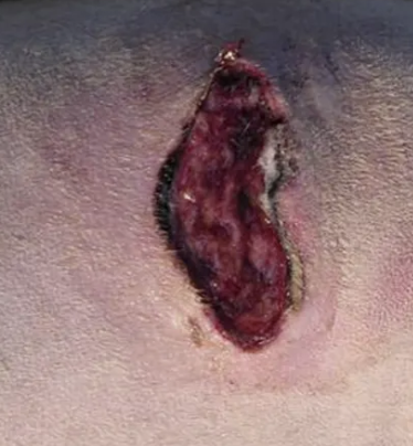
EXAMPLE: Dog flank laceration after running into a car door. Wound is only a couple hours old. There is no pus, but hair and dirt is present on the surface of the SC fat and muscle
Classify the wound
How do you proceed?
Clinical reasoning
Classification: Contaminated wound (traumatic, but <6hr and no pus)
Management: PRIMARY CLOSURE
Debride edges of the wound to leave healthy tissue for apposition (remove necrotic tissue)
Minimally debride and irrigate wound bed to remove dirt and contaminants
Place Penrose drain
Clinical Reasoning: Wound is large and would take a long time to heal by secondary intention, closure would be simple, a scar would be unsightly in a short coated breed
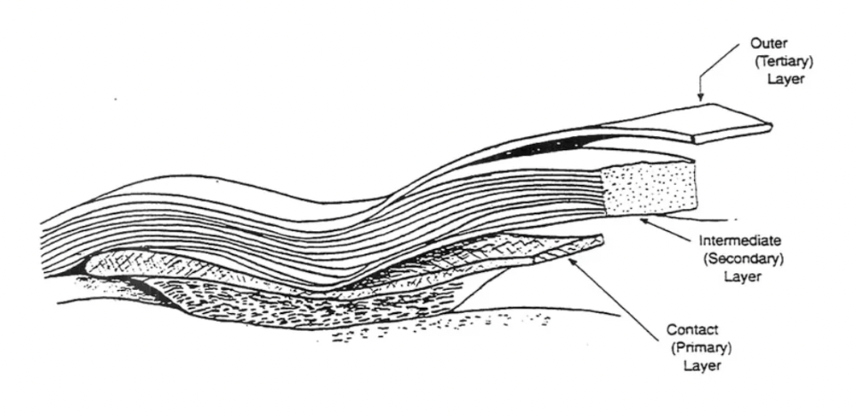
Dressing vs. bandage
Dressing = Layer of bandage in direct contact with wound
Bandage = Everything else
List 11 functions of bandages
Exert pressure to eliminate dead space
Pack a wound
Debride a wound
Absorb exudate/blood
Protect wound from environment
Protect environment from wound
Immobilise wound and support fractures
Provide comfort by reducing pain (eg. splint)
Vehicle for application of antibiotics and antiseptics
Indicator of wound secretions
Aesthetic appearance
5 Functions of the primary (contact) layer of a bandage
Most important layer in wound management
Function depends on stage of wound healing:
Debride necrotic tissue
Deliver medication (eg. analgesia)
Prevent infection or strike-through
Transmit wound exudates OR form occlusive seal over wound
Promote wound healing
Describe the classification of dressings (primary layer) + 2 descriptions
PASSIVE = Dressing does not change wound biology
Adherent
Non-adherent (semi-occlusive)
Absorbent
Vapour-permeable/barrier film
ACTIVE = Dressing interacts with wound biology to actively promote wound healing
Type of dressings required for 3 stages of wound healing
Inflammatory/debridement (dead tissue, bacteria, exudate) = Remove debris and fluid
Repair (granulation) = Protect and keep moist
Epithelialisation = Prevent trauma and drying out
Adherent dressings
Indication
Function
Type of material
2 Examples
Timeline of use
Indication: Inflammatory and debridement stage of wound healing
Dirty, infected, necrotic, sloughing wounds
Function: Mechanically rip away bacteria, necrotic tissue and debris
Exudate drawn out into moist gauze and dries → bacteria and dead tissue cannot retain water → they dry and stick → gauze pulls them away
Material: Woven gauze swabs (NOT pressed felt) to trap loose necrotic tissue and debris
Examples:
Wet-to-dry dressing
Dry-to-dry dressing
Timeline: Use for 3 - 5 days after injury ONLY (destroys granulation tissue)
Compare indications for wet-to-dry vs. dry-to-dry dressings
Wet-to-dry
Indication: Less exudate, high viscosity (thick)
Function: Sterile saline-soaked gauze dilutes exudate for easier absorption into secondary layer
Prevent wet swabs touching healthy tissue (maceration)
Dry-to-dry
Indication: Lots of exudate, low viscosity (thin)
3 Advantages and 4 disadvantages of adherent dressings
Advantages:
Cheap and readily available
Easy to apply
Effective mechanical debridement
Disadvantages:
Pain on removal (requires sedation or GA)
Bacteria multiply in wet environments
Requires daily bandage change
Non-selective debridement (injury to healthy cells and granulation tissue)
What is a “tie-over bandage”?
Bandage sutured in place when wrapping around a body structure is not possible
Non-adherent (semi-occlusive) dressings
Indication
Type of material
3 Examples
Indication: Early repair stages (granulation tissue, some exudates and no epithelialisation)
Protects fragile tissue while allowing fluid to escape
Clean wounds with mild exudate (eg. post-op)
Material: Perforated polyurethane (polyester) = Primapore and melolin
Examples:
Primapore
Melolin
Paraffin gauze
4 Advantages and 1 disadvantage of non-adherent (semi-occlusive) dressings
Advantages:
Cheap
Easy to use
Does not interfere with healing
Absorbs a little exudate
Disadvantages: No moist wound environment and open wounds dry out
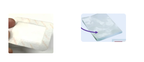
Compare primapore vs. melolin
BOTH have shiny and perforated polyurethane (polyester) which allows passage of blood/exudate away from the wound while remaining non-adherent
Primapore = Peripheral adhesive surrounding thin absorbent layer
Melolin = Thicker absorbent layer and no peripheral adhesive (needs bandage)
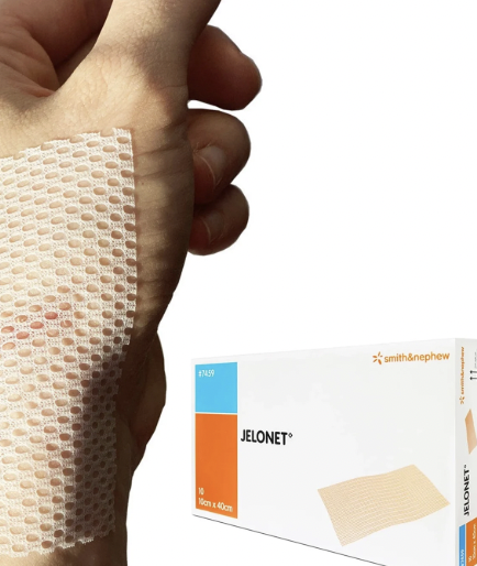
Jelonet (paraffin gauze)
Type of material
Indication/use
2 Disadvantages
Material: Cotton net impregnated with soft paraffin (petroleum)
Indication: Allows exudate to pass through to the secondary absorbent layer
Ideal for skin grafts
Disadvantages:
Difficult to remove hydrophobic paraffin
Excessive paraffin causes wound occlusion → fluid accumulation → inhibits epithelialisation
Vapour-permeable (barrier film)
Example
Type of material
Function
Indication
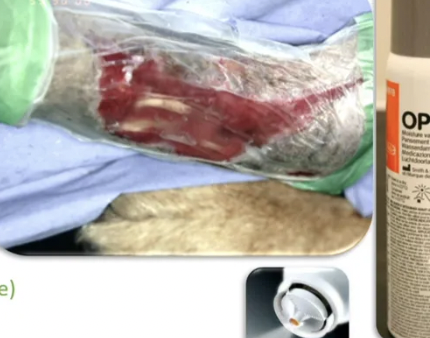
Example: Opsite flexigrid and opsite spray
Material: Thin adhesive film often used to stick primapore on
Function: Semi-permeable (aids in moist wound healing BUT impermeable to contaminants and bacteria)
Indication: Protective barrier against irrigation/urine/discharge
Clean wounds with NO exudate
Absorbent dressings
3 Examples
Indication
Examples:
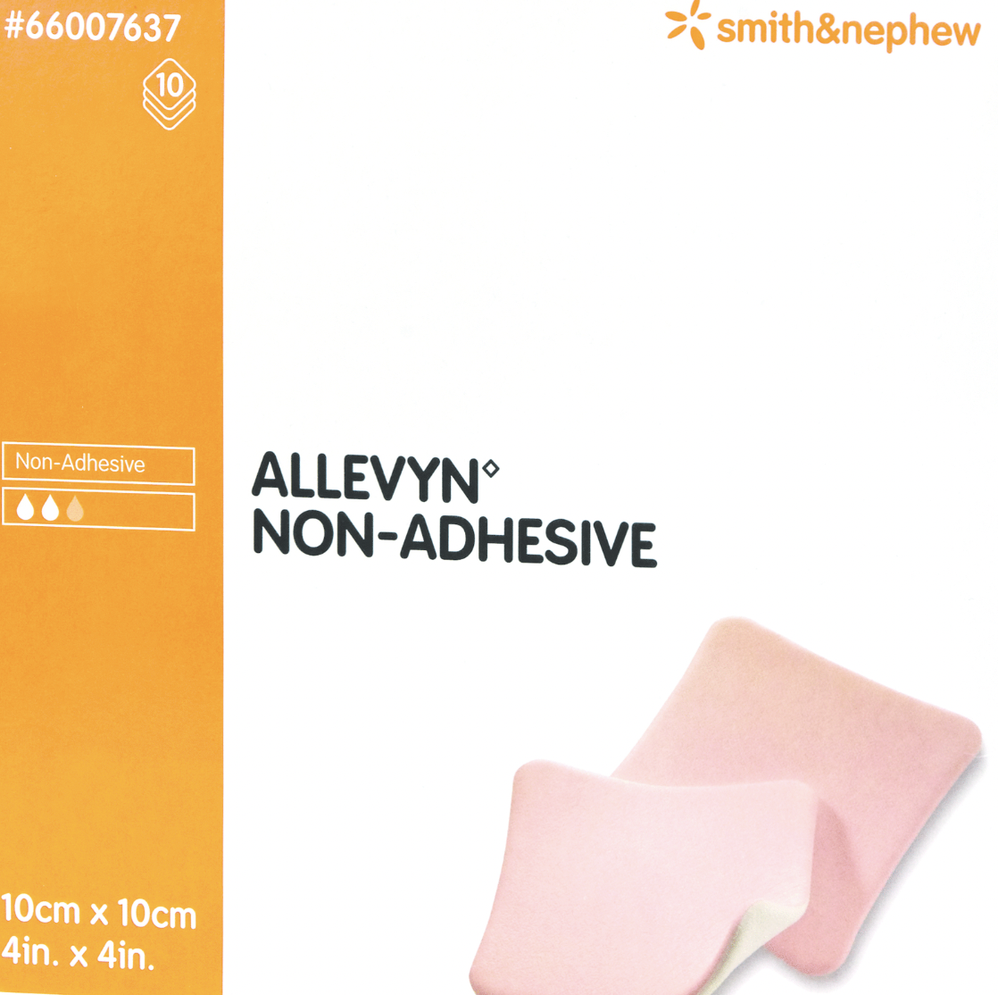
Allevyn
Nappies
Cotton wool
Indication: Highly exudative wounds which bandages cannot be changed
Allevyn (absorbent dressing)
Type of material
Use
Indication
3 Advantages
Material: TWO non-adherent polyurethane layers between a highly absorbent hydrophilic core
Use: White side down and pink side up; does NOT require secondary layer
Indication: Clean wound with marked exudate
Advantages:
Highly absorbent (10x own weight)
Moist (not wet) wound environment to promote granulation tissue formation
Outer layer prevents strike through
Nappies (absorbent dressing)
Type of material
2 Advantages
2 Disadvantages
Indication
Material: Hydrophilic gel sandwich
Advantages:
Cheap
Extremely absorbent
Disadvantages:
NOT sterile
Gel toxic if ingested
Indication: Open peritoneal drainage
Cotton wool (absorbent dressing)
2 Advantages
Disadvantage
Advantages:
Absorbent
Cheap
Disadvantage: May compact down and lose function (wound becomes WET not moist)
4 Types of active dressings
Examples
Structure
Function
Active dressings are uncommon as $$$
HYDROCOLLOIDS
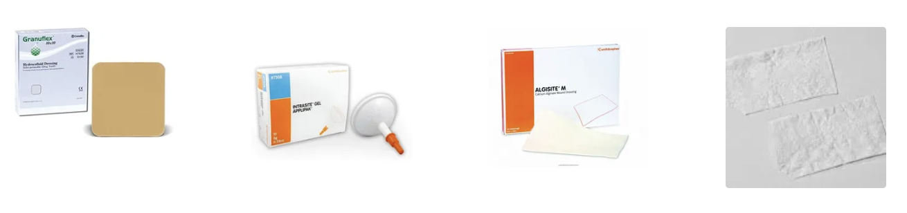
Examples: Granuflex, Tegasorb
Structure: Suspension of polymers eg. gelatin, pectin, carbomethylcellulose
Functions: Promotes moist wound healing and granulation tissue formation
Polymers absorb water in a hydrophilic matrix
Release H2O2 to oxygenate the wound
Dressing swells which applies pressure
NOT for infected wounds
HYDROGELS
Example: Intrasite
Structure: Inert cross-linked hydrophilic co-polymer (90 - 95% water)
Functions:
Rehydrates tissue for moist wound healing
Facilitates autolytic debridement
Analgesic (in fridge)
For open clean or infected wounds (eg. deep puncture)
ALGINATES
Example: AlgiSite M
Structure: Seaweed derivative
Functions:
Absorb exudate (moist wound healing)
Release Ca2+ to promote haemostasis
COLLAGENS
Examples: Collamend, Vet biosist, lyophilised collagen sheets
Disadvantage: No clinical trials show efficacy and only 1 human product (so why would it work?)
Function: Promote granulation and epithelialisation?
3 Functions of the secondary (intermediate) layer of a bandage
Bulky layer to:
Absorb and store blood, serum, exudates and necrotic debris
Wick bacteria from wound
Protect and splint lesion (padding)
Care not to apply excessive pressure → Reduced absorptive capacity
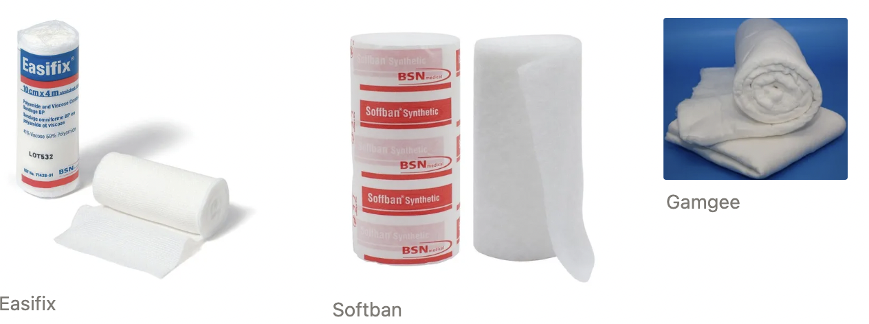
List 3 examples of secondary bandaging material (+ functions)
Softban = Non-absorbent cast padding material
Gamgee = 100% cotton gauze cover sheet keeping cotton fibres out of wound site
Easifix gauze = Conforming bandage to fix the primary layer dressing
4 Functions of the tertiary (outer) layer of a bandage
Holds the other layers in place
Stops minor haemorrhage by providing pressure
Prevents tissue oedema
Obliterates dead space
List 2 types of tertiary bandage material
Examples
Function
Indication
Method of application
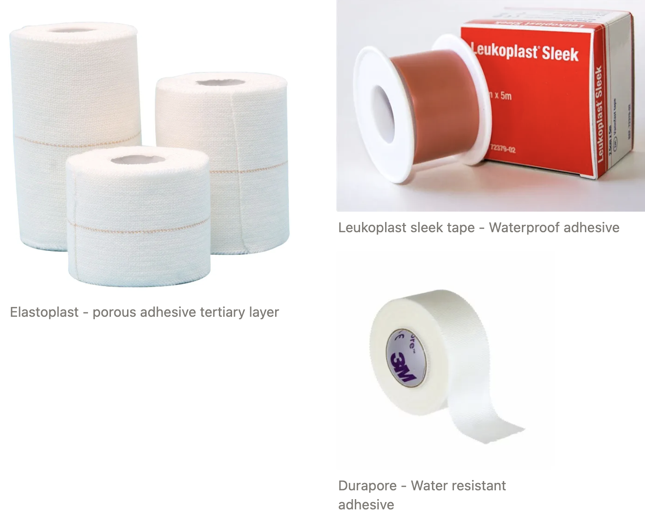
POROUS
Examples: Elastoplast (adhesive) and vetrap (self-adhesive)
Function: Evaporate fluid to create drier wound environment
Indication: Wet wounds and pressure application for minor haemorrhage, dead space obliteration and reduce passive oedema
Application:
Change if strike through occurs
Do NOT apply too tight
Expose 3rd and 4th digits to monitor for circulation and sensation
WATERPROOF
Examples: Sleek tape ad durapore
Function: Occlusive bandage to protect wound from environment
Indication: Dry wounds or at the distal aspect of the bandage which contacts the ground
Application: Do NOT use for effusive wounds → tissue maceration and bacterial infection
List 4 GOOD topical agents (+ why)
Silver sulfadiazine = Antibacterial Ag
Aloe vera = Contains acemannan which activates macrophages and stabilises fibroblasts → More rapid granulation
Maggots (Lucilia sericata) = Sterile maggots consume dead tissue to act as wound debriding agent (stage I larvae ONLY)
Sugar/honey = Antibacterial, debridement and anti-inflammatory
List 5 BAD topical agents (+ why)
Antiseptics (eg. chlorhexidine, iodine, H2O2) = Toxic and slow healing
Antibiotics = Diluted, toxic, bacterial resistances, poor contact time and penetration (systemic superior)
Cleansing agents (eg. Aserbine, Dermisol) = Low pH irritant
Poultice (eg. animalintex = boric acid) = Causes inflammation (banned in human medicine)
Zinc bacitracin = No evidence of improved healing (except for Zn deficiency)
3 Advantages and 2 disadvantages of Manuka honey
Advantages:
Antibacterial properties
Draws out lymphatic fluid into wound (inoculates with WBC and nutrients for healing)
Manages multi-drug resistant bacteria
Disadvantages:
Tasty for animal to eat
No evidence in vet medicine for superior healing
Negative pressure wound therapy (NPWT)
Mechanism
8 Functions
2 Advantages
4 Disadvantages
Mechanism: Large wounds packed with foam and a non-permeable film placed over op so vacuum can suck wound
Functions:
Decreases wound exudate, oedema, wound volume, bacterial contamination
Increases wound stimulation, contraction, granulation tissue formation, perfusion
Advantages: Only need to change bandage q3d
Less GA/sedation (animal can eat and recover from trauma)
Cheaper (no daily dressing changes)
Disadvantages:
NOT for infected wounds
Not over large exposed blood vessels
Risk of loss of vacuum
Gauze incorporated into healing wound
3 Functions of wound drainage
Obliterate dead space/anticipated fluid
→ Accumulation of serum and blood → increases infection risk
→ No skin adherence to underlying tissue (blood supply)
Channel harmful material away from wound/body cavity(eg. fluid in chest)
Flush solution/antiseptic/antibiotic through site after primary closure
→ Faster healing time
8 Indications of a drain
Impossible to completely debride wound as vital structures are contaminated (eg. tendons, bones)
Foreign material must be removed
Massive contaminated of wound is inevitable (eg. perianal)
Viability of wound is questionable (eg. bite wounds)
Dead space needs to be obliterated
Incomplete drainage (eg. abscess cavity that seals over)
Anticipated leakage (intestinal resection and anastomosis)
Monitor leaking into body cavity (eg. anticipated leakage from insecure closure of a hollow viscus eg. bladder or intestine)
Treatment of pneumothorax, recreate negative pressure in thoracic cavity after surgical intervention
Used for contaminated/dirty wounds (INCREASES likelihood of infection in clean or clean-contaminated surgery as it acts as a conduit for bacteria to enter)
7 Principles of drain placement
Place aseptically
Pass drain through stab wounds SEPARATE from incision line (never through incision as it can move and create more contamination)
Exit at most ventral/dependent site
ONE exit point (exit site at top will NOT drain and only act as entry point for bacteria)
Exit ports should be large enough for adequate drainage
Suture to skin and cover with sterile dressing ± E-collar
Record where and duration of drain placement
Indication of drain with TWO exit points
Inguinal or axillary area as single exit point acts as one-way valve and can draw in air when animal moves creating SC emphysema
2nd hole allows trapped air to escape
List 3 types of surgical drains
Examples
Advantages
Disadvantages
Indication
Unsutured skin (covered with absorbent bandage)
PASSIVE (open) DRAIN = Drainage depends on gravity and capillary action
Examples: Penrose, Yeates
Advantages:
ALWAYS works
Cheap
Disadvantages:
ALWAYS open = Risk of ascending infection
Approximate measure of fluid volume
Exit must be ventral to the wound
Indication: Contaminated/infected wounds that cannot be sealed
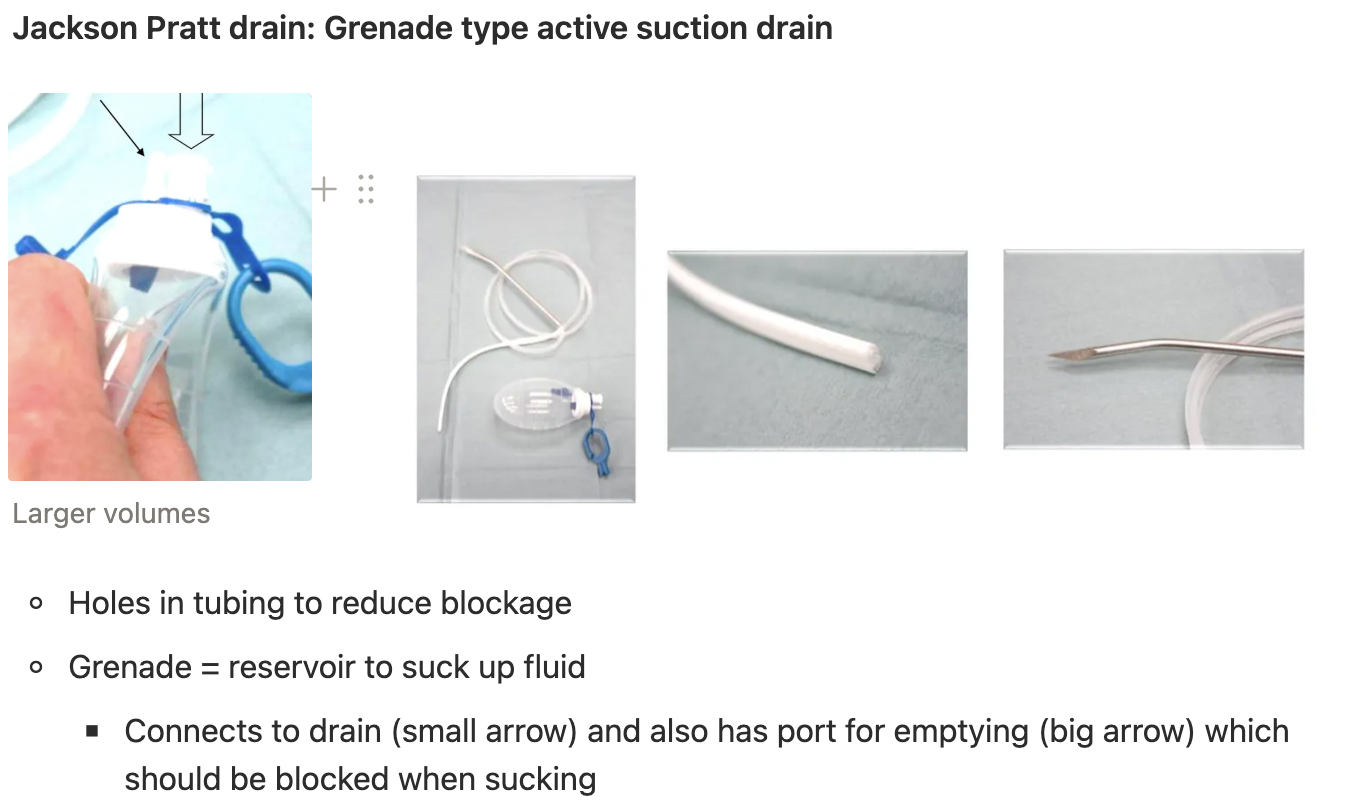
ACTIVE (closed) DRAIN = Actively removes fluid either intermittently OR continuously via a suction apparatus applied to a rigid fenestrated tube to create a negative pressure in the wound
Examples: Jackson Pratt, Uno, mini-vac
Advantages:
Exit at ANY point (can work against gravity)
Closed wound → Decreased risk of ascending infection
Accurate assessment of fluid produced
Disadvantages:
$$$
Failed drain due to premature loss of vacuum or obstructed tubing
Indication: Clean wounds which must be completely sealed
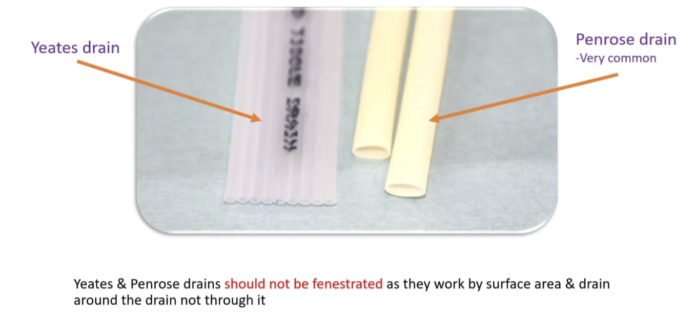
Compare the Penrose vs. Yeates passive drains
Penrose = Soft, thin and pliable latex rubber tubing
Most drainage occurs EXTRALUMINALLY
Efficient for draining soft tissue wounds
Yeates = Soft PCV, but more rigid than Penrose
Drainage occurs INTRA- and EXTRALUMINALLY
Useful for large volumes of fluid (increased SA available)
Increase flow rate by making single slit down tube (fenestration decreases SA and therefore, decreases drainage flow)
Maximum pressure of active drain
80mmHg
4 Indications of drain removal
Bacteria no longer present and inflammation reduced
Fluid level constant low level (<0.2mL fluid/kg/d)
Never get dry wound from drain presence (elicits an immune response)
72hr for abscess cavities
Remove when no longer beneficial
5 Complications fo drain placement
Nosocomial ascending infection (AB resistant bacteria)
Foreign body reaction to drain reduces # of bacteria required for infection
Esp. with obstruction
Can flush, but introduces contamination (not recommended)
Drains made from rigid materials may encroach upon adjacent structures, resulting in adhesions, ulceration and inflammation
A drain may slip into the wound and become lost
Drains may tempt the surgeon to close a wound that is better left open
4 Functions of sutures
Wound closure
Ligation
Attach tubes
Stay sutures
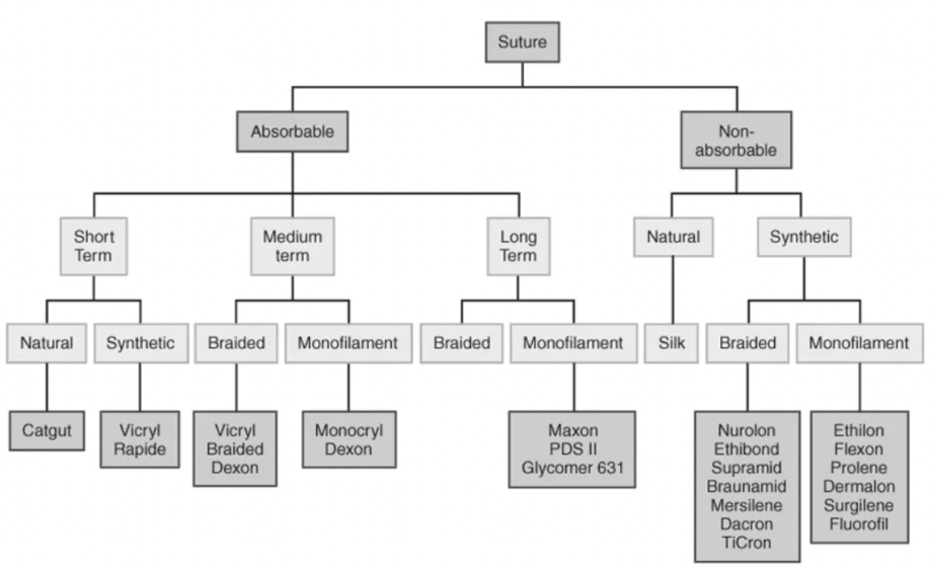
List 3 ways to classify suture materials (+ types)
Origin (natural OR synthetic)
Persistence (absorbable OR non-absorbable)
Structure (multifilament OR monofilament)
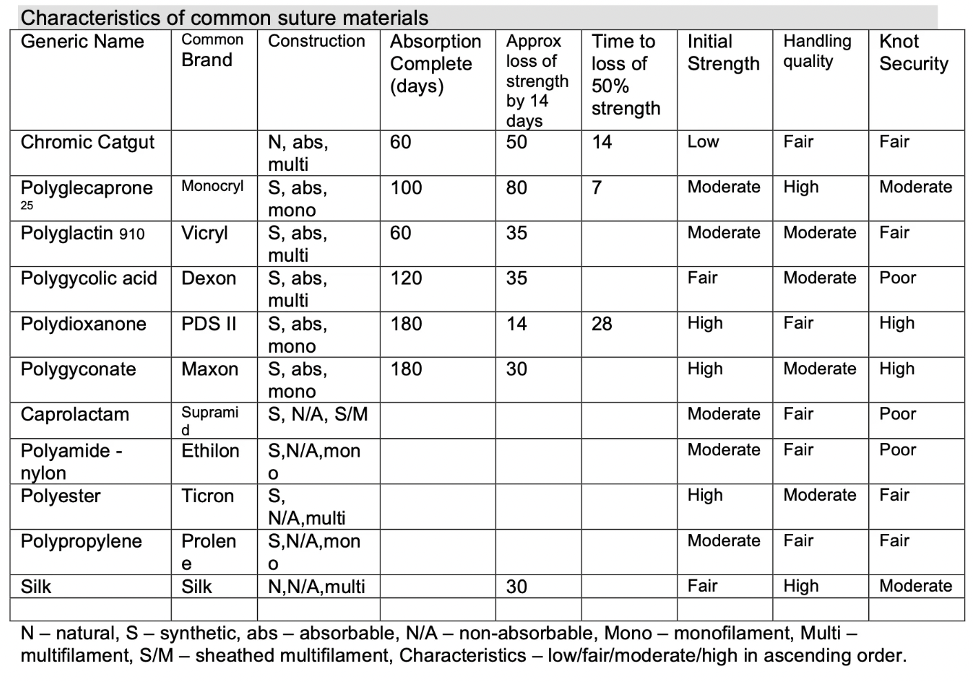
List 11 features of the ideal suture material
Maintain adequate tensile strength
Stimulate minimal inflammation
Discourage bacterial growth
Non-capillary but absorbable
Non-electrolytic (stainless steel 316L staples #1 as they do not rust or cause electrolysis)
Non-allergenic
Non-carcinogenic
Good handling properties
Good knot security
Inexpensive and readily available
Easily sterilised
Compare absorbable (+ types) vs. non-absorbable suture
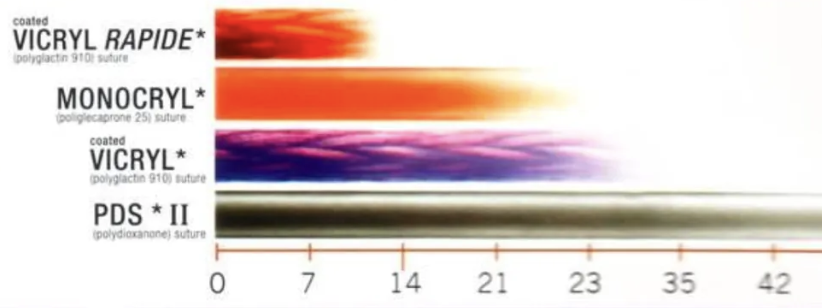
Absorbable = Temporary wound support <60d → Degrades predictably
<21d = Short duration
>21d = Long duration
Non-absorbable = Retains original strength for >60d
Encapsulated within body by fibrous capsule
Should NOT be used with infection (nidus for fistulation)
Compare natural vs. synthetic suture
Natural = Absorbed by phagocytosis → Granulation tissue and scarring
Provokes inflammation ± immune response
Variable resorption
Synthetic = Man-made polymers absorbed by hydrolysis (polymer + water → monomer)
Minimal inflammation
Predictable absorption
What does “PLUS” for suture material mean?
Antibacterial triclosan coating (eg. monocryl PLUS)
Compare advantages and disadvantages of multifilament vs. monofilament suture (5)
Multifilament
Nidus for infection (decrease # of bacteria required for infection)
Capillary = Wicks fluid (avoid for skin and inner luminal surfaces of hollow organs as suture can contaminate deeper tissue)
Easier to handle
Superior knot security
More tissue drag
Monofilament
Less likely to collect bacteria when dragger across the skin
Non-capillary = Does NOT absorb fluid (will not spread contamination into deeper tissues)
Difficult to handle due to memory (maintains new shape when stressed)
Poorer knot security
Less tissue drag (coatings reduce drag BUT also reduce knot holding)
What suture material is best for a heavily contaminated and infected wound?
Monofilament, non-absorbable
Catgut suture
Classification and absorption
Material
2 Advantages
4 Disadvantages
Indication
Classification: Natural, absorbable (short-duration), multifilament
Lost 50% tensile strength @ 14d (complete absorption by 60d)
Material: Collagen fibres derived from submucosa of sheep or subserosa of a cow
Chromic = Increased persistence and reduced tissue reactivity
Advantages:
Handles well compared to other multifilaments
Cheap
Disadvantages: Multifilament, natural
Unpredictable breakdown
NOT for infection
Inflammatory reaction
Capillary
Indication: Cheap closure of oral mucosa
Polyglecaprone
Trade name
Classification and absorption
3 Advantages
Disadvantage
Indications
Trade Name: Monocryl
Classification: Absorbable (short-duration), synthetic, monofilament
Lost 80% tensile strength by 14d (complete absorption by day 100d)
Advantages:
Smooth and atraumatic
Highest initial tensile strength of all absorbable sutures
Easier to handle than other monofilaments
Disadvantage: Expensive
Indications:
Soft tissue (eg. bladder, skin)
Infected tissue
Ligatures
NOT linea alba
Polydioxanone
Trade name
Classification and absorption
Advantage
Disadvantage
Indications
Trade Name: PDS II/PDO
Classification: Absorbable (long-duration), synthetic, monofilament
Lost 14% tensile strength by 14d (complete absorption by 180d)
Advantage: Longest retention of tensile strength of all absorbable sutures
Disadvantage: Cost
Indications:
Linea alba, fascia
Contaminated wounds
Ligatures
NOT intradermals, tendons, SC, bladder
Polyglactin
Trade name
Classification and absorption
Advantage
Disadvantage
Indications
Trade Name: Vicryl
Classification: Absorbable (medium-duration), synthetic, multifilament
Lost 35% tensile strength at 14d (complete absorption by 60d)
Advantages: Easier handling and secure knots
Disadvantages: NOT for skin or bladder (nidus for urinary calculi)
Indications:
SC
General tissue/muscle approximation
Pedicle ligation
Caprolactam
Trade name
Classification and absorption
2 Advantages
3 Disadvantages
Indications
Trade Name: Supramid
Classification: Non-absorbable, synthetic, multifilament
Advantages:
Cheap (available in reels)
Good knot tying
Disadvantages:
NEVER for internal structures as autoclaving does NOT make sterile
NEVER bury suture
Outer proteinaceous layer cracks and causes foreign body reaction
Indication: Skin closure with eyed needle
Polyamide/Nylon
Trade name
Classification and absorption
2 Advantages
3 Disadvantages
Indications
Classification: Non-absorbable, synthetic, monofilament
Advantages:
High tensile strength
Cheap
Disadvantages: Memory and poor knot security
Indications:
Interrupted skin closure (remove after 7 - 10 days)
Fine suture for ophthalmology and nerve repairs
Polypropylene
Trade name
Classification and absorption
2 Advantages
3 Disadvantages
Indications
Trade Name: Prolene
Classification: Non-absorbable, synthetic, monofilament
NEVER fully absorbed by body
Advantage: Permanent
Disadvantage: Most difficult to handle
Indications:
Cardiac surgery
Vascular anastomoses
Tendons
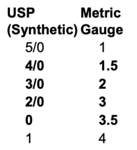
Describe the 2 suture sizing systems
USP
0 - 6 INCREASES
Added zeros (eg. 5/0 = 00000) DECREASES
Metric
Ascending scale in 1/10mm and linear
3M = 0.3mm