T3: Voice of the Genome
1/180
There's no tags or description
Looks like no tags are added yet.
Name | Mastery | Learn | Test | Matching | Spaced |
|---|
No study sessions yet.
181 Terms
How are cells organised in multicellular organisms?
Cells into tissues, tissues into organs and organs into systems, each specialised for a particular functions
What are eukaryotic cells?
Found in humans, contain a nucleus and membrane bound organelles incl. discrete membrane bound organelles e.g nuclei, mitochondria and chloroplasts. bigger than prokaryotic cells with diameters of 20 micrometers or more
What is the cell membrane?
External boundary of the cell, confines cell contents; regulates entry and exit of materials
What are lysosomes?
Throughout cytoplasm, vesicles, containing digestive enzymes, bound by a single membrane. Function is to digest invading cells or to break down old organelles
What is the mitochondria?
Scattered throughout the cell.
Usually oval shaped and bound by a double membrane called the envelope.
The inner membrane is folded to form projections called cristae, with a fluid matrix on the inside containing the enzymes needed for cellular respiration.
Controls the release of energy from foods; forms ATP
What is the ultrastructure?
A more detailed structure of a cell, which can be observed using a microscope
What is the Golgi Apparatus?
Within cytoplasm. A series of fluid filled flattened and curved sacs with vesicles surrounding the edges. Modifies and packages proteins for exocytosis (after budding off from reR), and also produces lysosomes.
What are centrioles?
Two rod shaped bodies near the nucleus. Hollow cylinders containing a ring of microtubules arranged at right angles to each other. Involved in cell division. They ‘spin’ the mitotic spindle
What is the smooth ER?
Within cytoplasm. A system of membrane bound sacs. Synthesises lipids and processes them
What are nuclear pores?
Within nuclear membrane. To allow mRNA to move into the cytoplasm
What is the rough Endoplasmic Reticulum?
Flattened membranous sacs located near the nucleus enclosed by a membrane with ribosomes on the surface. Folds and processes proteins made on the ribosomes. Synthesises proteins
What are ribosomes?
Attatched to membranes or scattered in the cytoplasm. Synthesises proteins
What are cillia?
Hair-like structures on the surface of animal cells that act collectively to move substances across the cell surface in one direction
What are microtubules?
Throughout cytoplasm; part of the cytoskeleton. Has Contractile protein (actin); moves cell or cell parts; core of microvilli
What is the nucleuolus?
Within the nucleus. Involved in the synthesis of ribosomes
What are 80s ribosomes?
Composed of 2 subunits. The site of protein synthesis
What is the nucleus?
Large body situated towards the centre of the cell.
Contains genetic info.
Surrounded by a double membrane called the envelope, which contains pores enabling molecules to enter and leave.
Contains DNA wrapped around histone proteins in a complex called chromatin, and a nucleolus, which is the site of ribosome production
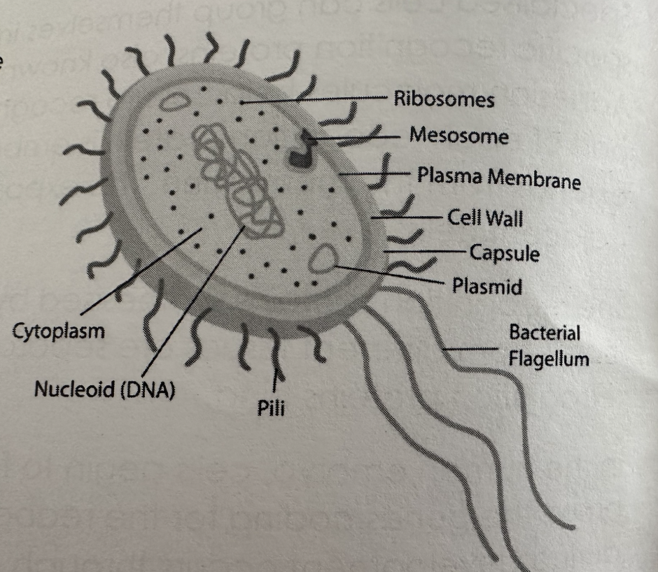
What are prokaryotic cells?
Incl bacteria and cyanobacteria. Don’t have nuclei or other membrane bound organelles. DNA is not associated with any proteins and lies free in the cytoplasm. Cell wall is always present
What are the 3 various types of prokaryotes?
Photosynthetic, disease causing and decomposers
What is the cell wall?
Cells rigid outer covering made of peptidoglycan. Provides cell with strength and support
What is the slime capsule?
Protective slimey layer which helps the cell to retain moisture and adhere to surfaces
What are plasmids?
Circular pieces of DNA
What is the flagellum?
A tail like structure, which rotates to move the cell
What are Pilli?
Hair like structures which attatch to other bacterial cells - allowing the exchange of plasmids
What are 70s ribosomes in prokaryotic cells?
Composed of 2 subunits and site of protein synthesis
What are mesosomes?
Infoldings of the inner membrane. Function is debated with many scientists believing they are just artefacts from the preparation process for microscopy, while others believe they contain enzymes required for respiration
How do specialised cells group themselves into clusters?
Have specific recognition proteins (adhesion molecules) on their cell surface membrane (CSM) .
Adhesion molecules helps cells to recognise other cells like themselves and stick to them.
Small part of each recognition protein is embedded in the CSM - larger part extends from membrane and exposed section binds to complimentary proteins on adjacent cells
What does recognition proteins do?
Recog proteins synthesised by a cell determine which cells it can and cannot attatch to. If cells from different tissues are separated and then mixed, they reform into the tissues as the recog proteins bind
How do cells begin to form tissues as they start their specialised functions?
The genes coding for the recognition proteins are switched on.
How does reorganisation of tissues during development occur?
Through changes in the type of recognition proteins produced by cells
Define what a cell is
Basic building blocks. Specialised for a particular function e.g muscle and epithelial cells
Define what tissues are
A group of specialised cells working together to carry out one function e.g muscle cells combining to form muscle tissue
What are organs?
A group of tissues working together to carry out one function e.g muscle, nerve and epithelium work together in the heart
What are organ systems?
Group of organs working together to carry out a particular function e.g circulatory system
How do you calc magnification?
Length of object in photograph / real length of object.
Remember I AM
What is a light microscope?
Uses beams of light, focused by lenses to achieve a magnified image.
Magnification is x1500.
Resolution is 200nm but objects closer together than the resolution of a microscope will appear to be one object.
Wide range of specimens can be viewed
What is a transmission electron microscope?
Magnification can be more than x1 million. 0.2nm resolution and produces 2D images
What is a scanning electron microscope?
Resolution is 0.2nm. Magnification is usually less than x500,000 and produces 3D images
What is an electron microscope?
Focused by lenses to achieve a magnified image. Use electron beams to achieve a magnified image
How do you stain samples of biological materials?
Add a stain, which is a chemical that binds to chemicals on or in the specimen, so details can be seen e.g acetic orcein stains DNA red
What is an eyepiece graticule and how is it used?
Piece of equipment that allows you to measure the size of a specimen.
It’s a glass disc placed on an eyepiece lens with a smaller ruler etched to it.
Scale of eyepiece represents diff lengths at diff magnifications.
Image of specimen looks bigger at higher magnifications, but actual specimen has not increased in size.
Eyepiece has to be calibrated.
What is the protein transport sequence more detailed vers?
1) Transcription of DNA to mRNA
2) mRNA leaves nucleus (through nuclear envelope)
3) Protein made on ribosomes enter rough ER
4) Protein moves through the ER assuming three dimensional shape en route
5) Vesicles pinched off the rough ER contain the protein
6) Vesicles from rough ER fuse to form the flattened sacs of the Golgi Apparatus
7)Proteins are modified within the Golgi Apparatus e.g carbs are added to form a glycoprotein
8) After the GA packages proteins Vesicles are pinched off the Golgi Apparatus to be transported and contain the modified protein
9) Vesicles fuse with cell surface membrane, releasing protein such as extracellular enzymes by exocytosis
Simplified protein transport sequence?
1- Proteins are prod on ribosomes
2- Proteins which are prod on the ribosomes on the surface on the rER are folded and processed in the rER
3- The proteins are then transported from the rER to the Golgi Apparatus (GA) in vesicles
4-They are then modified in the GA
5- GA packages proteins into vesicles to be transported around the cells to where they’re required . Some of the proteins, such as extracellular enzymes, leave the cell by exocytosis
What are gametes?
Sex cells that are specialised for their function to increase chances of fertilisation and successful developments of an embryo.
Females = ovum . Males = sperm
Fuse during fertilisation to form a zygote → they form during meiosis and only have one copy of each chromosome, so they are haploid cells
What is contained in the ovum?
-Zona pellucida
-Haploid nucleus
-Cortical granules
-Follicle cells
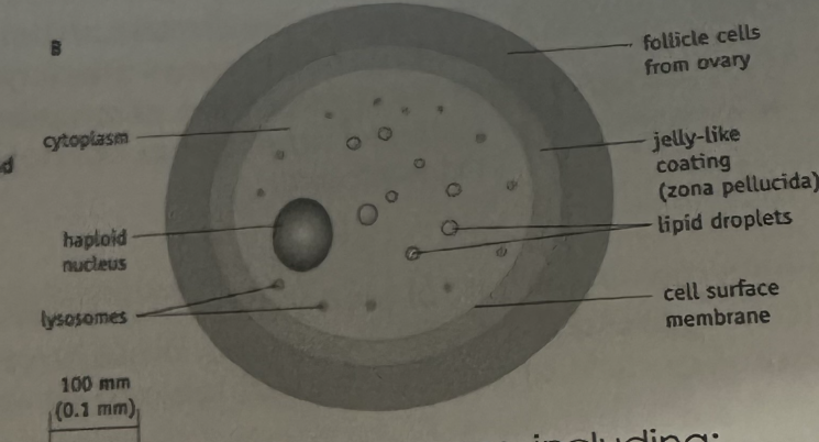
What is in the zona pellucida?
Protective coating,which the sperm have to penetrate in order for fertilisation to occur, hardens after the sperm has entered. Main purpose is to prevent polyspermy
What is the haploid nucleus in the ovum?
The ovum has a haploid nucleus that has a full set of chromosomes that is restored at fertilisation
What are cortical granules?
Release substances which cause the zona pellucida to harden
What are follicle cells?
Form a protective coating the egg
What is found within the spermatoza?
-Many mitochondria
-Acrosomes
-A long tail
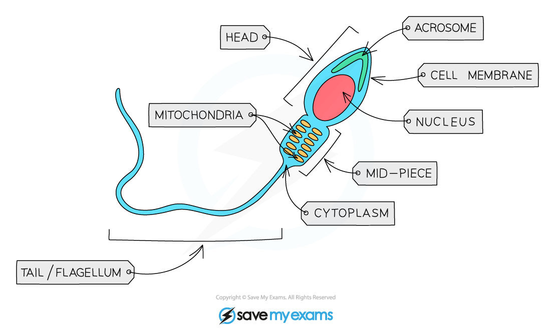
Why do sperm cells contain many mitochondria?
Provide energy for the flagellum which enables the cell to move. much smaller than ovum and mobile (can move) The long tail is powered by the energy released by mitochondria.
What are acrosomes?
(Type of lysosome involved in the breakdown of unwanted structures in cell). The acrosome contains digestive enzymes which break down the zona pellucida and allows sperm to penetrate the egg
How does sperm enter the ovum?
-Enter the vagina during intercourse + swim through the uterus, their passage assisted by muscular contractions of the uterus walls.
-If intercourse takes place at about the time of ovulation, sperm may meet the ovum in the oviduct.
-Sperm are attracted to the ovum by the chemicals released from it. To penetrate, the acrosome will release its digestive enzymes
What reactions take place during fertilisation?
Acrosome - the release of hydrolytic enzymes which softens the zona pellucida
Cortical- Hardening of the jelly coat post fertilisation to prevent potential polyspermy
What happens during the acrosome reaction?
1) When the sperm reaches the egg, the acrosome reaction allows the sperm to break through the zona pellucida. When the sperm makes contact with the zona pellucida, chemicals are released from the follicle cells
2) The acrosome swells and fuses with the sperm cell membrane
3) Digestive enzymes are released, and they digest the follicle cells and zona pellucida. Acrosin→ protease enzyme in the acrosome which hydrolyses/digests the glycoprotein layers of the zona pellucida + allows sperm to enter
4)The membrane of the egg and sperm then fuse and the sperm nucleus enters the egg
What happens during the cortical reaction?
1) Sperm cell fuses with egg cell membrane
2) Cortical granules from the egg, fuse with the egg cell membrane, release enzymes via exocytosis
3) These enzymes released into the jelly layer thicken and harden the glycoprotein matrix of the zona pellucida
4)This prevents other sperm from being able to penetrate the egg (polyspermy) ensuring the zygote formed is diploid
What is polyspermy?
When multiple sperm cells enter the egg it is usually pathological and results in an unviable zygote. Some species e.g pigs seem affected, lysosomes may break down extra genome
Summarise the whole process of fertilisation
1) The sperm head meets the protective jelly layer around the egg cell called the zona pellucida and the acrosome reaction occurs- enzymes digest the zona pellucida
2) The sperm head fuses with the cell membrane of the egg cell thus allowing the sperm nucleus to enter the egg cell
3) The cortical reaction occurs which causes the zona pellucida to harden and prevents polyspermy
4) The nuclei fuse and a full set of chromosomes is restored thus forming a diploid zygote
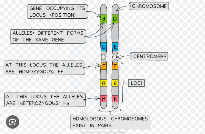
What is the locus of a gene?
Its location on a chromosome/ the position of a gene or chromosome
What does autosomally linked alleles mean?
Alleles on the same chromosomes and are inherited to a greater or lesser extent as if they were the same gene.
How do you know how closely linked genes are on a chromosome?
How close the loci of the genes are on a chromosome, the closer = more linked, this is because they are far less likely to be separated during recombination during meiosis
What does it mean if genes are sex-linked?
-Some genes are only present on the sex chromosome and not on the other
-Inheritance of these genes are dependent on the sex of the individual
-Mostly occur on the longer X chromosome, some genetic disorders are sex linked and therefore much common in men.
Why are sex linked diseases much more common in men?
Because the Y chromosome is smaller than the X chromosome, so men don’t have another copy of the allele for a particular characteristic. As a result, they only need one copy for the allele to be expressed e.g haemophillia and red-green colour blindness
What is the cell cycle? (mitosis)
-A process in which cells divide to produce 2 genetically identical daughter cells for growth, repair and asexual reproduction
-Since cells produced by mitosis are genetically identical, it doesn’t give rise to genetic variation
What are the 3 stages of the cell cycle?
-Interphase = cells duplicating their materical and synthesising proteins to prepare to divide
-Mitosis - PMAT
-Cytokinesis = Cytoplasm divides, producing 2 daughter cells
What are the 3 phases part of interphase?
G1= Cell grows and DNA replicates
S= Chromosomes are replicated, and begin to condense to form chromatin
G2=Cell prepares to divide replicating organelles for a full set in each new cell
=G1 + S + G2
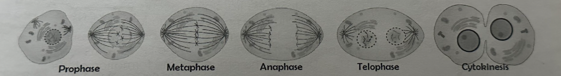
What are the stages in mitosis (PMAT)?
-Prophase, (prometaphase), metaphase, anaphase and telophase
-During these phases the nuclear envelope disappears, the mitotic spindle forms, chromosomes condense and are lined up at the metaphase plate, and separated by being pushed to each side of the cell
What happens during the final stages of the cell cycle?
Cytokinesis occurs where the cytoplasmic contents are separated into two daughter cells
What happens when the cell increases in mass and size during interphase?
-Cell increases in mass and size and carries out normal cellular functions e.g protein synthesis etc
-At some point during the G1 phase a signal is received telling the cell to divide again. DNA in the nucleus replicates→ resulting in each chromosome (consisting of 2 identical sister chromatids)
What happens after a signal is received during the G1 phase?
S phase, which is the synthesis of DNA, a relatively short phase
What is the gap between the previous cell division and the S phase called?
G1 phase - g stands for gap. Cells make the RNA, enzymes and other proteins required for growth during the G1 phase
When does the G2 phase occur?
-Between the S phase and the next cell division.
-During this phase the cell continues to grow and new DNA that has been synthesised is checked and errors are usually repaired.
-Other preparations for cell division are made e.g prod of tubulin protein, which is used to make microtubules for the mitotic spindle.
What happens after mitosis?
Process of nuclear division
2 identical daughter cells are produced that are identical to parent cell nucleus as they have the same number of chromosomes as the parent cell.
Most organisms contain many chromosomes in the nuclei of their cells
What happens during prophase?
-Chromosomes condense into visible structures that are visible when stained
-Chromosomes consist of 2 identical chromatids called sister chromatids (each containing one DNA molecule) that are joined together at the centromere
-The two centrosomes (replicated in the G2 phase just before prophase) move towards opposite poles (opposite ends of the nucleus)
-Spindle fibres (protein microtubules) begin to emerge from the centrosomes (which consist of two centrioles in animal cells)
-The nuclear envelope (nuclear membrane) breaks down into small vesicles
What happens during metaphase?
-Chromosomes reach opposite poles
-Spindle fibres (protein microtubules) continue to extend from the centrosomes
-Chromosomes line up along the equator of the spindle (metaphase plate) so they are equidistant to the two centrosome poles
-Spindle fibres reach the chromosomes and attatch to the centromeres
-Each sister chromatid is attatched to a spindle fibre originating from opposite poles
What happens during anaphase?
-Sister chromatids separate at the centromere (the centromere divides in two)
-Spindle fibres begin to shorten
-The separated sister chromatids (now called chromosomes) are pulled to opposite poles by the spindle fibres
What happens during telophase?
-Chromosomes arrive at opposite poles and begin to decondense (uncoil)
-Nuclear envelopes begin to reform around each set of chromosomes
-Spindle fibres break down
What is cytokinesis?
-Follows mitosis.
-Once the nucleus has divided into 2 genetically identical nuclei, the whole cell divides and one nucleus moves into each cell to create 2 genetically identical daughter cells.
-In animal cells, cytokinesis involves constriction of the cytoplasm between the 2 nuclei and in plant cells a new cell wall is formed
What are the 3 main reasons for mitosis?
-Growth of multicellular organisms
-Replacement of cells and repair of tissues
-Asexual reproduction
Why is mitosis important for the growth of multicellular organisms?
-The two daughter cells produced are genetically identical to one another (clones) and have the same number of chromosomes as the parent cell
-Enables unicellular zygotes(as the zygote divides by mitosis) to grow into multicellular organisms
-Growth may occur across the whole body of the organism or is confined to certain regions, eg meristems (growing points) of plants
Why is the replacement of cells and repair of tissues important?
-Damaged tissues can be repaired by mitosis followed by cell division
-As cells are constantly dying they need to be continually replaced by genetically identical cells
-In humans e.g cell replacement occurs particularly rapidly in the skin and the lining of the gut
-Some animals can regenerate body parts
Why is asexual reproduction important?
-Production of new individuals of a species by a single parent organism= the offspring are genetically identical to the parent eg for unicellular organisms such as amoeba, cell division results in the reproduction of a genetically identical offspring
-For multicellular organisms, new individuals grow from the parent organism (cell division) and then detatch from the parent in different ways e.g runners in strawberries
What does the phases of mitosis look like?
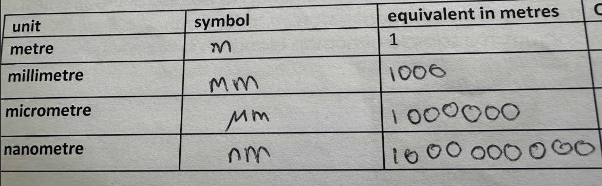
How do you convert the units of measurement?
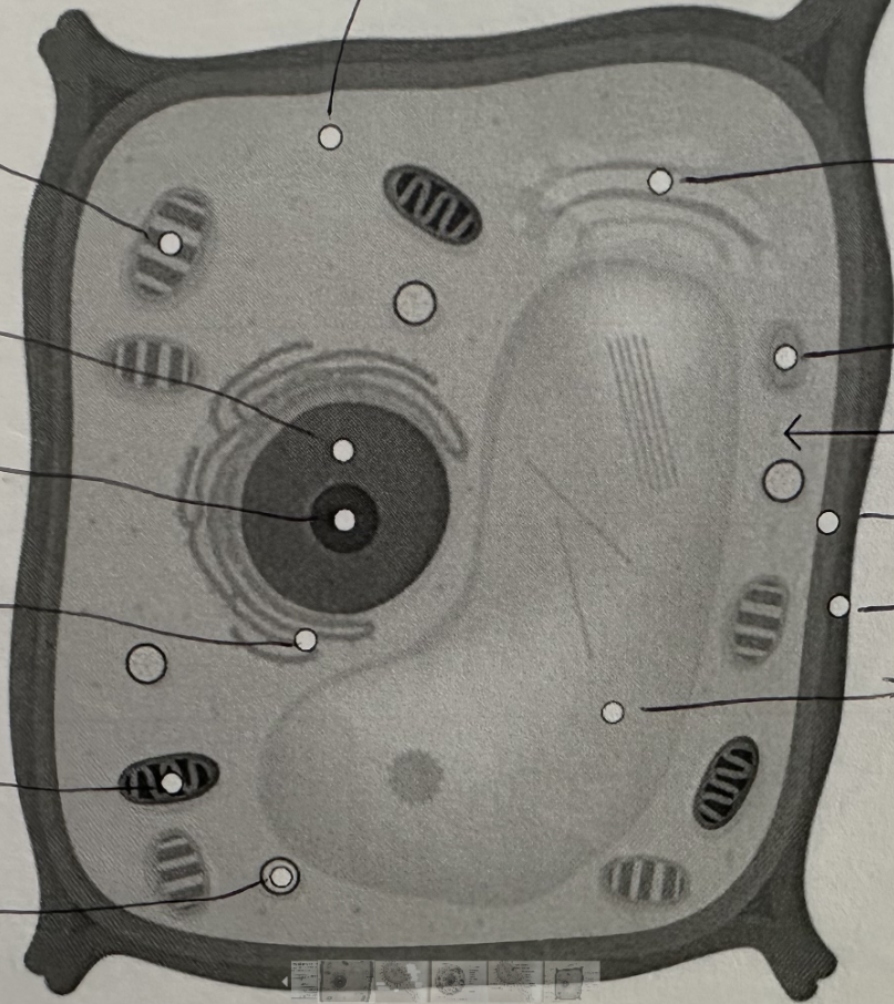
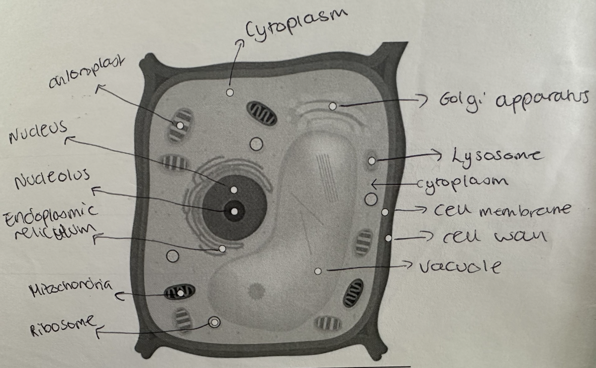
Label this plant cell (click on image to see full thing)
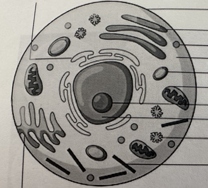
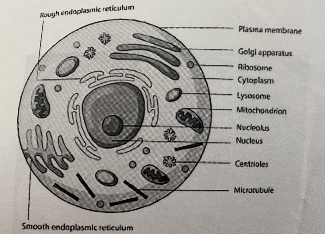
Label this animal cell
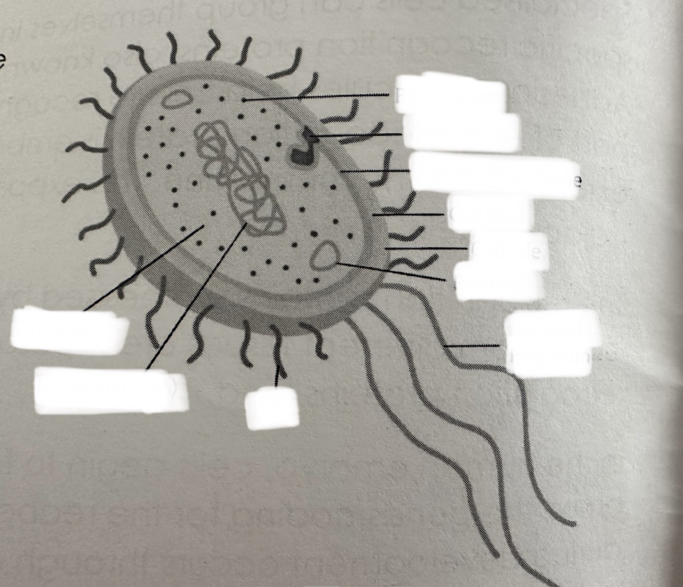
Label the bacterial cell
What is a chromosome?
A long DNA molecule that contains several hundred or even thousands of different genes coding for different proteins.
What is a gene?
A length of DNA that codes for a single polypeptide or protein. They can exist in two or more different forms called alleles.
How have scientists worked out the specific physical locations of genes on different chromosomes?
Through experiments and genetic mapping techniques
Why do genes occupy a specific locus?
Bc the gene for a particular characteristic is always found at the same position on a particular chromosome
Different alleles of a gene have slightly different nucleotide sequences but?
They can still occupy the same position (locus) on the chromosome
What is autosomal linkage?
-Occurs if two or more genes are located on the same autosome (non-sex chromosome).
Why do two or more genes on the same autosome do not assort independently during meiosis?
These genes are linked and they stay together in the original parent combo → these linked genes are passed on to offspring all together through gametes.
Genes are inherited independently ONLY if they are on separate chromosomes, or far apart on the same chromosome
What are the two sex chromosomes and what do females and males have?
-X and Y
-Females have 2 copies of the X chromosomes
-Whereas males have one X and one Y chromosome
Why are males much more likely to show sex linked recessive conditions?
Only have one X chromosome, whereas females have 2 copies of the X chromosome so they are likely to inherit one dominant allele that masks the effect of the recessive allele
How many pairs of autosomal chromosomes do humans have that are not involved in sex determination?
22 and then one pair of sex chromosomes is either X or Y
What is the difference between the X and Y chromosome?
Y = smaller than X therefore fewer loci and thus fewer genes than the X chromosome
X = most sex-linked traits are located on X chromosome with there being no equivalent locus on the Y
-Therefore, females will carry 2 alleles of a sex-linked gene, but males only a single allele, increasing susceptibility to sex-linked diseases
What is haemophillia?
A disease in which the blood clots slowly and there may be a slow+persistent internal bleeding, especially around the joints. It is caused by a change to DNA sequence resulting in a faulty protein being created
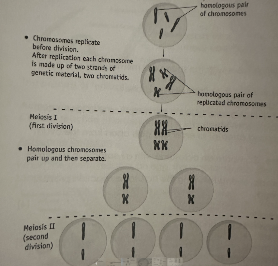
What is meiosis?
-A form of cell division that gives rise to genetic variation
-Produces genetically different cells