Upper GI Staging
 Call Kai
 Learn
Learn Practice Test
 Spaced Repetition
 Match
Match Flashcards
 Knowt Play
1/53

There's no tags or description
Looks like no tags are added yet.
Name | Mastery | Learn | Test | Matching | Spaced | Call with Kai | Chat |
|---|
No analytics yet
Send a link to your students to track their progress
54 Terms
What surgical specimens may be received for esophageal cancer?
Esophagectomy
Esophagogastrectomy
What are the two major histologic types of esophageal carcinoma?
Squamous cell carcinoma (SCC)
Adenocarcinoma
Where along the esophagus is SCC most commonly found according to the lecture?
Most common in the middle to lower third
Can occur throughout the esophagus
Where does esophageal adenocarcinoma usually originate?
Near the esophagogastric junction (EGJ)
What gross patterns can esophageal tumors demonstrate?
They can be:
Exophytic
Circumferential
Obstructive
Ulcerative
Constricting/strictures
Viable tumors may have sharply demarcated borders
What gross appearance may be seen after treatment response in an esophageal tumor?
A white, flattened appearance with a fibrous cut surface may indicate treatment response

What should be submitted/identified in an esophagogastrectomy specimen?
Esophageal margin
Gastric margin
Adventitial margin
Lymph nodes
Uninvolved tissue
What is this showing?

Ulcerative SCC of the esophagus
Verrucoid SCC of the esophagus
What is this showing?

Adenocarcinoma of the esophagus
What determines the T category for esophageal carcinoma?
Primarily the depth of invasion through the esophageal wall and into adjacent structures

What is pTis for esophageal carcinoma?
High-grade dysplasia/carcinoma in situ with malignant cells
Confined to the epithelium by the basement membrane
What defines pT1 esophageal carcinoma?
Tumor invades the:
T1a — Lamina propria or muscularis mucosae
T1b — Submucosa
What defines pT2 esophageal carcinoma?
Invasion into the muscularis propria
What defines pT3 esophageal carcinoma?
Invasion through the muscularis propria into the adventitia
What defines pT4 esophageal carcinoma?
Invasion into adjacent structures
T4a vs T4b
NOT CONSIDERED DISTANT METS
What is pT4a for esophageal carcinoma?
A resectable tumor invading structures such as:
Pleura
Pericardium
Azygos vein
Diaphragm
Peritoneum
What is pT4b for esophageal carcinoma?
Tumor invasion into other adjacent structures such as the:
Aorta
Vertebral body
Airway
What is pN1 for esophageal carcinoma?
Metastasis in 1–2 regional lymph nodes
What is pN2 for esophageal carcinoma?
Metastasis in 3–6 regional lymph nodes
What is pN3 for esophageal carcinoma?
Metastasis in 7 or more regional lymph nodes
According to the lecture, how many lymph nodes should ideally be examined based on pT category for esophageal carcinoma?
pT1 — 10 nodes
pT2 — 20 nodes
pT3/pT4 — 30 nodes
What sites are identified as distant metastatic sites for esophageal carcinoma?
Nonregional lymph nodes
Organs
Liver
Lung
Adrenal glands
What is pM1 esophageal carcinoma?
Distant metastasis
What is an important distinction regarding direct extension in esophageal cancer?
Direct extension into structures such as the:
Pleura
Pericardium
Diaphragm
Aorta, or
Vertebral bodies
is NOT automatically considered distant metastasis
These are considered in the T category
What major anatomic regions of the stomach should you recognize?
Cardia
Fundus
Corpus/body
Antrum
Pylorus
How is a tumor involving the EGJ classified when its epicenter is ≤2 cm below the EGJ?
It is staged according to the esophageal protocol
How is a tumor involving the EGJ classified when its epicenter is >2 cm into the proximal stomach?
It is classified/staged as a gastric cancer
What is the key measurement for determining whether an EGJ tumor is staged as esophageal or gastric?
The location of the tumor epicenter relative to the EGJ
Particularly the 2-cm cutoff
Where is this tumor located?
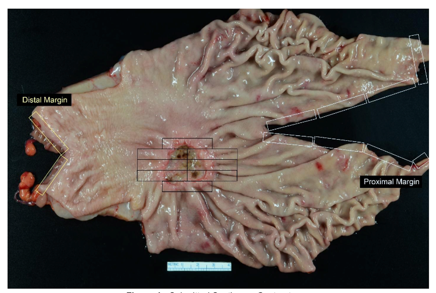
The stomach
What is pTis for gastric tumors?
Carcinoma in situ
Intraepithelial tumor WITHOUT invasion of the lamina propria
High-grade dysplasia
What defines gastric pT1?
Tumor invades the lamina propria, muscularis mucosae, or submucosa
T1a — Lamina propria or muscularis mucosae
T1b — Submucosa
What defines gastric pT2?
Tumor invades the muscularis propria
What defines gastric pT3?
Tumor penetrates the subserosal connective tissue WITHOUT invading the visceral peritoneum or adjacent structures
What defines gastric pT4?
Tumor invades:
T4a — The serosa (visceral peritoneum), or
T4b — Adjacent structures/organs
What are the regional lymph node categories for gastric carcinoma?
pN0 — No regional node metastasis
pN1 — 1–2 regional nodes
pN2 — 3–6 regional nodes
pN3 — ≥7 regional nodes
pN3a — 7–15
pN3b — ≥16
What regional lymph node groups are illustrated for gastric carcinoma?
The diagram includes:
Perigastric nodes along lesser curvature
Perigastric nodes along greater curvature
Right/left paracardial nodes
Suprapyloric nodes
Infrapyloric nodes
Hepatoduodenal nodes
Common hepatic nodes
Celiac nodes
Splenic artery nodes
Splenic hilum nodes
Left gastric nodes
MAKE SURE TO LOOK FOR LYMPH NODES

What is gastric pM0 vs pM1?
pM0 — No distant metastasis
pM1 — Distant metastasis
What distant metastatic sites are emphasized for gastric cancer?
Liver
Peritoneal surfaces
Lung
Adrenal gland
Ovary
Krukenberg tumor
Sister Mary Joseph nodules
CNS
Nonregional/distant lymph node
Virchow node
Irish node
What is a Krukenberg tumor?
Bilateral ovarian metastases
Often from diffuse-type gastric carcinoma
What is a Sister Mary Joseph nodule?
A periumbilical metastatic nodule
What is a Virchow node in the context of gastric cancer?
A left supraclavicular lymph node representing nonregional/distant lymph node involvement
What is an Irish node?
A left axillary lymph node representing nonregional/distant nodal involvement.
What is this?
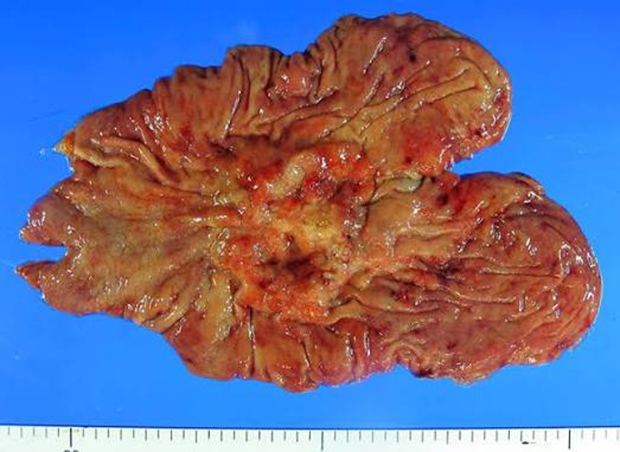
Gastric carcinoma
What three factors does the lecture emphasize when evaluating GIST?
Tumor site
Mitotic rate
Grade
What mitotic rate corresponds to GIST G1?
≤5 mitoses/5 mm²
What mitotic rate corresponds to GIST G2?
>5 mitoses/5 mm²
What is the GIST pT staging system based on?
Tumor size
What are the GIST pT size categories given in the lecture?
pT1 — ≤2 cm
pT2 — >2–5 cm
pT3 — >5–10 cm
pT4 — >10 cm in greatest dimension
(SIZE MATTERS)
What is T suffix (m) for GIST?
Multiple primary synchronous tumors in a single organ
How is GIST pN staged?
pN0 — No regional lymph node metastasis
pN1 — Regional lymph node metastasis
What is GIST pM1?
Distant metastasis
What is no LNs are present in GIST specimens?
An N category is not assigned/reported
What gross appearance should you recognize as a GIST?
A large, well-defined mass involving the GI tract

What type of cellular appearance is illustrated in the GIST microscopic image?

A spindle-cell morphology is illustrated