Neural Crest Cells and Axonal Specificity
1/23
There's no tags or description
Looks like no tags are added yet.
Name | Mastery | Learn | Test | Matching | Spaced |
|---|
No study sessions yet.
24 Terms
What do the neural crest cells give rise to?
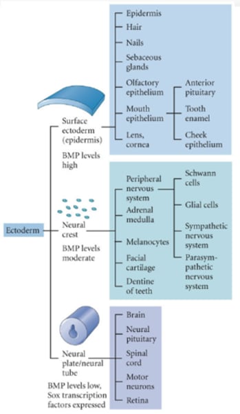
Peripheral nervous system, adrenal medulla, melanocytes, facial cartilage and the dentine of the teeth.
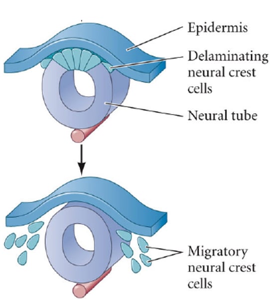
Where are the neural crest cells located?
Between the neural tube and the presumptive epidermis.
What are neural crest cells?
Migratory, multipotent mesenchymal cells induced at the dorsal-most part of the neural tube, in an interaction between the neural plate and presumptive epidermis, both of which contribute to the neural crest.
What do the axonal growth cone and the neural crest cells have in common?
They are governed by the same guidance cues.
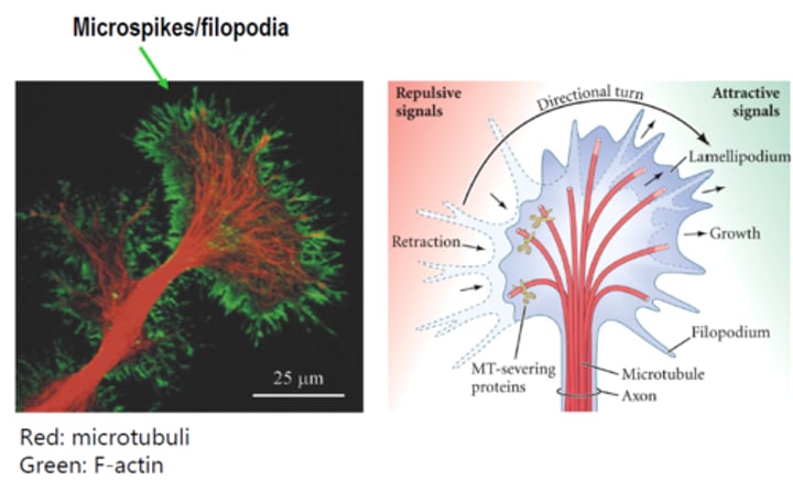
Actin microspikes
Mediate neuronal pathfinding
Microtubules
Mediate axon elongation
What do guidance cues do?
They regulate the cytoskeleton. Guidance cues can be soluble factors or glial cells that lay out tracks for the neurons to migrate on. They can also be located in the ECM. Neural crest cells and neurons express integrins, allowing them to migrate on specific parts of the ECM.
Guidance cues tell the neurons/neural crest cells where to go by altering their cytoskeleton.
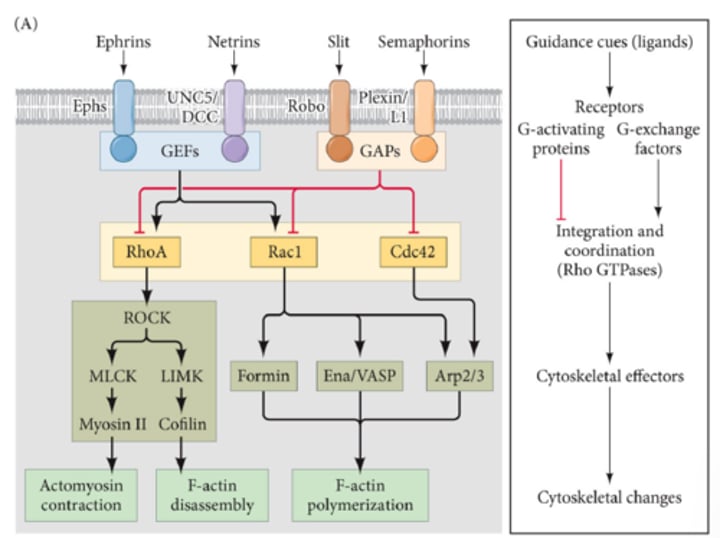
How is the axonal growth cone initiated?
Guidance cues initiate the activation of Rho GTPases RhoA, Rac1 and Cdc42, allowing the axon to form either filopodia (Rac) or lamellipodia (Cdc42) and facilitate cellular contractility (Rho).
The axonal growth cones move in the direction
How does the axonal growth cone move in the right direction?
The axonal growth cones move in the direction that attracts them and this is mediated by chemoattractants. The chemoattractant gradient activates a stimulatory pathway, which allows actin branching out into the cone, moving it in the direction towards the gradient.
Opposite, repulsive signals cause the growth cone to retract and activation of MT severing proteins, cleaving off MT fragments and shortening the branches.
How are dorsal root ganglions repulsed in the posterior parts of the sclerotomes?
Repulsion of dorsal root ganglions is facilitated by ephrins, found in the posterior sclerotome and interact with Eph receptors. Ephrin is an inhibitory factor of the dorsal root ganglion --> ensures that the dorsal root ganglion only moves into the anterior part, regulating patterning.
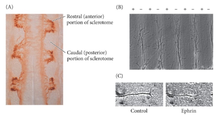
Ephrins
Membrane proteins which interact with Eph receptors. This interaction has a repulsive effect on the dorsal root ganglion.
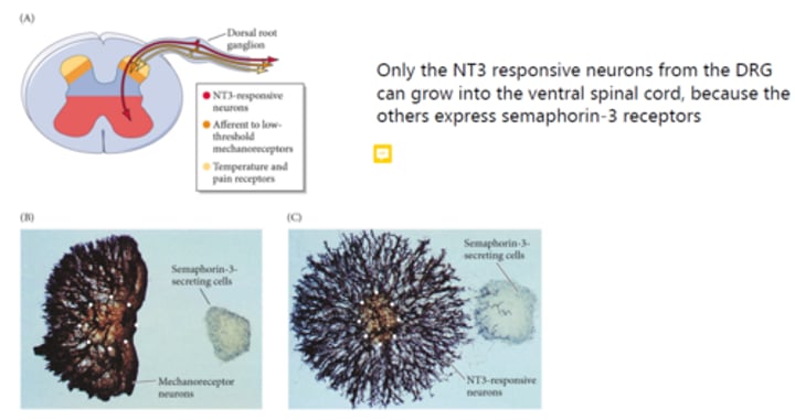
Semaphorin 3
Inhibits axon outgrowth from mechanoreceptor neurons in the ventral spinal chord. Only NT3 responsive neurons from the dorsal root ganglion can grow into the ventral spinal cord.
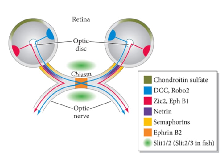
How do guidance cues control retinal ganglion neurons?
Retinal gangions must migrate through the optic disc and grow out either ipsilaterally (same side, red) or contralaterally (over to the other side, blue).
* Netrins are stimulatory, which ensures that the ipsilateral neurons migrate into the optic nerve.
* Semaphorins ensure that the contralateral neuron grows out into the opposite optic nerve and guides it into the direction of the chiasm.
* Robo's and Slits inhibit the ganglions that do not have to cross the chiasm.
* Slit 1 and 2 ensure that the crossing of the chiasm is done in an orderly fashion.
What are limitations to what neural crest cells can differentiate into?
The neural crest cells of the trunk have lost the ability to give rise to cartilage.
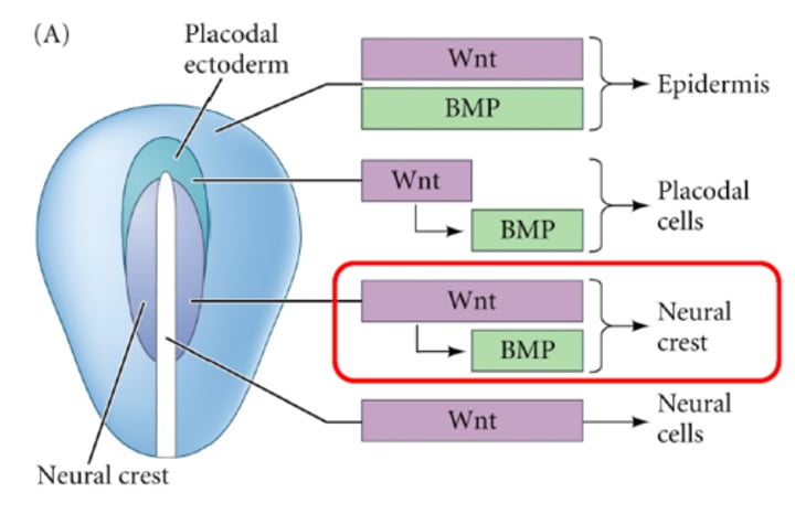
What determines neural crest cell specification?
It is determined by Wnt and BMP exposure patterns.
Wnt is secreted from the ventral ectoderm and paraxial mesoderm, after BMP activation. Increase in BMP occurs when Noggin (BMP inhibitor) levels are reduced.
What does delamination require?
Contact inhibition between cells expression E-cadherin (surface ectoderm), N-cadherin (neural tube) and cadherin-6B (top of neural tube, neural crest cells). Contact inhibition is facilitated by Sox2 and Snail2 --> cells can come apart and migrate.
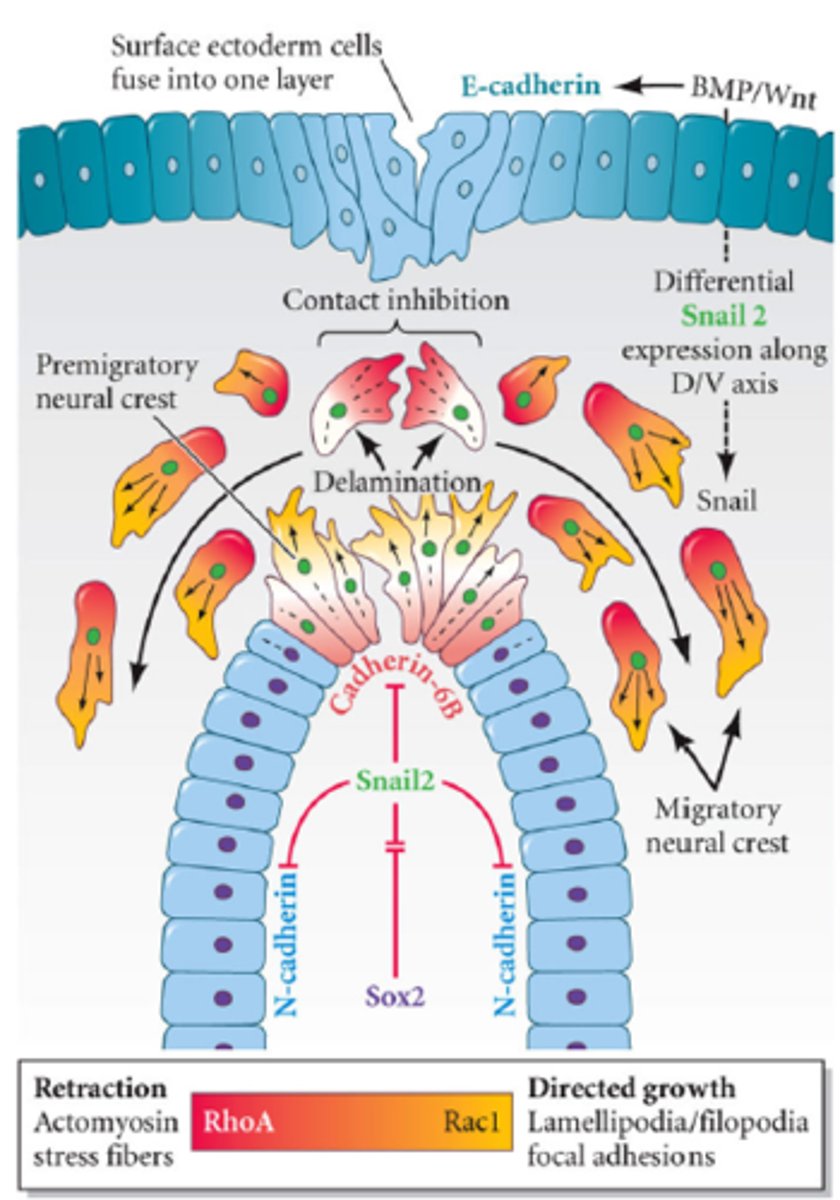
What regulates neural crest cell migration?
Contact inhibition of locomotion: facilitated by RhoA and Rac1 signaling. Has a repulsive effect; RhoA expression closest at the site of contact between the cells --> contraction via actin-myosin interplay. Rac1 expressed on the other side of the cell --> lamellipodia formation, allowing the cell to move away.
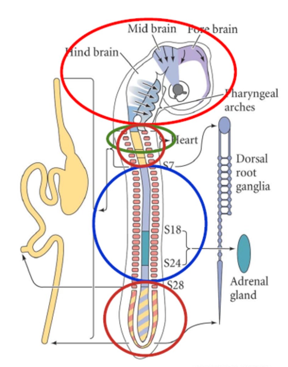
What do the cranial neural crest cells give rise to?
They move into the pharyngeal arches --> cartilage, bone, connective tissue, neurons, and glia of the head
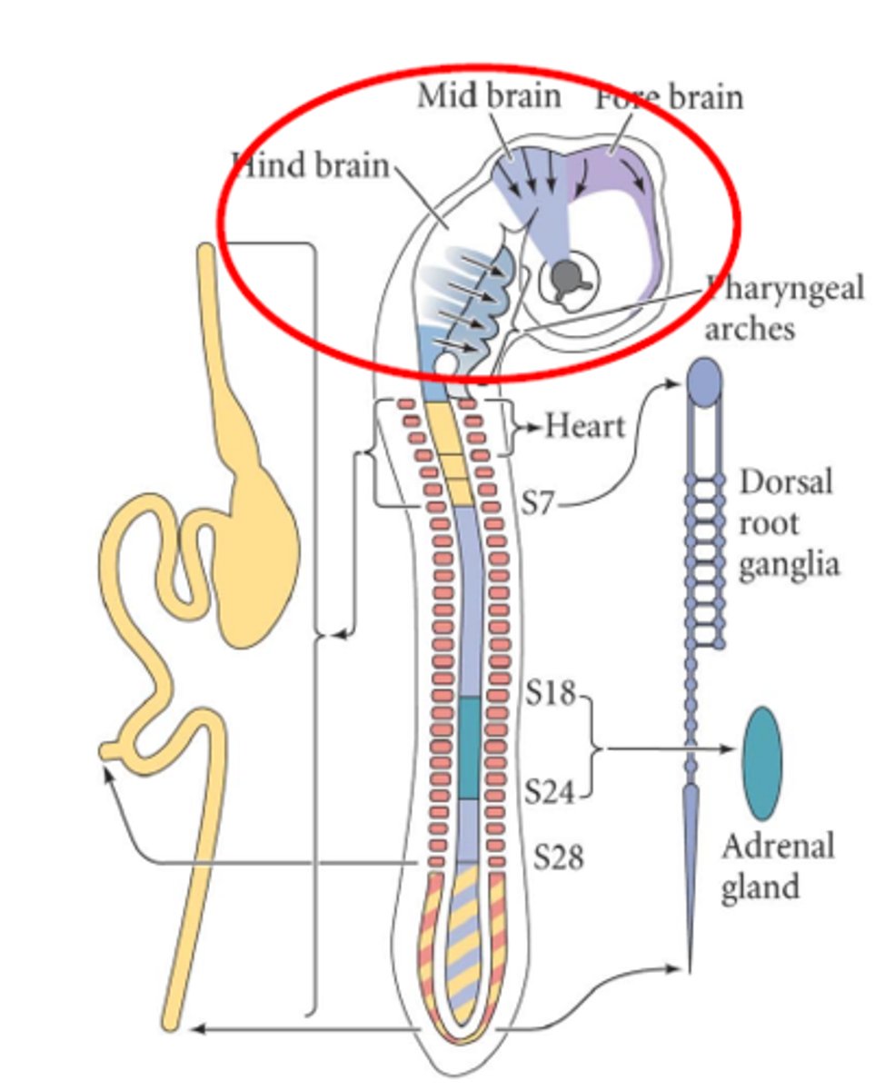
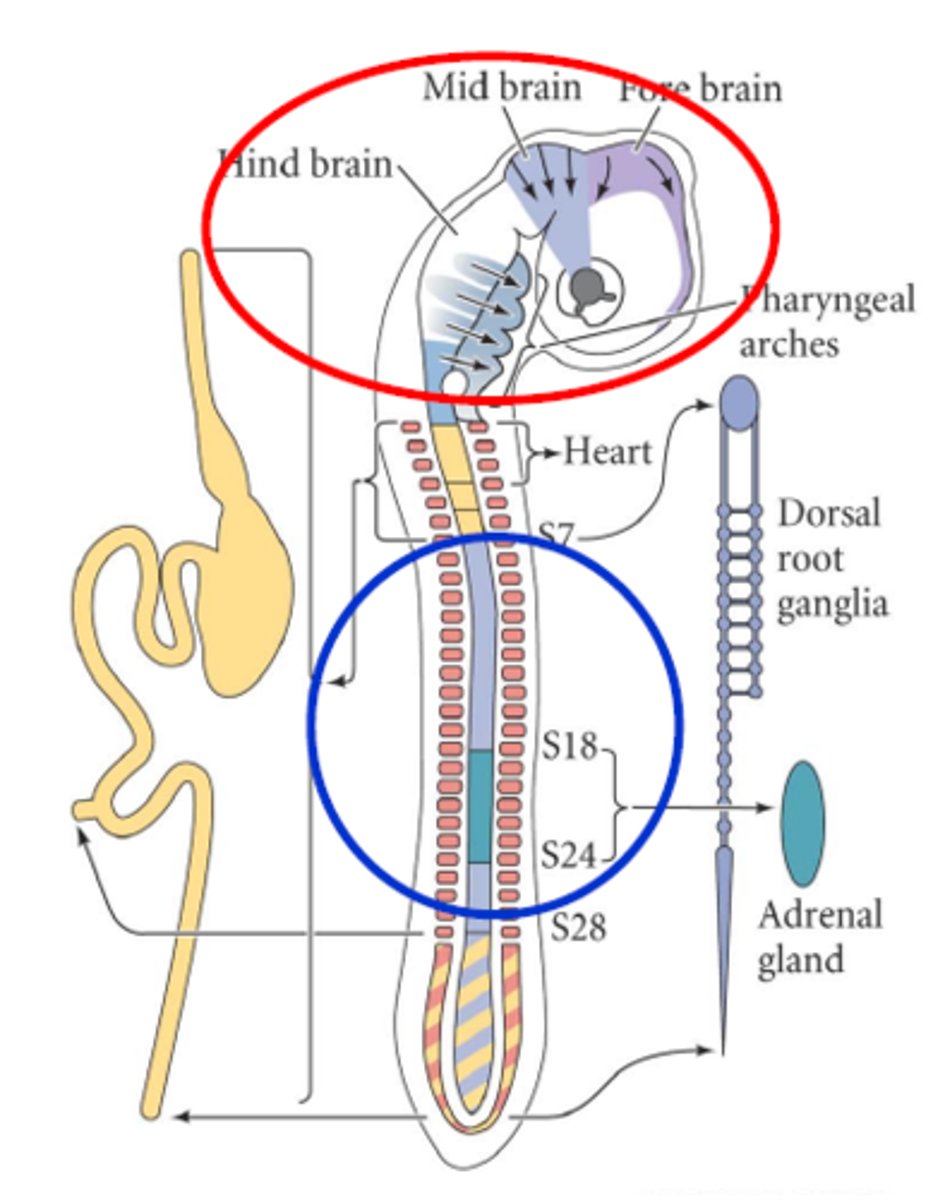
What do the trunk (from somite 6 to tail) neural crest cells give rise to?
- Dorsal root ganglia (sensory neurons)
- Sympathetic nervous system
- Adrenal medulla (somite 18-24)
- Melanocytes
What do the cardiac neural crest cells give rise to?
- Cartilage, connective tissue, melanocytes,
neurons
- Separation of aorta and pulmonary artery
- Large arteries muscular-connective tissue wall
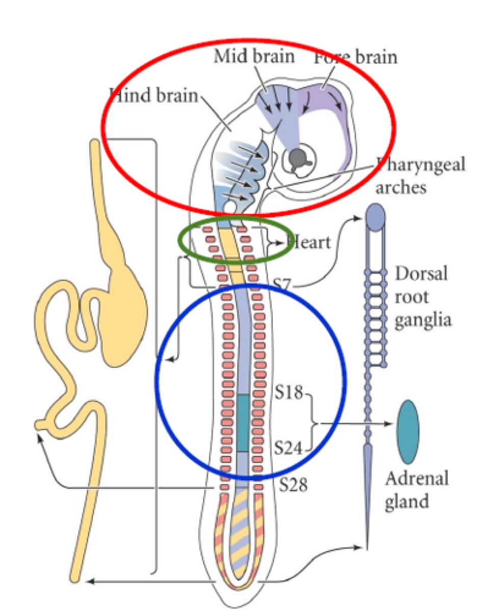
What do the vagral and sacral neural crest cells give rise to?
Parasympathetic nerves of the gut
(enteric nervous system)
Which diseases are associated with abnormal neural crest cell function?
Piebaldism; disruptive mutation in KIT --> aberrant neural crest cell proliferation.
What do the pharyngeal arches give rise to?
cartilages, bones, and muscles of the head, face, and neck
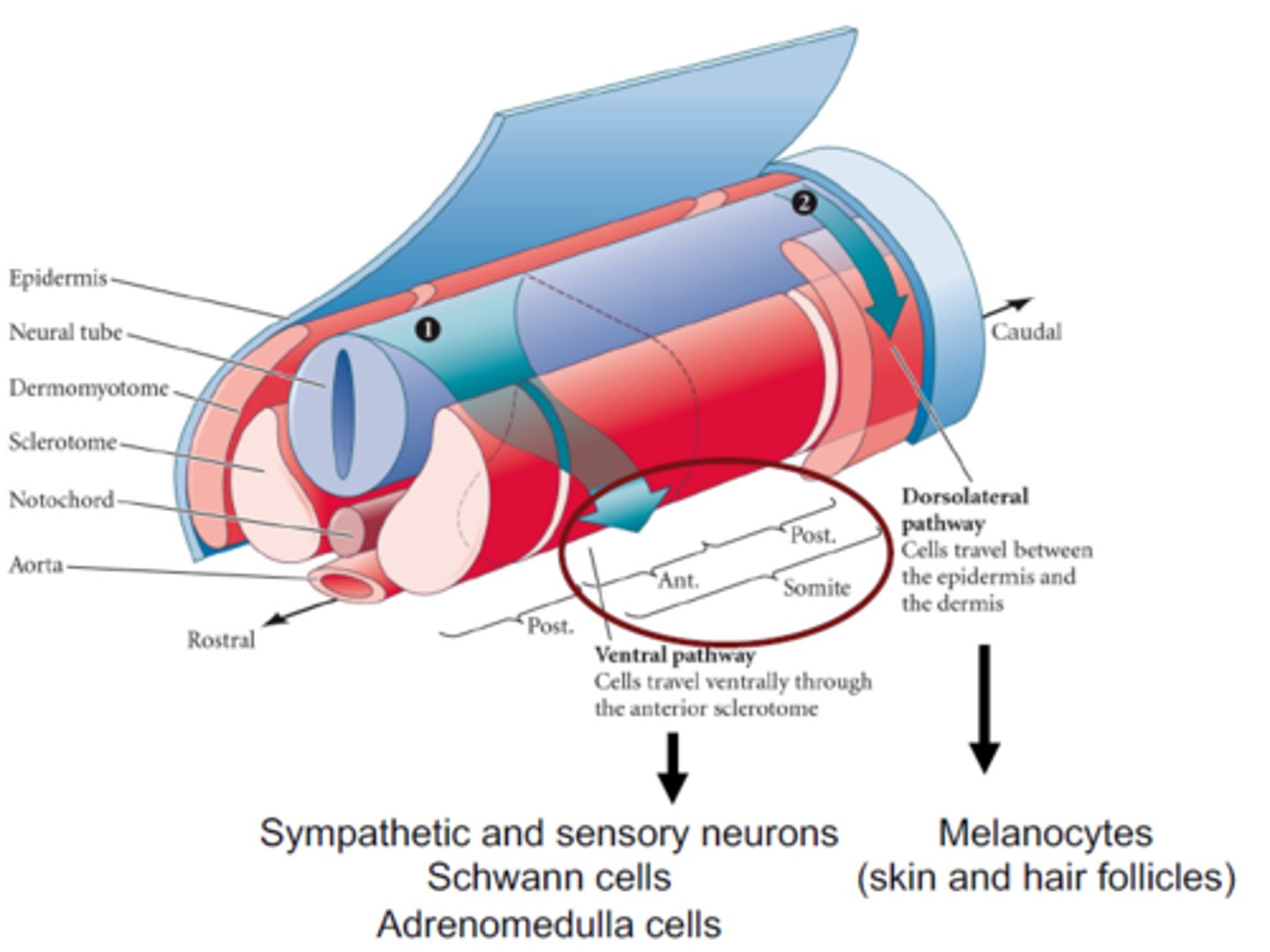
How do trunk neural crest cells migrate?
They migrate in two waves;
- Ventral movement first; down along the NT and under the somite (sclerotome). Can only move in the anterior part due to repulsive effects from the ephrins.
- Dorsolateral movement: these neural crest cells move between the epidermis and the dermis. Become the melanocytes. Ephrins are stimulatory here.