NEURO COMPLETE
 Call Kai
 Learn
Learn Practice Test
 Spaced Repetition
 Match
Match Flashcards
 Knowt Play
 Podcast
1/106

There's no tags or description
Looks like no tags are added yet.
Name | Mastery | Learn | Test | Matching | Spaced | Call with Kai | Chat |
|---|
No analytics yet
Send a link to your students to track their progress
107 Terms
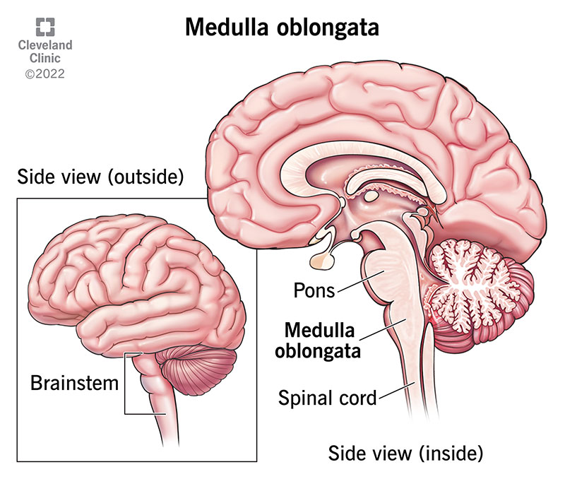
Medulla
A brain region that transmits information from the spinal cord to higher brain centers and controls autonomic activity.
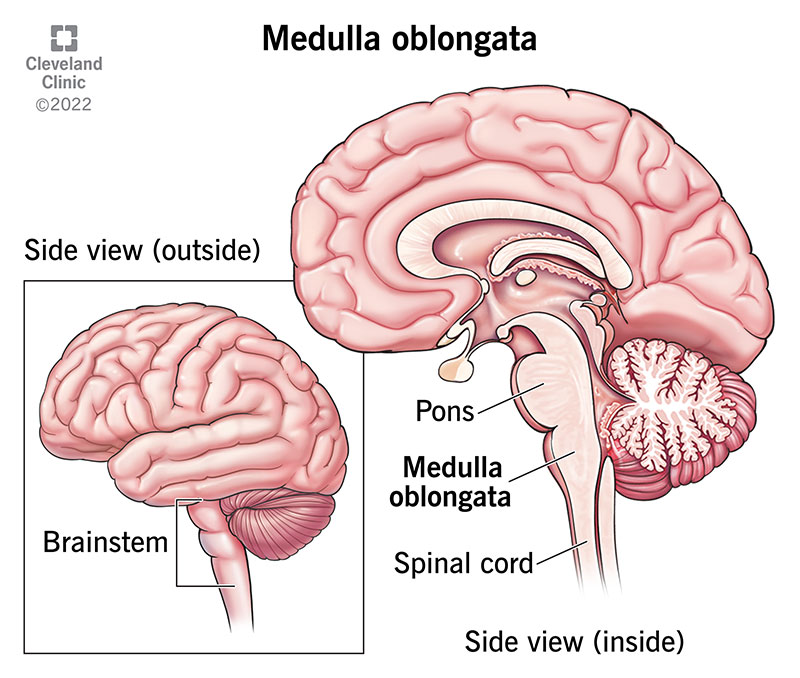
Pons
Relay center that connects cerebral hemispheres and controls breathing.
Medulla and Pons clinical signs
Depression
altered respiration
spastic tetraparesis/plegia
ataxia
multiple CN deficits.
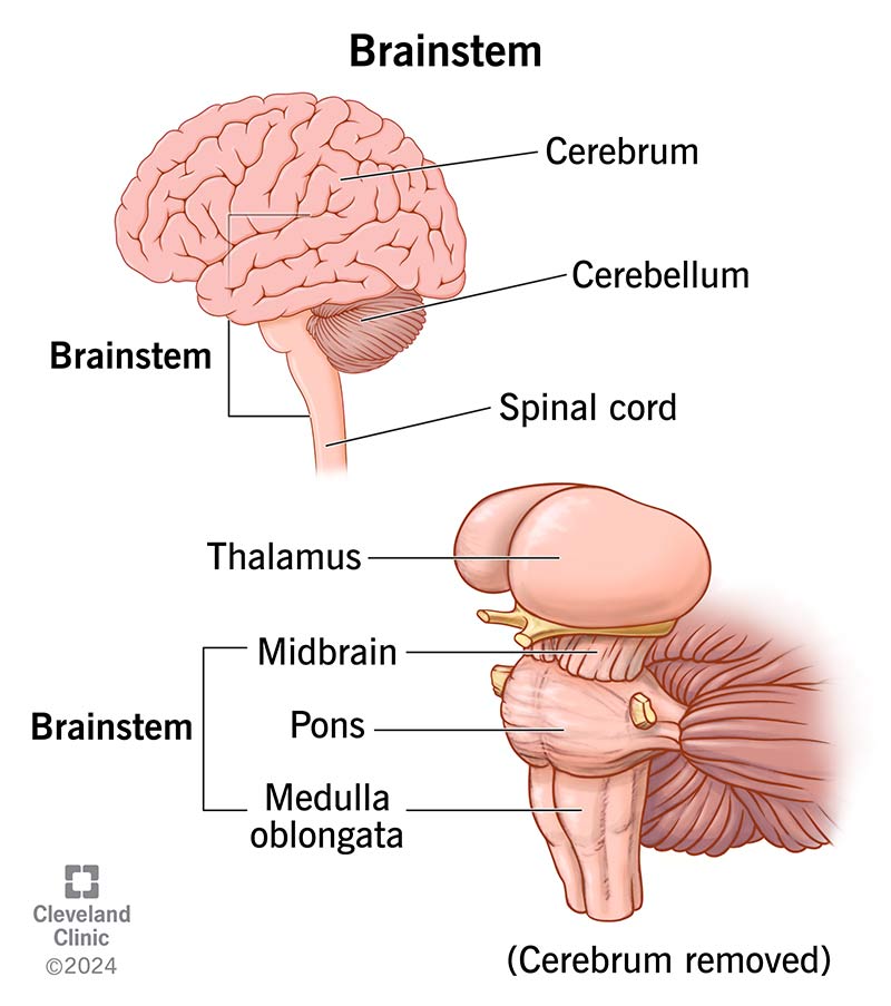
Cerebellum
The brain region responsible for controlling voluntary movement and posture.
Hypermetria
A clinical sign of cerebellar lesions described as high stepping.
Decerebellate rigidity
A sign of severe cerebellar lesions (such as brain herniation) involving opisthotonus and extensor rigidity ×4.
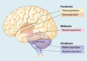
Mesencephalon (Midbrain)
Region that connects the hind- and forebrain and performs visual and auditory processing; lesions can result in CN III and CN IV deficits.
Decerebrate rigidity
A clinical sign of a severe mesencephalon lesion characterized by opisthotonus and extensor rigidity ×4.
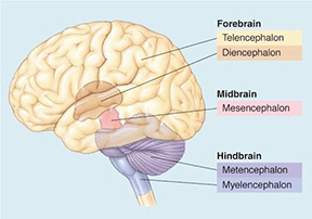
Diencephalon (thalamus, hypothalamus)
Region involved in endocrine/autonomic control and regulation of consciousness; the optic chiasm is located here.
Diencephalon lesion signs
Blindness with absent pupillary light reflex (PLR) (if bilateral)
circling towards the lesion
seizures
changes in thermoregulation, thirst, or appetite.
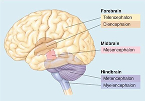
Telencephalon (cerebrum)
The largest region of the brain involved in most brain activity.
Telencephalon lesion signs
Blindness with normal PLR
seizures
abnormal behavior
depression
lethargy
movement disorders.
Cranial nerve (CN) deficits
Clinical findings that suggest lesions in the more caudal brain or brainstem.
What constitutes the Central Nervous System (CNS) and what are its primary roles?
The brain and spinal cord; it receives sensory input (afferent), produces motor output (efferent), and coordinates nervous system activity.
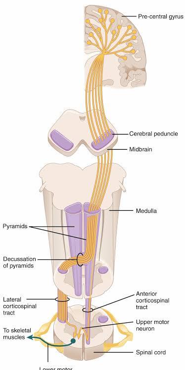
What is the definition of an Upper motor neuron (UMN)?
The 1st neuron in the motor pathway with its cell body in the CNS (cerebral cortex), which synapses on the lower motor neuron in the brainstem or spinal cord.
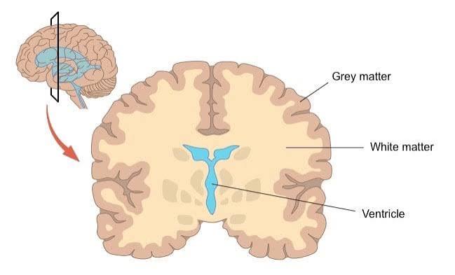
What are the characteristics of gray matter within the CNS?
An unmyelinated region of the CNS that contains cell bodies and dendrites.
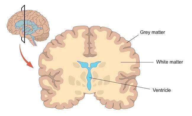
What does white matter in the CNS consist of?
The myelinated region of the CNS containing myelinated axons, which in the spinal cord are interconnecting axons.
What defines the Peripheral nervous system (PNS)?
All parts of the nervous system excluding the brain and spinal cord.
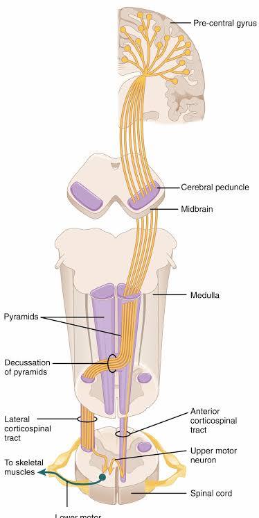
What is a Lower motor neuron (LMN) and where does it connect?
The 2nd neuron in the motor pathway that begins in the spinal cord or brainstem and connects to skeletal muscle or a gland.
What is the function and origin of a Cranial nerve (CN)?
Paired nerves that emerge directly from the brainstem and provide motor and sensory function to the head and neck.
What functional systems are controlled by the Somatic NS?
Proprioception, voluntary movement, and reflexes.
What is the role of the Visceral/autonomic NS?
Controlling internal organ function through the sympathetic and parasympathetic systems.
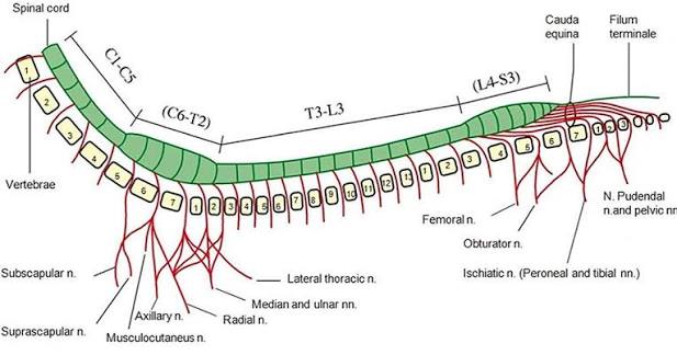
List the number of spinal cord segments in the canine/feline spinal regions.
8 cervical, 13 thoracic, 7 lumbar, 3 sacral, and 5 caudal (variable number of vertebrae).
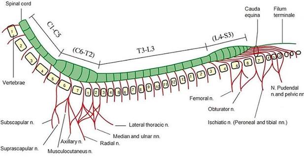
At what vertebral level does the canine spinal cord typically end?
L6−7 vertebrae.
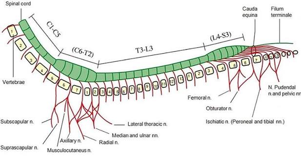
At what vertebral level does the feline spinal cord typically end?
L7−S3 vertebrae.
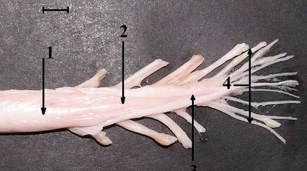
What is the cauda equina?
Long spinal roots and nerves located caudal to L7.
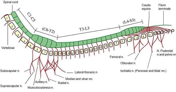
How do cervical spinal nerves C1−7 exit relative to their vertebrae compared to other spinal nerves?
C1−7 spinal nerves exit the intervertebral foramina cranial to the vertebra of the same number; all other nerves (except C8) leave foramina caudal to the vertebra of the same number.
Where does spinal nerve C8 exit?
Cranial to T1, between vertebrae C7 and T1.
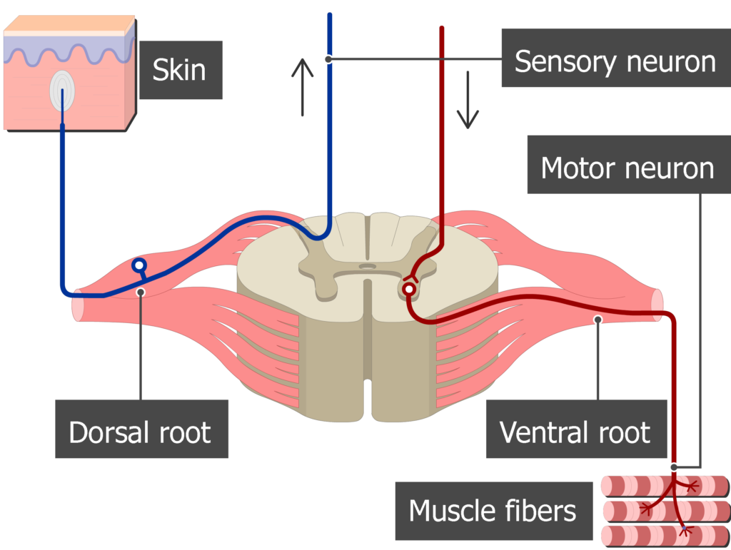
What is the functional distribution of nerves within the spinal cord horns?
Motor tracts are located in the ventral horn (descending/efferent), while sensory tracts are in the dorsal horn (ascending/afferent).
Under what condition does a lesion in the spinal canal cause LMN signs?
When the spinal nerve is affected, as spinal nerves are considered LMN/part of the PNS once they leave the spinal cord.
What structures are included in the Upper Motor Neuron (UMN)?
The Brain and Spinal cord.
What components make up the Lower Motor Neuron (LMN)?
Peripheral nerves, neuromuscular junction, and muscle.
What is the general physiological outcome of an Upper Motor Neuron (UMN) lesion?
Loss of inhibition.
What is the general physiological outcome of a Lower Motor Neuron (LMN) lesion?
Loss of function.
How is paresis defined in canine and feline neurology?
Abnormal gait generation (UMN) or the inability to bear weight (LMN).
What is the definition of paralysis?
Loss of voluntary movement.
What is the definition of ataxia?
Disordered movement and loss of proprioception or postural reactions.
What distinguishes a reflex from a reaction?
Reflex → is an immediate motor response to sensory input not requiring higher integration from the brain (patellar reflex)
Reaction → requires conscious or unconscious cerebral integration (menace response)
What is nociception, and what is its alternative name?
Also known as “deep pain,” it is a conscious reaction to noxious stimuli, distinct from the withdrawal reflex.
How does muscle tone differ between UMN and LMN lesions?
UMN lesions → hypertonia
LMN lesions → hypo- or atonia
How do spinal reflexes differ between UMN and LMN lesions?
UMN lesions result in normal to hyperreflexia, while LMN lesions result in hypo- or areflexia.
Compare the gait characteristics of UMN vs. LMN dysfunction.
UMN gait involves delayed generation and long-strided movements; LMN gait involves weak, short-strided movements and an inability to support weight.
Under what condition does neurogenic atrophy occur in LMN lesions?
If the lesion persists for more than 7d.
Which spinal segments comprise the ventral branches of the brachial plexus?
C6−T1(2).
Which spinal segments comprise the ventral branches of the lumbosacral plexus?
L4−S3.
What types of paralysis are characteristic of UMN versus LMN lesions?
UMN lesions cause spastic paresis/paralysis, while LMN lesions cause flaccid paresis/paralysis.
How do postural reactions/proprioception findings differ between UMN and LMN lesions?
They are abnormal or absent in UMN lesions, whereas they are typically normal in LMN lesions unless the lesion is severe.
Spinal cord regions (Neurological grouping)
The spinal cord is separated into four distinct regions based on the neurologic signs resulting from lesions in those specific areas.
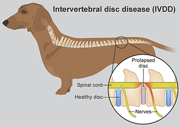
Intervertebral disc disease (IVDD)
A very common cause of spinal cord injury in dogs, particularly in predisposed breeds like the Dachshund.
Lumbosacral intumescence
Located at L4-S3, this region contains the cell bodies of spinal nerves for the pelvic limb and perineal region.
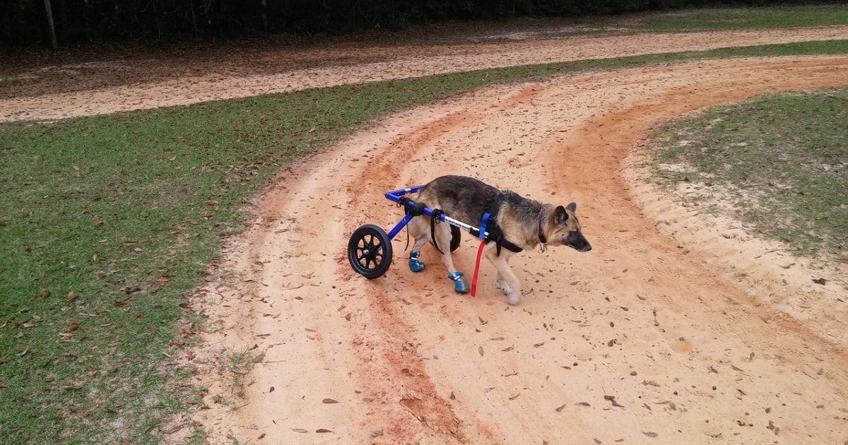
LMN deficits (Caudal to L4 lesion)
Lower Motor Neuron deficits seen in the pelvic limbs, featuring flaccid paresis or paralysis, while thoracic limbs remain normal.
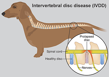
T3-L3
The most common location for intervertebral disc degeneration (IVDD), characterized by thoracic limbs being normal and pelvic limbs showing UMN deficits.
Cutaneous trunci cut-off
A diagnostic indicator where the reflex cut-off is typically 2 vertebral bodies caudal to the actual spinal lesion.
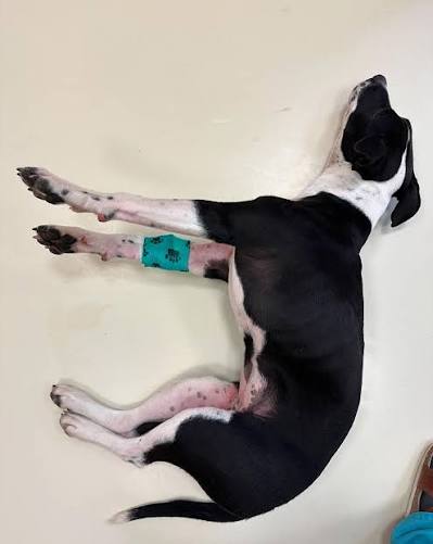
Schiff-Sherrington syndrome
A condition caused by an acute, severe transverse T3-L3 lesion (e.g., spinal fracture) resulting in pelvic limb flaccid paralysis and thoracic limb hyperextension.
Spinal shock
A state resulting from severe acute lesions involving flaccid paralysis that usually spontaneously resolves in 10−14 days; it is not a prognostic indicator but indicates lesion severity.
C6-T2 (cervical intumescence)
A site where incomplete lesions cause LMN deficits in thoracic limbs and UMN deficits in pelvic limbs.
Two-engine gait
A specific gait observed with C6-T2 lesions where the thoracic and pelvic limb movements are distinctly different.
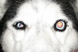
Horner syndrome
A clinical sign that may be present if a spinal cord lesion involves the T1-T3 segments.
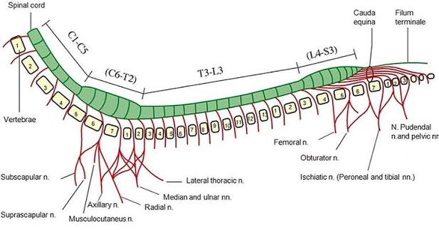
C1-C5 (cranial cervical lesion)
A localization where lesions cause UMN deficits to all limbs (tetraparesis/plegia), often with pelvic limb deficits being more severe.
UMN bladder
A condition resulting from lesions cranial to L4 where the bladder is distended and firm, urethral sphincter tone is increased, and expression is difficult.
LMN bladder
A condition resulting from lesions at the lumbosacral intumescence, LS plexus, or pelvic nerve where the bladder is distended and soft, sphincter tone is decreased, and expression is easy.
Descending inhibition
Motor control input that, when lost in a T3-L3 lesion, results in increased pelvic limb tone and reflexes.
Respiratory failure
The outcome of a complete spinal cord lesion at the C1-C5 or C6-T2 levels.
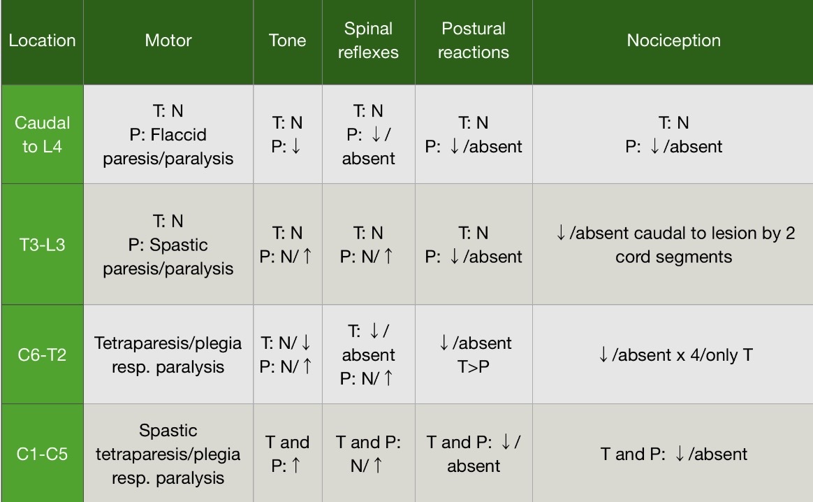
Localisation of Spinal Cord Lesions
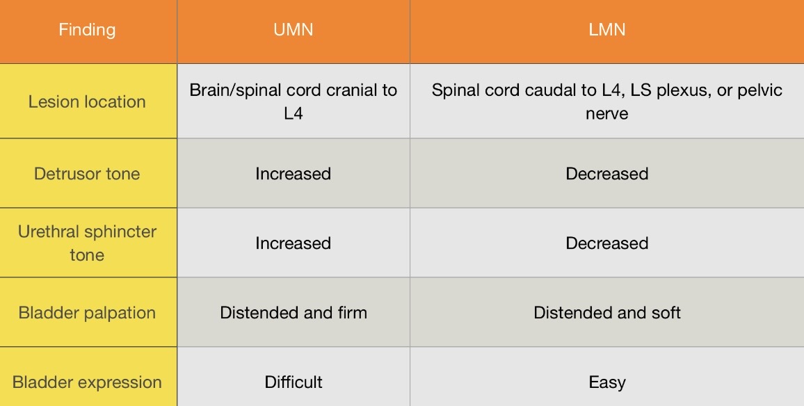
Bladder Function Based on Lesion Location
Vestibular disease
A common small animal presentation involving the disruption of CNVIII.
Menace response
A test assessing the visual pathway (CNII, retina, and visual cortex) and motor response (CNVII) by moving a hand towards the eye while avoiding air disturbance.
Pupillary light response (PLR)
An assessment of pupil size and symmetry using sensory input (CNII and retina), midbrain integration (Edinger-Westphal nucleus), and motor output (CNIII).
Central blindness
Blindness caused by a visual cortical lesion where the Pupillary Light Response (PLR) remains intact.
Peripheral blindness
Blindness accompanied by an absent Pupillary Light Response (PLR) due to a lesion in the retina, CNII, optic tract, or optic chiasm.
Horner syndrome
The disruption of sympathetic innervation to the eye, involving a pathway from T1−T4 to the cranial cervical ganglion and through the tympanic cavity.
Miosis
A constricted pupil, which is a classic clinical sign of Horner syndrome.
Ptosis
The narrowing of the palpebral fissure, noted as a sign of Horner syndrome.
Enophthalmos
The recession of the eyeball into the orbit, often seen in cases of Horner syndrome.
Strabismus
The deviation of eye position caused by the disruption of extraorbital muscle innervation.
Physiologic nystagmus
Normal involuntary eye movement during head rotation where the fast phase is in the same direction as head movement and the slow phase is opposite.
Peripheral vestibular lesion
A lesion of CNVIII where the fast phase of nystagmus is always away from the lesion, and postural reactions remain normal.
Central vestibular lesion
A lesion of the medulla where the nystagmus fast phase direction is unpredictable, and postural reactions are abnormal.
CN V (Trigeminal)
The cranial nerve responsible for motor innervation to the muscles of mastication and sensory innervation to the nose and ears.
CN VII (Facial)
The cranial nerve responsible for motor innervation to the facial muscles; dysfunction results in facial asymmetry, lip droop, or ear droop.
Palpebral reflex
A test of CNV (sensory) and CNVII (motor) where touching the medial or lateral canthus of the eye elicits a blink.
Decussation
The crossing of neuronal fibers that allows for a bilateral blink or consensual Pupillary Light Response (PLR) when only one side is stimulated.
Swallowing/Gag reflex
An assessment of CNIX, CNX, and CNXII performed by touching the caudal oropharynx.
Neuromuscular Exam Goal
To determine if an animal has a neurological deficit and, if so, the anatomic location of the lesion.
Sensorium
The first component of the neurologic exam relating to the patient's behavior and reaction to its owner and the environment.
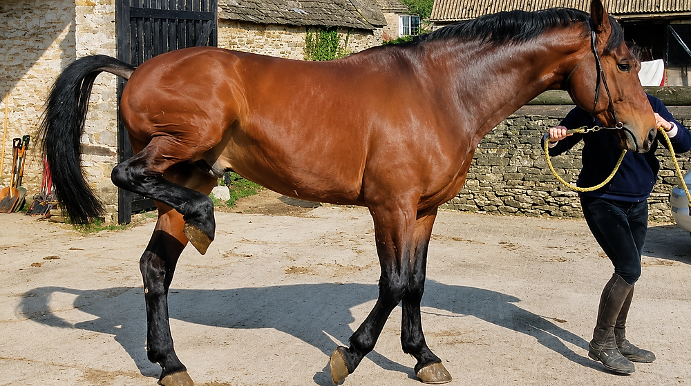
UMN (Upper Motor Neuron) Gait
Abnormal gait generation characterized by lengthened stride and delayed protraction in the affected limb(s).
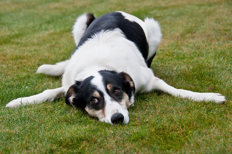
LMN (Lower Motor Neuron) Gait
Muscle weakness characterized by shortened stride/shuffling gait and collapse/weakness in the affected limb(s).
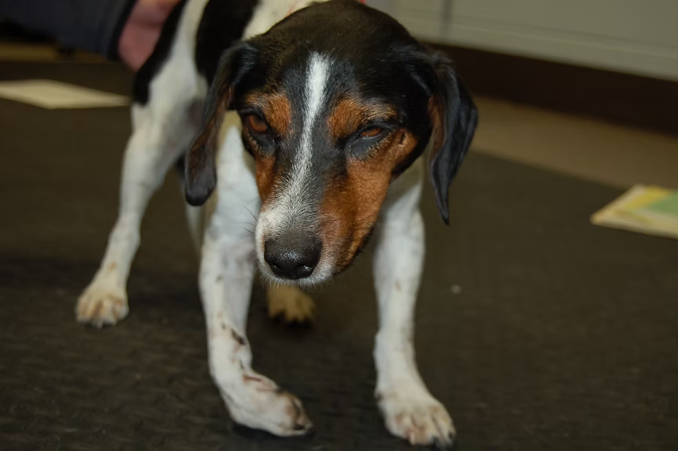
Proprioceptive Ataxia
Incoordination caused by a spinal cord lesion where the animal is unaware of limb position in space and may bear weight on the dorsal surface of the paw.
Vestibular Ataxia
Incoordination involving a head tilt and stumbles or falls due to a loss of balance.
Cerebellar Ataxia
Incoordination involving a delay in protraction and an exaggerated gait with limb flexion, also known as high-stepping.
Gait Grade 0
no voluntary movement.
Gait Grade 1
Slight movement with supports
Gait Grade 2
voluntary movement and ataxia, but the patient cannot stand without support.
Gait Grade 3
paresis and ataxia, but the patient stands with support.
Gait Grade 4
Mild Paresis/Ataxia
Gait Grade 5
Normal Function (Horse Grading is nearly opposite)
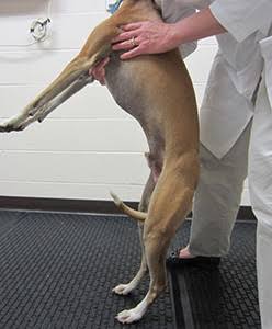
Postural Reactions
The third component of the neuro exam that tests upper motor neuron (UMN), lower motor neuron (LMN), motor, sensory, and proprioception.
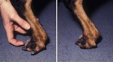
Conscious Proprioception (CP)
The assessment of placing the dorsal aspect of the paw on the ground and measuring the time it takes for the pet to replace it to a normal position.
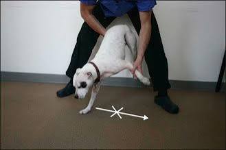
Hopping Response
The most reliable postural reaction test where the patient's body weight is supported to assess their ability to hop laterally.
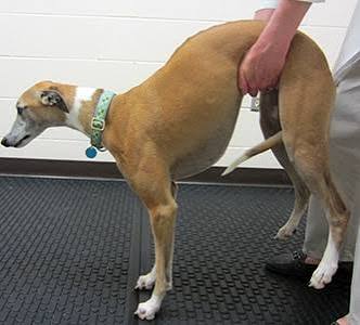
Hemiwalking
An alternative to hopping in large dogs where the thoracic and pelvic limbs on one side are picked up while the pet is pushed away to step sideways.