biochem unit 1 post quiz 1
 Call Kai
 Learn
Learn Practice Test
 Spaced Repetition
 Match
Match Flashcards
 Knowt Play
1/89

There's no tags or description
Looks like no tags are added yet.
Name | Mastery | Learn | Test | Matching | Spaced | Call with Kai | Chat |
|---|
No analytics yet
Send a link to your students to track their progress
90 Terms
pka of amino group
10-12
pka of COO- group
2-3
for all amino acids besides proline and glycine, the alpha carbon is bonded to:
acidic carboxyl group
a basic amino group
hydrogen
unique side chain
chirality
cannot be superimposed on mirror image
all amino acids besides glycine are chiral
how do you determine L and D forms
COO- to top, R group to bottom, where is amino?
amino acids in natural proteins are in what configuration
L
where do D amino acids occur
small peptides in peptidoglycan of bacterial cell walls
peptide antibiotics
neurotransmitters (D-glutamate)
platypus venom
all three aromatic amino acids absorb UV light at
270-280 nm
side chains in what class of amino acids can form H bonds
polar, uncharged
where do cysteines form disulfide bridges with each other
extracellular environment to stabilize individual proteins and protein complexes
cytosol is what kind of environment
reducing
pka of lysine
10.53
basic, positively charged
pka of arginine
12.48
basic, positively charged
pka of histidine
6.00
basic, positively charged
pka of aspartate
3.65
negatively charged, acidic
can bind Mg, Ca, Mn, Zn, ionic interactions
pka of glutamate
4.25
negatively charged, acidic
can bind Mg, Ca, Mn, Zn, ionic interactions
what form of amino acids, carbs, and nucleic acids do living organisms use
L amino acids
D carbs
D nucleic acids
ionization of amino acids
-amino acids are weak polyprotic acids, each amino acid has at least two titratable groups
-at acidic pH, carboxyl group is protonated and the amino acid is in the cationic form
-at neutral pH, carboxyl group is deprotonated but the amino group is protonated. the net charge is zero, zwitterion
-at alkaline pH, the amino acid group is neutral -NH2 and the amino acid is in the anionic form
diprotic amino acid
pk1- carboxyl group
pk2- amino group
isoelectric point of amino acid
pH where there is an equal amount of positive and negative charges
isoelectric point for glycine
5.97
In his, the imidazole side chain also contributes a
titratable group
therefore is triprotic
if pH is greater than pka
deprotonated
if pH is less than pka
protonated
what is the only AA that can be an effective buffer at physiological pH
histidine
its R group has a pka of 6
no other AA side chain has a pka near neutral pH
peptide bonds
peptides form covalent bonds between the alpha-COOH and alpha-NH2 groups of two amino acids, resulting in loss of water molecule
O=C-N-H
peptide definition
two or more amino acids joined covalently by a peptide bond
polypeptide definition
many AA joined together by peptide bonds (MW less than 10,000) (~15-50 AA)
protein definition
macromolecule with one or more polypeptide chains (50+ AA)
numbering and naming starts from which terminus
N
other relevant small peptides
oxytocin (9 AA)- stimulates uterine contractions
Bradykinin (9 AA)- causes tissue inflammation
Polypeptide examples
insulin- pancreatic hormone, needed for sugar metabolism, 2 polypeptide chains (30 AA and 21 AA)- not a dipeptide
Glucagon- pancreatic hormone, opposes insulin (29 AA)
average sized protein
hemoglobin with 574 AA and MW of 64,500
protein sequence logos
graphical representations of patterns within a multiple sequence alignment
provide a richer and more precise description of sequence similarity than consensus sequences and can rapidly reveal significant features of the alignment otherwise difficult to perceive
height of stack indicates sequence conservation at that position, measured in bits
height of symbols within stack reflect relative frequency of corresponding amino or nucleic acid at that position
native fold
specific three-dimensional conformation that enables a protein to fulfill a specific biological function
protein conformation is stabilized by
disulfide bonds
weak, non covalent interactions
hydrophobic effect relating to protein folding
release of water molecules from the structured solvation layer around the molecule as protein folds increases the net entropy
correctly position hydrophobic side chains depending on environment
hydrogen bonds relating to protein folding
interaction of N-H and C=O of the peptide bond leads to local regular structures such as alpha helices and beta sheets
side chain side chain interactions
van der waals relating to protein folding
weak attraction between all atoms contributes significantly to the stability in the interior of the protein
electrostatic interactions/ionic bonds relating to protein folding
stronger interactions between permanently charged groups
salt bridges especially buried in the hydrophobic environment strongly stabilizes protein
primary structure
amino acid sequence, includes all covalent bonds between amino acids and locations of disulfide bonds
spatial arrangement is unspecified (high degree of flexibility and rotation about bonds, except peptide)
clearly distinguishes one protein from another
amino acid sequence and protein function are closely linked, but difficult to tell function at this level
sickle cell mutation
single AA substitution
abnormal hemoglobins crystallize, deforming red blood cells and leading to clogs in tiny blood vessels
paralogs
same species
orthologs
different species
secondary structure
regular, recurring arrangements in space of adjacent amino acid residues in a polypeptide chain
most prominent secondary structures are alpha helices (coils) and beta sheets (folds)
irregular arrangement of the polypeptide chain is called a random coil
any one protein can be all one type of secondary structure, or a mixture
peptide bond rigidity
rigid and planar constraining the protein to certain allowed conformations
no rotation around peptide bonds BUT
rotation around C alpha bonds is permitted
phi bond
Calpha-amide nitrogen
psi
Calpha to carbonyl carbon
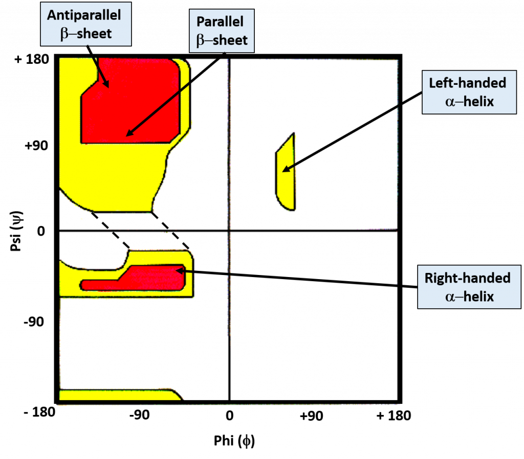
Ramachandran plot
shows common secondary structural elements and the acceptable range of rotation
the alpha helix
-helical backbone is held together by hydrogen bonds between the backbone amide of an “n” and carbonyl group of the n+4 amino acid
-right handed helix with 3.6 residues (5.6 A*) per turn
-hydrogen bonds are aligned roughly parallel with the helical axis
-side chains point out and are roughly perpendicular with the helical axis, H bonds are mostly parallel with helical axis
-residues 1 and 8 align on top of each other, heptad repeat
-the inner diameter of the helix (no side chains) is about 4-5 A*
-the outer diameter of the helix (with side chains) is 10-12 A*, fits well into the major groove of dsDNA
-some amphipathic alpha helices can form coiled coil dimers
in an alpha helix, where are hydrophobic AA found?
in A and D positions
primary sequence affects helix stability
small hydrophobic residues such as Ala and Leu are strong helix formers
pro acts as a helix breaker because the rotation around the N-alphaC bond is impossible
gly acts as a helix breaker because the tiny R group supports other conformations, too flexible
attractive or repulsive interactions between side chains 3-4 AA apart will affect formation
residues near N and C ends don’t necessarily H bond like the rest, but they can stabilize the helix (helix capping)
beta sheets
sheet-like arrangement of the backbone is held together by hydrogen bonds between the backbone amides and carbonyl groups in different strands
side chains protrude from the sheet alternating in up and down direction
which AA will or will not be found in beta sheets
found: large, aromatic (Tyr, Phe, Trp) branched (Leu, Val, Ile)
not found: Gly, Pro
parallel beta sheets
in parallel beta sheets the H bonded strands run in the same direction
results in bent, weaker H bonds
individual strands can be close, or distant, in the primary structure
antiparallel beta sheets
H bonded strands run in opposite directions
resulting in stronger, linear H bonds
individual strands can be close, or distant, in the primary structure
ribbon diagrams of secondary structure
most proteins are globular in shape as a result of frequent turns or loops in the polypeptide chain that connects beta strands and alpha helices
strands, and sometimes helices, will have arrows to indicate N and C termini of the secondary structural element
beta turns help organize secondary structures
beta turns occur frequently whenever strands in beta sheets change direction
the 180 degree turn is accomplished over four amino acids
the turn is stabilized by a hydrogen bond from a carbonyl oxygen to amide proton three residues down the sequence.
common AA in beta turns
proline in position 2 or glycine in position 3
proline in cis conformation forms a tight turn, most peptide bonds are a trans conformation
tertiary structure
overall 3D arrangement of all atoms in a protein
includes long range contacts between AAs in a single polypeptide chain
stabilized by numerous weak interactions between AA side chains (largely hydrophobic and polar interactions, can be stabilized by disulfide bonds, side chain with backbone interactions are also possible)
major classes of tertiary structure
fibrous, globular, membrane, intrinsically disordered
fibrous proteins
often structural rather than dynamic and are water insoluble
typically contain high proportions of alpha helices (keratin) or beta pleated sheets (silk fibroin)
underlying structure is relatively simplistic
high proportion of hydrophobic AA
extensive supramolecular complexes
secondary structures and properties of fibrous proteins- alpha helix
tough, insoluble protective structures of varying hardness and flexibility
e.g. alpha keratin of hair, feathers, and nails
secondary structures and properties of fibrous proteins- beta conformation
soft, flexible filaments
e.g. silk fibroin
secondary structures and properties of fibrous proteins- collagen triple helix
high tensile strength, without stretch
e.g. collagen of tendons, bone matrix
alpha keratins
typical right handed alpha helices
super twisting helices, in a left handed fashion, wrap around each other to make a very tight coiled coil
cross linked by disulfide bonds
real life example using alpha keratins
permanent waving of hair
reduce disulfide bonds
moist heat breaks H bonds and causes uncoiling of alpha helix
remove reducing agent, add oxidizing agent, new S-S bonds
collagen
provides strength and structure
connective tissue (tendons, cartilage, organic matrix of bone, cornea)
each protein folds into left handed helix (alpha chains, NOT alpha helix)
coiled coil of three separate alpha chains supertwisted around each other in a right handed manner to provide strength (like a rope)
covalent cross links
globular proteins
enzymes, transport proteins, motor proteins, regulatory proteins, immunoglobins, etc
tight spherical structure, hydrophobic to middle
alpha helices, beta sheets, beta turns, etc. can all be found
arrangement of different secondary structural elements
compact conformation
folding provides structural diversity
general rules
bury nonpolar AA R groups
distant segments may come together, but not the norm
optimize number of weak interactions
globular proteins: motifs
specific arrangement of several secondary structure elements
all alpha helix
all beta sheet
combination
sometimes considered supersecondary structures
motifs can be found as reoccurring structures in numerous proteins
proteins are made of different motifs folded together
weird globular protein motifs
not secondary structures in themselves
twisted beta sheet
alpha alpha corner
beta alpha beta loop
beta barrel
globular proteins: domains
can have multiple domains on one protein, can have motifs in domains, domains by themselves are stable
-a domain is a relatively stable, independently folded region within the tertiary structure of a globular protein
-each domain may encompass one or more motifs
-domains having more than 30% of their AA seq in common normally adapt same folding pattern
-a single protein can have several domains with each domain performing a different function (enzyme, docking, regulatory, etc)
quaternary structure
results from the association of two or more polypeptide subunits into a larger functional cluster
side chain-side chain and side chain-polypeptide backbone interactions drive these associations. can have stabilizing H bonds
collagen is a fibrous protein of three polypeptides that are supercoiled like a rope
hemoglobin is a globular protein with two copies of two kinds of polypeptides
what drives quaternary association?
stability: reductio of surface to volume ratio
genetic economy and efficiency
bringing catalytic sites together
cooperativity
biological function may be regulated by complex interactions of multiple subunits
how can proteins be denatured
strong acid or base, organic solvents (more nonpolar than water), detergents, reducing agents, salt concentration, heavy metal ions, temperature, mechanical stress
denatured protein (inactive form)
lost enough structure, can no longer function. does not have to go all the way back to primary
denaturation does not disrupt primary structure
Levinthal’s paradox
it is mathematically impossible for protein folding to occur by randomly trying every conformation until the lowest energy one is found.
search for the minimum is not random because the direction toward the native structure is thermodynamically most favorable
nonpolar inside, polar outside
assistance to ensure proteins fold properly
small polypeptides (<100 AA) often form with no intermediates
larger polypeptides often require several intermediates (molten globules)
chaperones
ribosome brings partially folded or misfolded protein to GroEL barrel, ATP and GroES cap join, ADP + P, GroES cap, and properly folded protein emerge
how do chaperones assist protein folding
prevent inappropriate protein-protein interactions
provide a more isolated environment
help folding occur rapidly and precisely
two major classes: Hsp70s Hsp60s (chaperonins)
PDI- protein disulfide isomerase
reduces disulfide bonds
PPI- peptide protyl cis-trans isomerase
moves from cis to trans if needed
intrinsically disordered proteins
composed of AA whose higher concentration forces less-defined structure: Lys, Arg, Glu, Pro (all charged, no hydrophobic effect)
NPC is one example
overview of globular protein functions
storage of ions and molecules- myoglobin, ferritin
transport of ions and molecules- hemoglobin, serotonin transporter
defense against pathogens- antibodies, cytokines
muscle contraction- actin, myosin
biological catalysis- chymotrypsin, lysozyme
protein function: binding to ligands
function relies on interactions with other molecules
binding of molecules to proteins is reversible
ligand: any molecule that a protein can bind to in a reversible manner; often a small molecule
binding site: site on protein where ligand binds, specific
ligand binds via same noncovalent forces that dictate protein structure- allows interactions to be transient
reversible binding of ligands is essential
specificity of ligands and binding sites, high specificity, only certain ligands bind
ligand binding is often coupled to conformational changes, sometimes quite dramatic (induced fit)
in multi subunit proteins, conformational changes in one subunit can affect the others (cooperativity)
interactions can be regulated
high specificity can be explained by the complementarity of binding site and ligand
size, shape, charge, hydrophobicity
lock and key model- rigid interaction
induced fit- both protein and ligand can change conformations upon binding, makes binding site more complementary to ligand
myoglobin
compact globular protein composed of a single polypeptide chain 153 AA in length
carries and stores oxygen for muscles
contains a heme prosthetic group- a porphyrin ring complexed with iron ion
oxygen binds to Mb via the heme as amino acids alone cannot bind O2
porphyrin ring- heme
6 coordination sites: 1-4 by Ns, 5=AA, 6=O2
hemes are also used in ETC