Bones and Bone structure
 Call Kai
 Learn
Learn Practice Test
 Spaced Repetition
 Match
Match Flashcards
 Knowt Play
1/56

There's no tags or description
Looks like no tags are added yet.
Name | Mastery | Learn | Test | Matching | Spaced | Call with Kai | Chat |
|---|
No analytics yet
Send a link to your students to track their progress
57 Terms
What does the skeletal system have?
– Bones of the skeleton
– Cartilages, ligaments, & other connective tissues
Primary functions of the skeletal system
– Support
– Storage of minerals and lipids
– Blood cell production (RBC/WBC/Platelets)
– Protection
– Leverage
What classifies bones?
shape & structure
Bone shapes
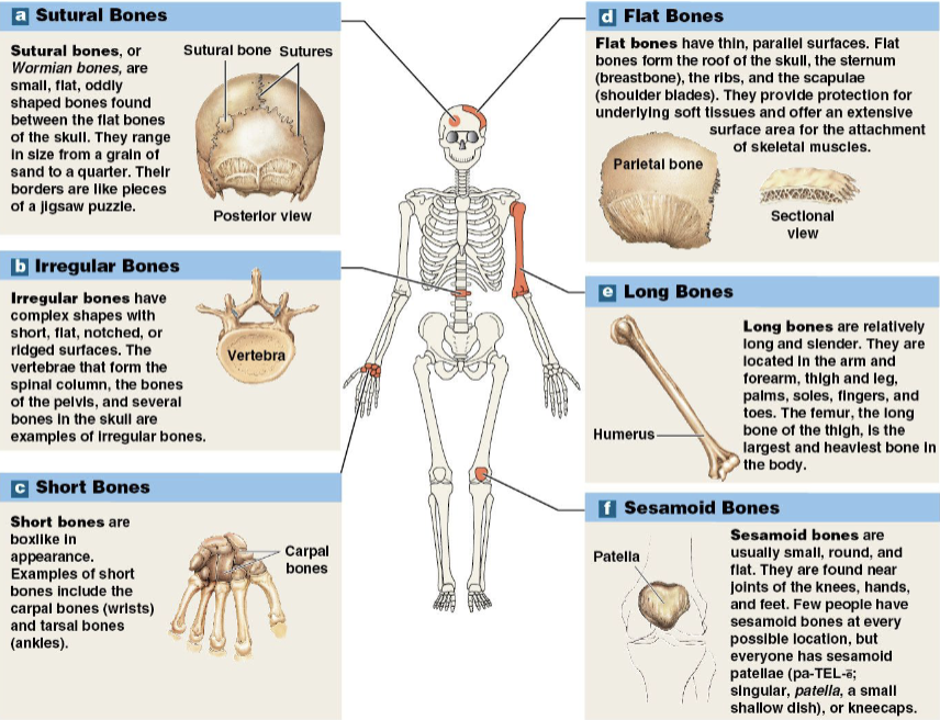
– Sutural
– Irregular
– Short
– Flat
– Long
– Sesamoid
Sutural bones
(Wormian bones)
– Small, flat, irregularly shaped bones
– Between flat bones of the skull
pieces of jigsaw puzzle*
(# varies among individuals)
Irregular bones
– Have complex shapes w/ short, flat, notched, ridged surfaces
– Examples: spinal vertebrae, pelvic bones, many bones in skull

Short bones
– Boxy
– ex: carpal bones and tarsal bones
Nemonic:
carpals: “so long to pinky here comes the thumb” OR “some lovers try positions that they can’t handle”
tarsals: “tiger cubs need milc”

Flat bones
– Thin w/ parallel surfaces
– ex: bones of skull roof, sternum, ribs, & scapulae
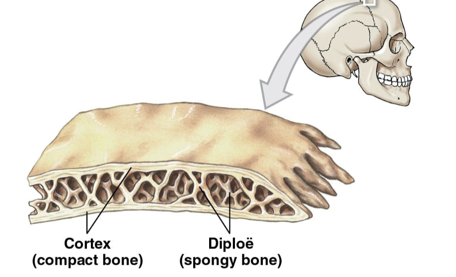
(protect soft underlying tissues; think about the cortex/diple/cortex sandwich)

Long bones
– Long & slender
– Found in arms, legs, palms, soles, fingers, toes

Sesamoid bones
– Usually small, round, and flat
– within tendons near joints of knees, hands, & feet
– Location & number vary between people
– ex: patella/patelle

Bone markings
(surface features)
– Projections
• Where muscles, tendons, & ligaments attach
• were articulations w/ other bones take place
– Openings and depressions
• For passage of blood vessels & nerves
Long bone structure
– Epiphysis (wide part at each end)
• Mostly spongy bone/trabecular bone
– Metaphysis
• Where diaphysis and epiphysis meet
–Diaphysis (shaft)
• Wall of compact bone
• Central space inside is medullary cavity (marrow cavity)
Flat bone structure
– Made of spongy bone between two layers of compact bone; cortex
– Layer of spongy bone is called the diploë
cortex→ diploë→cortex (bone sandwhich)
ex: parietal bones of skull
Bone tissue
– Dense, supportive connective tissue
– has specialized cells (vascularized
– Solid extracellular matrix w collagen fibers (perforating fibers in the periosteum!!!)
Characteristics of bone
Dense matrix: calcium salts make bone hard/dense
Osteocytes: bone cells located in lacunae, around blood vessels (vascularized=growth)
Canaliculi: tiny passageways that allow nutrients, wastes, & gases to be exchanged (w osteocytes & blood cells)
Periosteum: covers the outer surface of bone (except at joints)
Outer layer: fibrous; hardness and protection
Inner layer: cellular; growth
Bone matrix physiology
Calcium phosphate makes up 2/3 of bone mass (main substance) (other 1/3=collagen fibers)
Combines with calcium hydroxide → forms hydroxyapatite crystals (makes stronger bones!!)
no calcified matrix, bone looks normal but becomes very flexible
*constant remodelation
Bone cells
– 2% of bone mass
• Osteogenic cells
• Osteoblasts
• Osteocytes
• Osteoclasts

Osteogenic cells
(osteoprogenitor cells)
– stem cells that divide to produce osteoblasts
– Located in inner (where made) cellular layer of periosteum & in endosteum
– helps in fracture repair

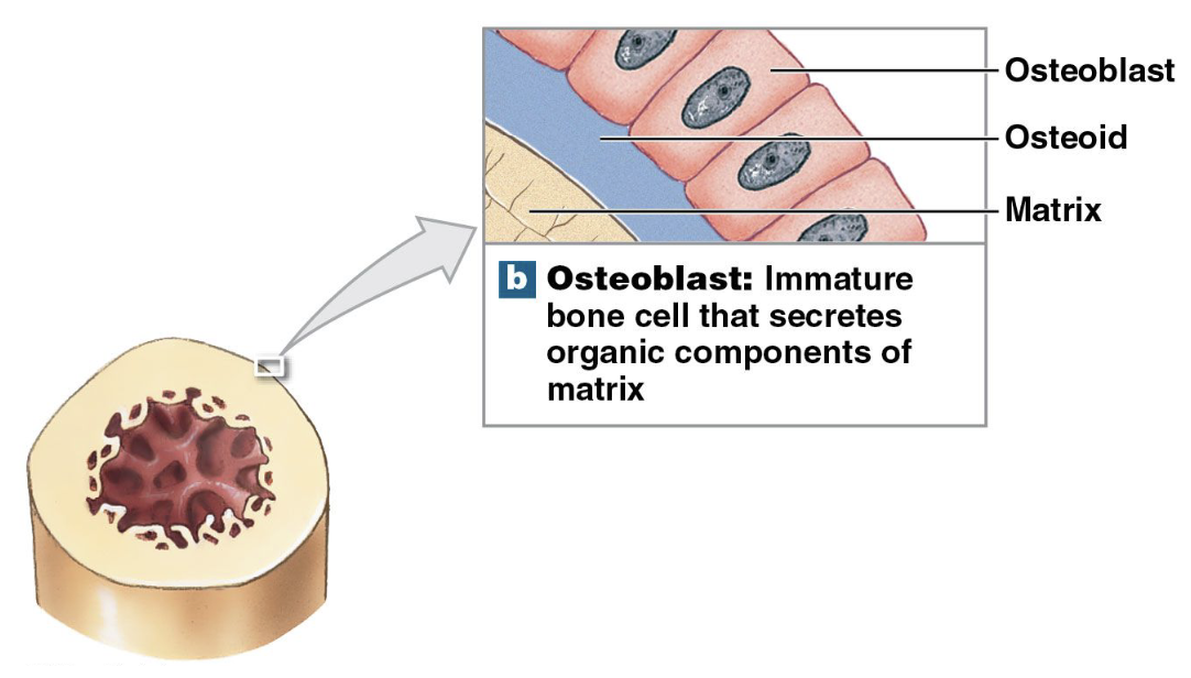
Osteoblasts
– Immature cells that make new bone matrix during osteogenesis (ossification; make cells)
– Osteoid: matrix made by osteoblasts that hasn’t been calcified yet (w/ calcium salts so not strong)
(after mineralization w/ calcium salts= bone matrix)
– Osteoblasts surrounded by bone matrix become osteocytes
basically make their own matrix, to become osteocytes
Osteocytes
– Mature bone cells that don’t divide
– Live in lacunae
–cell extensions pass through canaliculi (passageways)
Two major functions
• Maintain protein & mineral content of matrix; maintain the bone
• Help repair damaged bone

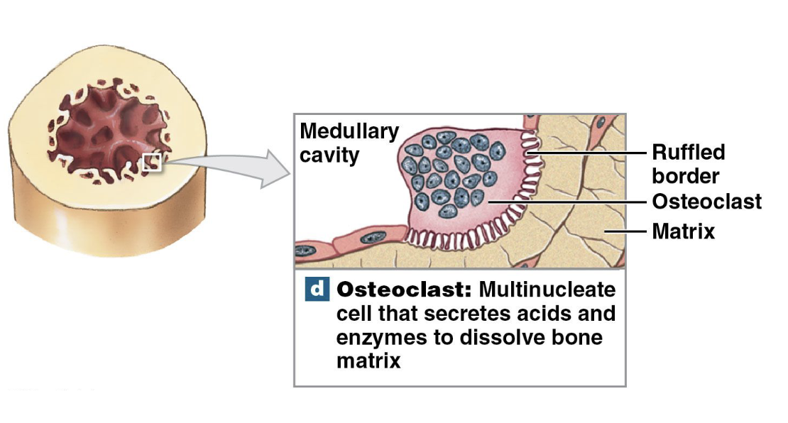
Osteoclasts
– break down osteocytes
– Secrete acids & protein-digesting enzymes to break down bone
• break down bone matrix & release minerals
• Osteolysis: breakdown of bone; to regulate calcium important in homeostasis
Lacunae
a small, bubble-like space or cavity located within the hard bone matrix; cuna for the osteocytes/home; around blood vessels that branch through the bone matrix
What is an osteon, and what is found inside it?
Osteon = functional unit of compact bone (Haversian system)
What found inside:
Central canal → has the blood vessels
Lamellae → layers of bone matrix
Perforating canals

What are the 3 types of lamellae and what do they do?
layers of bone matrix around central canal
Concentric lamellae → surround the central canal
Interstitial lamellae → fill spaces between osteons
Circumferential lamellae → found at outer & inner surfaces of bone
What are perforating canals?
run perpendicular to the osteons to connect them
carry blood vessels deep into bone & marrow
Spongy bone
Doesn’t have osteons
matrix made of open network of trabeculae
Red bone marrow
-Fills spaces between trabeculae; makes blood cells
-Has blood vessels (vasuclarize) that provide nutrients to osteocytes by diffusion=(spread out; not quick)
Yellow bone marrow
-Found in other areas of spongy bone; stores fat
-Energy reserves; triglycerides: 3 fatty acids w/ 1 glycerol backbone
weight-bearing bones
Trabeculae found in the epiphysis (end) of the femur
Transfers forces from the pelvis → compact bone of the femur shaft
Medial side of the shaft = compressed
Lateral side of the shaft = tension/stretching
compact + spongy working tgt
-OPPOSITE FORCES
lateral impact→ medial force
Periosteum
membrane that covers outside of bones; except within joint cavities
Outer = fibrous layer (protection & strength)
Inner = cellular layer (growth & maintenance)
– Fibers connect periosteum to tendons
–Perforating fibers extend into (within) the bone → strengthen tendon-to-bone attachments
Functions of periosteum
isolates bone from surrounding tissues (bc around)
pathway for blood vessels & nerves
Helps w/ bone growth & repair (bone cells )
Endosteum
Endosteum = thin, incomplete layer of cells that lines the inside of bone; medullary cavity
Covers trabeculae of spongy bone
Lines central canals of compact bone
Made of thin layer of osteogenic cells
Active in bone growth, repair, & remodeling
What is bone development & ossification?
Ossification (osteogenesis) = formation of bone
Calcification = buildup of calcium salts in bone
-Happens during ossification
2 types of ossification:
-Endochondral ossification
-Intramembranous ossification
Some bones continue growing until about age 25
Bone remodeling
happens throughout life
Maintains bones by recycling & renewing bone tissue
Involves Osteocytes, Osteoblasts, & Osteoclasts
bone breakdown & replacement are balanced (influx/eflux)
More breakdown than replacement → bones weaken
More bone building than breakdown → bones strengthen
Effects of exercise on bone
Bones adapt to stress bc of mineral recycling
More stress → bones become thicker & stronger
Weight-bearing exercise stimulates osteoblasts (bone-tissue building cells)
What happens to bones during inactivity?
Bones can degenerate quickly
Lack of stress = bone loss
Up to 1/3 of bone mass can be lost in few weeks of inactivity
What minerals & vitamins are important for healthy bones?
Calcium + phosphorus → required in the diet
Small amounts of magnesium, fluoride, iron, & manganese are also needed
Vitamin D3 (cholecalciferol) → used to make calcitriol
Calcitriol → made in the Kidneys
-Helps digestive tract (stomach) absorb calcium & phosphate
-Essential for normal bone health
How do vitamins affect bone health?
Vitamin C
-Needed to make collagen
-Helps osteoblasts develop (division)
Vitamin A → stimulates osteoblast activity
Vitamins K & B12 → needed to make bone proteins
How do hormones affect bone growth and calcium levels?
Growth hormone + thyroxine → stimulate bone growth
Estrogen + testosterone (sex hormones) → stimulate osteoblasts (build bone)
Parathyroid hormone + calcitonin → help maintain calcium ion balance in the blood (homeostasis)
How do bones act as a calcium reserve?
Bones store about 99% of the body’s calcium; & other minerals (39% calcium but osteoclast breaking it down provides 99% of what we get in body) HYDROXYAPPEPTIE
Calcium is most abundant mineral in the body
Calcium ions are important for many body processes
How do hormones regulate calcium ion balance?
Calcium levels in body fluids must be closely regulated
Parathyroid hormone (PTH) + calcitonin help control calcium levels by affecting:
-Bones → calcium storage (to breakdown)
-Digestive tract → calcium absorption (if needed, if not go to kidney)
-Kidneys → calcium excretion (when enough)
What does parathyroid hormone (PTH) do?
Made by parathyroid glands in the neck
Increases blood calcium levels by:
-Stimulating osteoclast activity→ releases calcium from bone
- Increasing calcium absorption in the intestines
By increasing calcitriol production in the kidneys
-Decreasing calcium excretion by the kidneys

What does calcitonin do?
Made by C cells in the thyroid
Decreases blood calcium levels by:
-Preventing osteoclast activity → less calcium released from bone
-Decreasing calcium absorption in the intestines
-Reducing calcitriol secretion by the kidneys
-Increasing calcium excretion by the kidneys

Fractures
– Cracks/breaks in bones bc of physical stress
Open (compound)
Closed (simple)
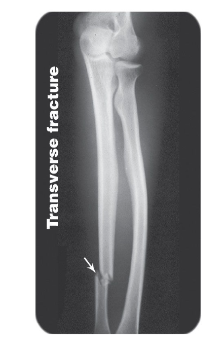
Transverse Fracture
break goes straight across the bone, broke in half along horizontal axis
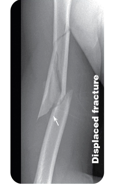
Displaced Fracture
broken bone pieces are out of normal alignment; into 2 of more pieces
Compression fracture
bone is crushed/compressed, often affecting vertebrae; osteoporosis; bone thinning

Spiral fracture
fracture twists around the bone, usually caused by a twisting force; long bone

Epiphyseal fracture
fracture happens through the epiphysis/growth plate area of a bone (end/head of bone)

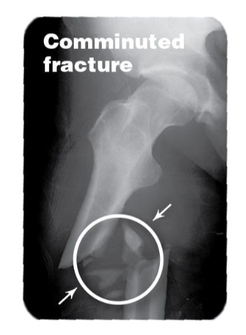
Comminuted fracture
shattered bony fragments broken into multiple pieces (3+)
Greenstick fracture
bone partially breaks and bends; not fully broken (most common in kids)

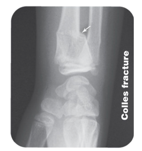
Colles fracture
fracture of the radius near the wrist (distal end), w broken end typically displaced backward
Pott's fracture
fracture involving the lower leg near the ankle (mallelar), typically involving the fibula & often the tibia too

4 steps of fracture repair?
Fracture hematoma formation → blood clot forms at the break
Callus formation → tissue forms around and stabilizes the break
Spongy bone formation
Compact bone formation
(not talk about yet)
What happens during fracture hematoma & callus formation?
Fracture hematoma formation
-A large blood clot forms; makes fibrous network
-Some bone cells near the fracture die
Callus formation
- endosteum & periosteum cells divide & move to fracture
-Calluses stabilize broken bone
Internal callus → forms inside the medullary cavity
External callus → cartilage & bone form around the break
(not talk about yet)
What happens during the final 2 steps of fracture repair?
1. Spongy bone formation
Osteoblasts replace the cartilage in the external callus w/ spongy bone
2. Compact bone formation
Spongy bone is replaced w/ compact bone
repaired bone may be slightly thicker & stronger than normal
(not talk about yet)
What is osteopenia, and how does aging affect bone mass
Osteopenia = reduced bone mass bc of lack of bone formation (ossification)
-Usually begins around ages 30–40
Bone loss each decade:
Women: about 8%
Men: about 3%
Most affected: epiphyses, vertebrae, & jaws
-Cause fragile bones, reduced height, & tooth loss
(not talk about yet)
What is osteoporosis?
Osteoporosis = severe loss of bone mass
bones weak & fragile
interferes w/ normal bone function
More common w/ increasing age
Sex hormones help maintain bone mass
-In women, bone loss increases after menopause
(not talk about yet) more severe
How can cancer cause bone loss?
cancerous tissues release osteoclast-activating factors
factors stimulate osteoclasts
Increased osteoclast activity → more bone breakdown
leads to bad osteoporosis
(not talk about yet)