Auditory System
1/89
Earn XP
Description and Tags
Midterm 2
Name | Mastery | Learn | Test | Matching | Spaced |
|---|
No study sessions yet.
90 Terms
Sound
audible sinusoidal fluctuations in air pressure : speed, frequency, amplitude, timbre
frequency
#cycles of air pressure per second, measured in Hertz (Hz)
pitch: perception of frequency
timbre
distinctive character of a sound based on relative intensities of different frequencies
each note has a “fundamental frequency” - lowest frequency produced → determines pitch we hear
harmonics - higher frequencies (harmonics & their timing determine the unique qualities of sound)
amplitude
magnitude of air pressure changes (decibels dB)
loudness: perception of amplitude
Outer ear anatomy
Pinna
Auditory canal
Tympanic membrane

Middle ear Anatomy
ossicles
eustachian tube
oval window

inner ear anatomy
cochlea
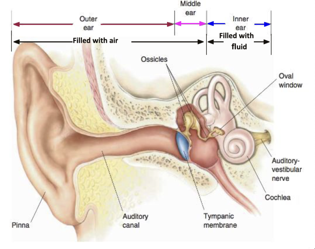
Functions of Outer Ear
Pinna ● Collects and funnels sound into auditory canal ● Vertical sound localization
Auditory canal ● Channels sound waves to the tympanic membrane
Tympanic membrane (eardrum) ● Vibrates in sync and at the same frequency as sound waves entering the ear
Which of the following would Picasso have trouble with if he lost his pinna?
Localizing sound in the vertical plane
Middle Ear Functions
Ossicles ● Amplify and transfer vibrations from the tympanic membrane to the oval window of the cochlea
Eustachian tube ● Equilibrates air pressure between both sides of the tympanic membrane
Oval window ● Transfers vibrations from the middle ear to the inner ear
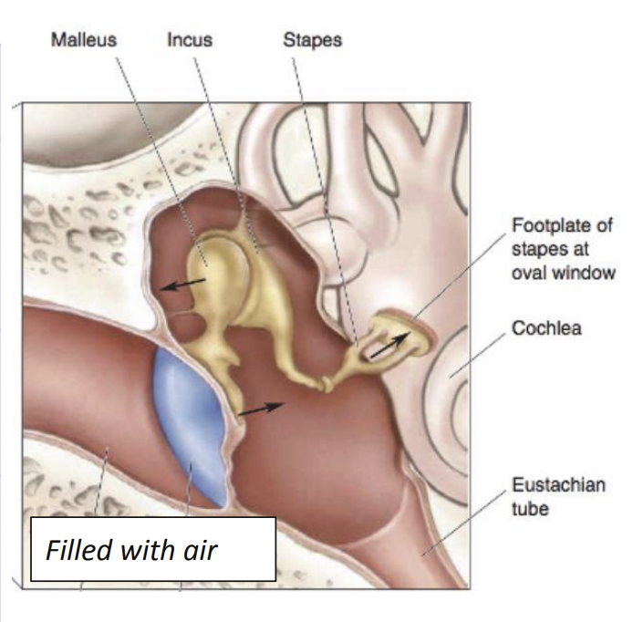
Ossicles
bones in middle ear
Malleus - hammer
Incus - anvil
Stapes - stirrup → Acts as a piston that moves in and out of the oval window to displace cochlear fluid
Fluid filled cochlea
the fluid is much harder to displace than air
Fluid in cochlea solution
To make sure sound waves don’t die out, need to amplify them (AKA impedance matching)
Impedance of air is low: easy to displace
Impedance of fluid is high: hard to displace How? → Ossicles!
Impedance Matching
We concentrate the force of the vibrations onto a small area on the footplate of the stapes (oval window) in comparison to the large area of the tympanic membrane → higher pressure
Lever action of ossicles increases force on oval window
Which of the following is NOT true about sound?
Sound waves will directly move the oval window
Attenuation Reflex
Loud sounds cause a reflexive contraction of two muscles
○ Tensor tympani: attached to the malleus
○ Stapedius muscle: attached to the stapes
Contraction of muscles → ossicles become more rigid (lose power to act as lever) → reduced sound conduction to cochlea
Useful bc→ Adapts ear to continuous loud noise: protects delicate ear machinery (hair cells)
Limits→ delay of 50-100ms: cannot protect against sudden loud sounds → effective at lower frequencies
Cochlea function
Auditory transduction: Sound waves → electrical signals
Cochlea anatomy
Scala vestibuli (assoc. w/ oval window)
Reissner’s membrane separates scala vestibuli and scala media
Scala media
Basilar membrane separates scala media and scala tympani
Scala tympani (assoc. w/ round window)
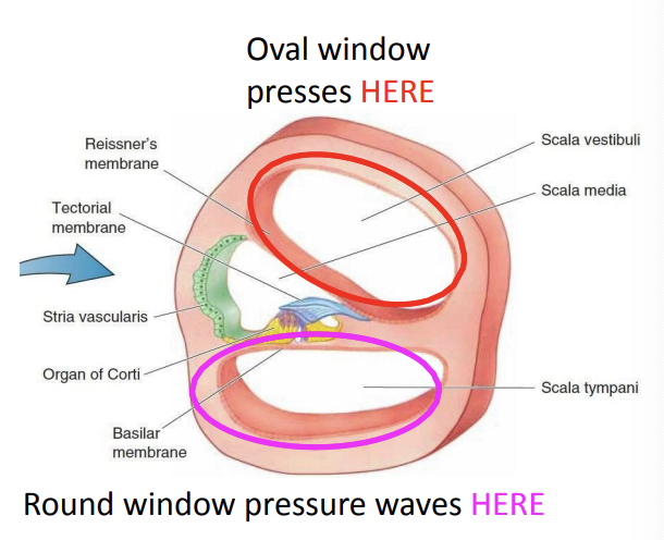
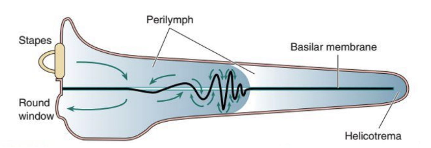
Cochlea flow of fluid
Oval window moves → fluid in scala vestibuli moves → goes down thru helicotrema → fluid in scala tympani moves → fluid pushes against round window
Cochlear Fluids
Perilymph - in scala vestibuli and tympani (low [K+], hi [Na+])
Endolymph - in scala media (hi [K+], low [Na+])
Endocochlear potential = +80 mV → Endolymph is 80 mV more positive than perilymph
Concentration gradients are maintained by stria vascularis → Absorbs Na+ from and secretes K+ into endolymph
![<ul><li><p>Perilymph - in scala vestibuli and tympani (low [<span style="color: blue;">K+]</span>, hi [<span style="color: red;">Na+]</span>) </p></li><li><p>Endolymph - in scala media (hi <span style="color: blue;">[K+]</span>, low <span style="color: red;">[Na+]</span>) </p></li><li><p><span style="color: purple;">Endocochlear potential = +80 mV</span> → Endolymph is 80 mV more positive than perilymph </p></li><li><p>Concentration gradients are maintained by <span style="color: green;">stria vascularis</span> → Absorbs Na+ from and secretes K+ into endolymph</p></li></ul><p></p>](https://knowt-user-attachments.s3.amazonaws.com/7d104dad-506f-46c4-a339-9c434e6c74b7.png)
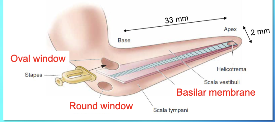
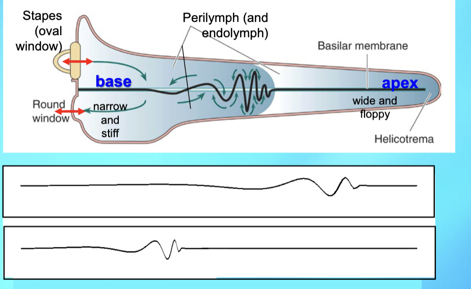
Basilar Membrane structure
Base: narrow, stiff
Apex: wide, floppy
organ of corti: sits on top of BM, contains auditory receptors (hair cells)
tectorial membrane: hangs over Organ of corti
Movement of Basilar Membrane
Stapes movement evokes a traveling wave on the basilar membrane.
Low frequency: vibrations greatest at apex
High frequency: vibrations greatest at base (tonotopy more accurate)
If frequency too low → membranes don’t move & fluid flows from SV to ST, goes thru helicotrema
Path of Basilar Membrane movement
Oval window displaces perilymph in scala vestibuli → displaces flexible Reissner’s membrane → displaces endolymph in scala media → displaces BM
tonotopy - tones (frequencies) are represented by their location on the BM
What does the Organ of Corti contain?
auditory receptors called hair cells
Hair cells
each hair cell has 10-300 stereocilia
sound waves → BM moves → stereocilia bend → neural signal
hair cells are NOT neurons (no axons, no AP) → they are epithelial cells
release excitatory transmitter (glutamate) onto spiral ganglion neurites
Inner hair cells
1 row
between modiolus & rods of Corti
stereocilia move in endolymph
provide 90% of input to SGC
outer hair cells
3 rows
further out from rods of corti
stereocilia stick to tectorial membrane
provide 10% of input to SGC
Hair cells release glutamate onto…
spiral ganglion cells
→ SGC are bipolar neurons that get input from hair cells and extend to CNS, somas located in SG in modiolus
→ SGC send axons to auditory nerve → cochlear nuclei in medulla Paradoxical Connections
One IHC connects to…
15-20 SGCs
→ auditory transduction
Many OHC connect to …
one SGC
→ cochlear amplification
IHC Stereocilia are unique:
Actin filaments make it so they only bend at the base (near the top of the hair cell)
Tip-link K+ channels at the tip of each
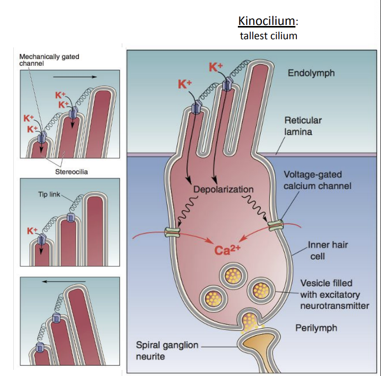
IHC Tip-link K+ channels are mechanically gated:
Tips point straight up → channels half open, half closed
Tips bend toward kinocilium → channels open more → K+ enters → depolarization
Tips bend away from kinocilium → more channels close → no depolarization

What causes K+ entry and depolarization in IHC
No [K+] gradient (high in endolymph and in cell), BUT… large voltage difference will drive K+ into cell
Endocochlear potential = +80 mV, which is 140 mV higher than inside cell
in age-related degeneration of stria terminalis - the K+ concentration of endolymph is insufficient → deafness results w/o endocochlear potential
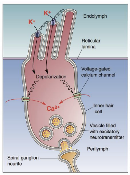
Effect of depolarization in IHC?
Depolarization → VG Ca++ channels open → Ca++ enters → IHCs release Glu onto spiral ganglion cells
Graded synapse: amt. of depolarization determines how much Glu is released
Sound waves → displacing BM → stereocilia bend back and forth → alternating depolarization and hyperpolarization
What are OHCs doing?
OHCs amplify movement of BM, particularly so we can hear low intensity stimuli
OHC length changes → BM movement amplified → V m changes amplified (pos feedback)
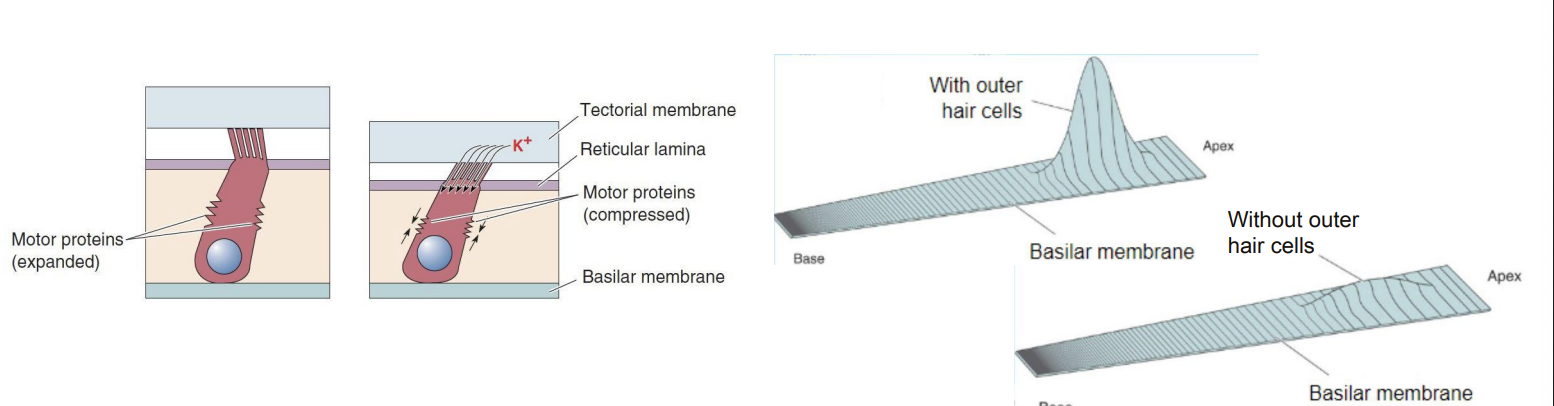
Prestin
motor protein in OHC membrane; can change length in response to changes in Vm
Full Summary of Hair Cell path
Sound waves cause tympanic membrane to vibrate
Sound amplified from tympanic membrane via ossicles to oval window
Displacing oval window membrane displaces perilymph in scala vestibuli
Displacing perilymph displaces Reissner’s membrane → displaces endolymph
Displacing endolymph displaces BM → makes stereocilia bend towards kinocilium
BM displacement amplified by OHCs
Tip link K+ channels open and let K+ into IHC due to endocochlear potential difference
In IHCs, Depolarization by K+ opens VG Ca2+ channels
Ca2+ triggers Glu release from IHCs a. Graded synapse - voltage level determines amount of Glu released
Glu affects postsynaptic spiral ganglion cells → AP!
auditory transduction is faster than would be possible with ion diffusion and faster than visual transduction that uses G-proteins
What non-bony structure does the stapes directly push on?
Oval window
How is a receptor potential generated in a cochlear hair cell when sound makes stereocilia bend?
The fluid surrounding the cilia is more positive than the inside of the hair cell and this potential difference allows K+ ions to flow inward
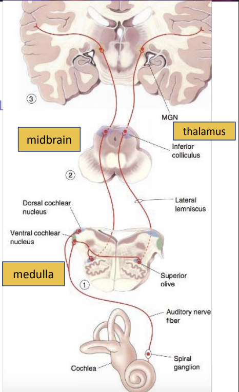
Auditory Pathway
1) Cochlea
2) Spiral ganglion neurons
Auditory nerve
3) Ventral and dorsal cochlear nuclei (Medulla)
4) Ventral cochlear nuclei (Decussation)
5) both superior olive
lateral lemniscus
6) Inferior colliculus
7) MGN (in thalamus)
Acoustic radiation
8) Auditory cortex
Tonotopy: characteristic frequency
The frequency of sound at which a given neuron is most responsive
→ neurons near each other have similar characteristic frequencies
*tonotopy does not extend to frequencies <200Hz bc neurons do not have this low of characteristics
Tonotopy on Basilar Membrane
A specific point on the BM is displaced maximally by a specific frequency; frequencies are represented by their location on the BM
Tonotopy on Auditory Nerve
Each auditory nerve fiber is sensitive to a range of frequencies around a characteristic frequency; which neurons are active indicates frequency
Tonotopy on Cochlear Nuclei
Auditory axons synapse in cochlear nuclei in an organized pattern based on characteristic frequency
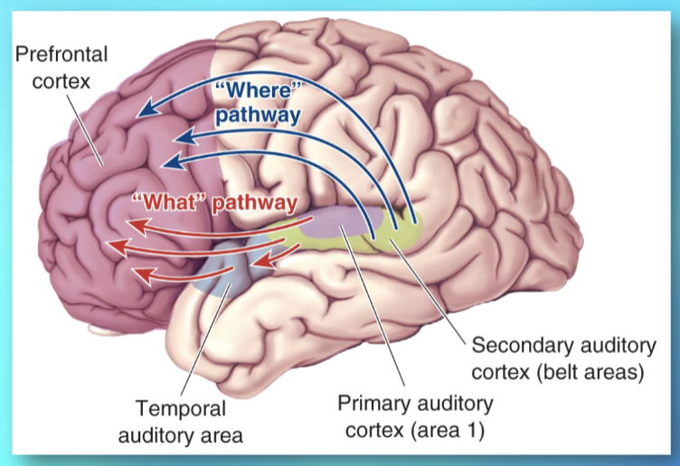
Primary Auditory Cortex (A1)
Tonotopic organization on basilar membrane is preserved in A1!
Isofrequency bands: neurons organized in columns according to frequency
Similar to orientation columns in V1
Both A1 cortices get input from both ears → lesioning one side impairs sound identification and localization BUT doesn’t cause unilateral deafness
Dorsal Stream
pure tones and sound localization (“where?”) → areas above A1 toward parietal and frontal lobes

Ventral stream
complex sound identity (“what?”) → areas below A1 toward temporal and frontal lobes (ie monkey vocalizations)
A lesion of which of the following would produce deafness in the left ear?
Left cochlear nucleus
Auditory Coding Pathway
Sound Vibrations → Hair Cells → Auditory Nerve Fibers (ANFs) → Auditory Centers
What does the Auditory Pathway encode?
1) Intensity (loudness)
2) Frequency (pitch)
○ At high, intermediate, and low frequencies
3) Location
○ At high and low frequencies ○ Vertical and horizontal localization
How does the Auditory System encode intensity of sound?
At higher sound intensities, the BM vibrates with a greater amplitude, which causes…
● hair cells to depolarize and hyperpolarize at a higher frequency, which causes auditory neurons to fire APs at a higher frequency
● more hair cells to be depolarized and hyperpolarized, which then causes more auditory neurons to fire APs
What is Tonotopy?
A specific point on the BM is displaced maximally by a specific frequency
Range of 200-20,000 Hz (high & intermediate frequencies)→ because neurons do not have characteristic frequencies < 200 Hz
What is Phase locking (spike timing)?
Neuron fires consistently at same phase of a sound wave
A frequency might be represented by one cell alone or by multiple cells working together (volley principle)
Range of 20-5,000 Hz (intermediate & low frequencies)→ because neurons can only fire so often
What is Volley principle?
take a group of phase-locked neurons, we can make up for the fact that they might not fire every cycle
Some neurons will fire when others don’t, so with a good enough group, we will have at least 1 neuron firing every cycle
Very helpful for intermediate frequencies that might be a little too fast for neurons to fire every cycle
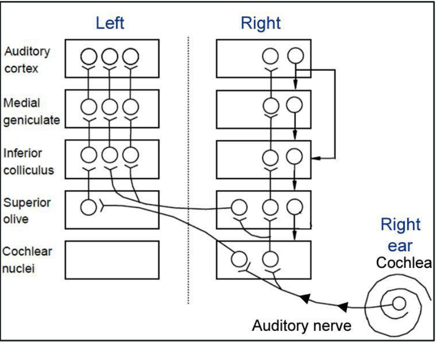
Horizontal Location
AKA azimuth
Starting at the superior olive, ascending auditory structures get input from both ears (i.e. binaural input) → inputs from ears combined for sound localization
Interaural Intensity Difference
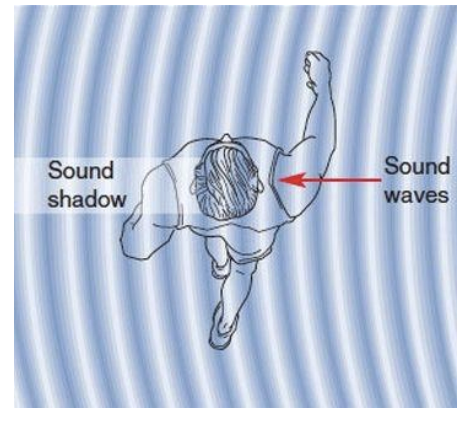
higher frequency sounds (>1.5 KHz), the head casts a “sound shadow” that dampens intensity of sound at the other ear
moving head around detects interaural intensity difference → infer sound direction
Interaural Time Delay
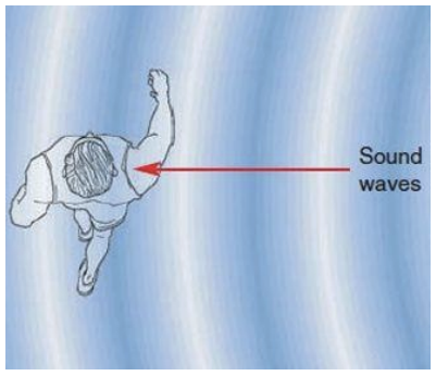
Lower freq. sounds do not produce a sound shadow due to diffraction (sound waves bend around head)
Sound wave peak reaches each ear at different times (right ear hears sound from right side first)
What parts of the superior olive are used for Azimuth Sound Localization?
MSO: medial superior olive nucleus
Neurons in the MSO are tuned for interaural timing difference (delay) at lower frequencies (20 - 2000 Hz)
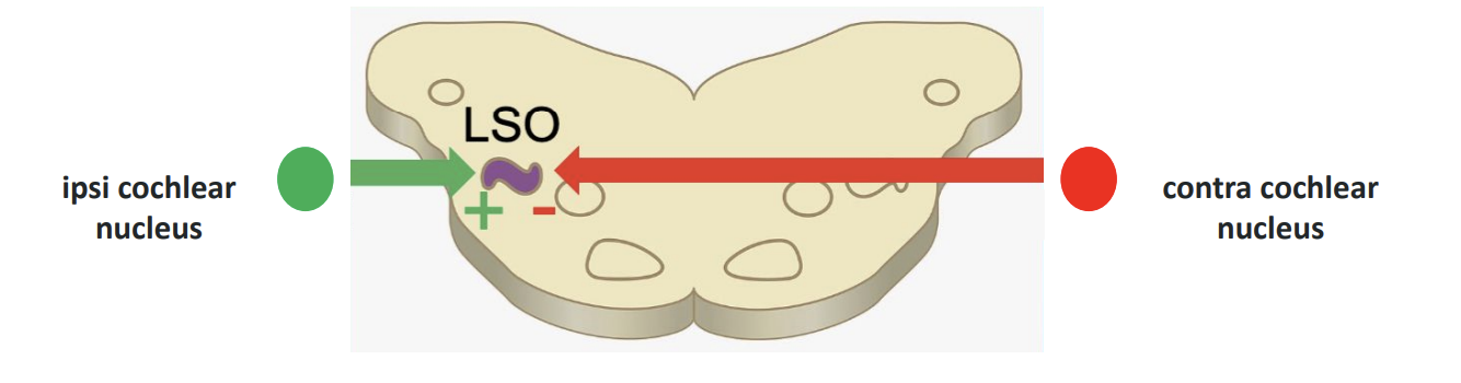
LSO: lateral superior olive nucleus
Neurons in the LSO are tuned for interaural intensity difference at higher frequencies (2,000 - 20,000 Hz)
What are some MSO neurons sensitive to?
delays in time → arranged in delay lines and tuned for specific time delays
(Negative delay = sound reached left ear first ; Positive delay = sound reached right ear first)
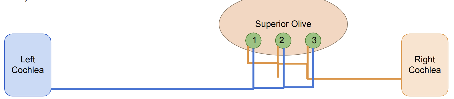
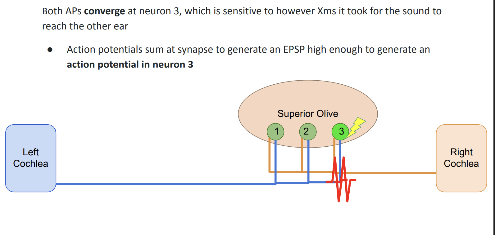
Interaural Time Delay example pathway (MSO)
sound comes from left side → reaches left cochlea first → creates AP in left auditory nerve → cont down left auditory nerve → X ms later, sound reaches right cochlea → right cochlea generates AP down right auditory nerve ——→ both AP converge at neuron 3 (sensitive to amt of Xms it took for sound to reach other ear)
AP sum at synapse to generate an EPSP high enough to generate an AP in neuron 3
Interaural Intensity Difference example pathway (LSO)
Excitatory input from the ear ipsilateral to LSO
Inhibitory input from the ear contralateral to LSO
— Spike rate varies continuously as sound direction moves:
More intense sound to ipsi ear → gradual decrease in spiking
More intense sound to contra ear → steeper decrease in spiking
Vertical Auditory Coding
Pinna directs sound to auditory canal
→ better at higher frequency sounds (esp from high elevations) into canal
→ can enter directly or indirectly (sound reflected towards canal, cause intensities of diff freq to inc or dec based on origin)
direct & indirect paths interfere → spectral shaping → brain can tell original elevation of sound
monaural - each pinna get own unique input
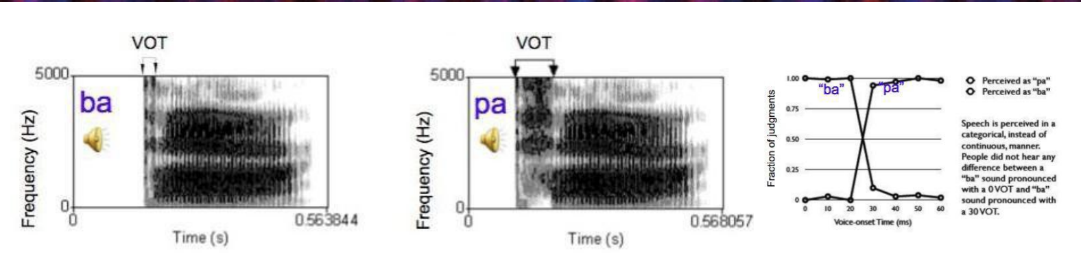
Timing in Speech Interpretation (graphs)
Axes: frequency vs time (darker color = more intense sound)
VOT: voice onset time = Time btwn consonant release and vocal cord vibration (difference in VOT can make the difference in the sound we hear)
Otoacoustic Emissions
Ear is both a detector and emitter
Otoacoustic emission: A sound generated by the ear == Caused by OHC amplification of BM
What are the 2 types of Otoacoustic Emissions?
Spontaneous Otoacoustic Emissions: No stimulus needed -occurs in about 50% of people - sometimes associated with damage to ear
Evoked Otoacoustic Emissions: CLICK played into ear, after short delay, sound will be played back. Used for testing hearing noninvasively in babies
Causes of Congenital Deafness
Genetics (some associated w down or usher)
Prenatal infection (in mother or fetus - rubella, cytomegalovirus, meningitis)
Ototoxins in utero (hair cell loss - certain antibiotics)
Causes of Acquired Hearing Loss
Excessive exposure to loud noise
Infection
Autoimmune disease
Aging
Ototoxins: aspirin, certain antibiotics, diuretics (e.g. furosemide), chemotherapy drugs
What is the pathology cause of most hearing issues?
Hair cell damage or inner ear damage.
very fragile, and over time they can be damaged by loud noises, infection etc. → goes undetected till serious
OHCs are more vulnerable to damage
Hair cells responding to high frequencies are more vulnerable to damage
Hair cells DO NOT regenerate
Causes of conductive hearing loss (outer or middle ear damage)
Bacteria or virus causes inflammation in middle ear
Abnormal bone growth in the middle ear
Occlusion of ear canal
Developmental defects
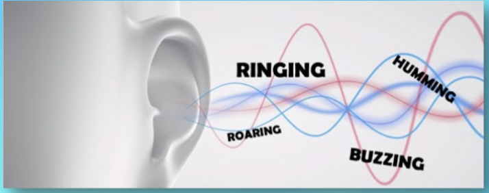
Tinnitus
Sound perception in absence of stimulus
Linked to hyperexcitable and elevated spontaneous activity in auditory system
Associated with: hearing loss, anxiety, exposure to loud noise, ototoxic drugs
no cure, treat w white noise or sound masking machines
Amusia
Tone deafness
Issues with perceiving, memorizing, and producing music, pitch
discrimination, discriminating instruments, rhythm, timingUsually accompanied by other deficits (cognitive, aphasia)
right hemi considered dominant (opp of language)
Wernicke’s Aphasia
poor language comprehension
Wernicke’s area: higher order auditory area, involved in word interpretation (damage of this area), may be specialized for storing memories of sounds that make up words
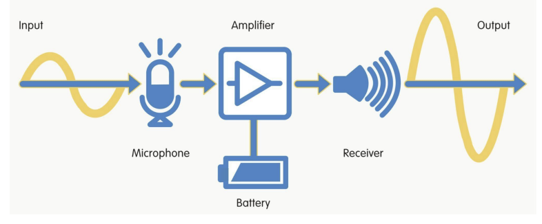
Hearing aids
Amplify sound from microphone and deliver to speaker in the ear
Frequencies amplified can be tailored
Cochlear Implants
Takes sound wave frequencies and stimulates the respective spiral ganglion cells directly
Bypasses hair cells and exploits the tonotopy of the cochlea → stimulating different
places on the basilar membrane (base...apex) to evoke different sensations of pitch
has about 12-24 electrodes stretching along length of scala tympani
The following statements about sound localization are correct EXCEPT:
High frequency sounds do not generate sound shadows
What is meant by the “cochlear amplifier”?
Outer hair cells amplify movements of the basilar membrane
Which nucleus in the auditory system receives input only from the ipsilateral ear?
Cochlear nucleus
Which is the most important factor used to represent the frequency of a 50 Hz sound?
Phase locking
The auditory system determines the horizontal location of a 10 kHz sound (azimuth) based on
Interaural intensity difference
what is the speed of sound?
340 m/sec (770 mph)
what is hearing damage related to?
sound intensity (not loudness)
why do we use the ossicles?
Because the cochlea is filled with fluid. Without the
ossicles, 99.9% of sound energy would be reflected off the
eardrum and back into the environment.
Large, low-pressure eardrum vibrations become small, high-pressure movements at the oval window
The stapes footplate is about 30× smaller than the tympanic membrane
Ossicles amplify force through lever action
Movements are extremely tiny—only a few nanometers at hearing threshold
What neurons are sensitive to sound elevation?
Neurons in the MGN and A1 are sensitive to sound elevation
This is probably based on sensitivity to spectral shape
(i.e. the ratio of different sound
frequencies)
how do owls hunt in the darkness?
with precise azimuth and elevation information (their ears are at diff heights)
cocktail party effect
ability to focus attention on one stimulus and filter out others
→ based on sound frequencies & location
What vs where in human auditory cortex
Pictures and sounds simultaneously presented
Where= pictures appear on L or R side, sound came from L or R side of headphones
What= sound and picture semantically consistent (dog barking) or inconsistent (horse picture with dog barking)
subjects asked to report:
1. Location task: did visual and auditory stimuli
appear on the same side?
2. Recognition task: were visual and auditory stimuli
semantically consistent?
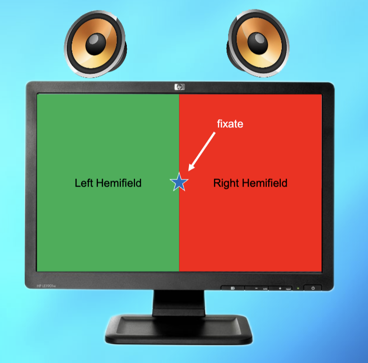
Brain activation in what vs where tasks
Higher parietal lobe activity in location task than recognition task
Higher temporal lobe activity in recognition task than location task
How loud is safe?
below 60 dB is safe
• Avoid prolonged exposure above 70 dB
• Club concerts are often 120 dBMaximum earbud/airpod output is typically ≥ 100 dB
• Volume loud enough to block out ambient
sound is typically 80 dB or higher
• Limit exposure to 90 min at 80% max volume
• Noise isolating or canceling headphones help