Anatomy Lecture Exam 2 Study
1/65
There's no tags or description
Looks like no tags are added yet.
Name | Mastery | Learn | Test | Matching | Spaced | Call with Kai |
|---|
No analytics yet
Send a link to your students to track their progress
66 Terms
Name the cerebral arteries
Posterior cerebral artery
Middle cerebral artery
Anterior cerebral artery
Where do each of the cerebral arteries supply blood to the brain?
Posterior: Temporal and occipital
Middle: Frontal, parietal, temporal
Anterior: Frontal, parietal, and occipital lobes
Describe the Pineal body
Produces melatonin
Helps maintain the circadian rhythm
When the environment is dark, more melatonin is produced; when its light less melatonin is produced.
The cycle can be disrupted by too much light at night, too little in the day.
Describe the Medulla
Inferior portion of the brain stem.
Contains the cardiac, respiratory, and vasomotor centers.
Autonomic functions of maintenance of heart rate, breathing, and blood pressure.
Describe the Pituitary gland
Known as the “master gland.”
Smaller than most glands that regulate hormones.
Produces hormones that influence the thyroid gland, the adrenal gland, and the gonads (thyroid stimulating hormone, adrenocorticotropic, and gonadotropic hormone).
What does the pituitary gland produce?
Hormones: thyroid stimulating hormone, adrenocorticotropic, and gonadotropic hormone
Describe Stroke Traits as opposed to brain tumors
Lack of blood, lack of oxygen to brain tissue; possible death of brain tissue.
Four arteries supply all blood to the brain - vertebrals and internal carotids.
Rapid onset.
Implies death of brain tissue due to lack of oxygen.
Describe Brain tumors traits as opposed to strokes
Slower onset (weeks or months).
Implies brain compression by a “space occupying lesion.”
Why might a partially occluded vertebral artery cause fainting?
The vertebral artery is partially confined by neck (cervical) vertebrae, and twisting these vertebrae may cause distortion and further occlusion of the vertebral artery. Which means lack of blood flow, which means lack of oxygen.
Transient Ischemic Attack (TIA)
Some call it a mini-stroke
Usually passes quickly with little residual defect.
Can be a sign of an increased risk of stroke
Give an Example of Functional deficits
Posterior cerebral arterial occlusion could result in oxygen deprivation of the occipital area, resulting in a stroke and possible blindness
What is Anastomosis
The joining of two arteries.
Communication between blood vessels by means of collateral channels, especially when usual routes are obstructed
Advantage of the circle of willis
Availability of blood from the unblocked side being reverted to the blocked site; creates redundancies and helps to avoid symptoms of ischemia
Bell’s Palsy
Sudden weakness in muscles on one side of the face.
Temporary weakness.
½ of face appears to droop.
Smiles are one sided.
Eye on affected side resists closing.
Significantly improves over weeks.
Some may have bell’s palsy for life (rare)
Exact cause is unknown - May be due to a viral infection.
Know Cranial Nerves of the Eye
Superior oblique: trochlear nerve (CN4)
Lateral Rectus: Abducens (CN6)
All the rest (Superior Rectus, medial rectus, inferior rectus, inferior oblique): oculomotor (CN3)
Medial rectus action and innervation
innervated by CN lll, rotates eye inward
Lateral rectus
Innervated by CN VI, rotates eye outward
Superior rectus
innervated by CN III, rotates eye upward and inward
Inferior rectus
innervated by CN III, rotates eye downward and inward
Superior oblique
innervated by CN IV, rotates eye “down and out”
Inferior oblique
innervated by CN III, rotates eye upward and outward
Trigeminal Dermatomes
V1: Ophthalmic Division
V2: Maxillary Division
V3: Mandibular Division
V1: Ophthalmic Division
Sensory to skin of forehead, nose, nasal mucous membranes, cornea of eye, other structures.
V2: Maxillary Division
Passes into foramen rotundum.
Main branch exits through the infraorbital foramen. Sensory to skin of cheek, lower eyelid, upper jaw, teeth, maxillary sinus, and adjacent structures.
V3: Mandibular Division
Passes through the foramen ovale.
Sensory to skin of lower jaw, teeth, and gums, external ear, temporomandibular joint.
A motor division of this nerve passes along with V3 to innervate muscles of mastication (chewing), namely the masseter muscle and temporalis muscle.
Trigeminal Neuralgia
Nerve damage, inflammation of the trigeminal nerve.
Sudden, intense pain on one side of the face; pain can feel like an electric shock.
Cause is not clear.
Mumps
Communicable viral disease.
Parotid (salivary) gland (inflammation and swelling).
Pain with chewing, swallowing, fever, muscle aches, fatigue.
Can cause deafness, inflammation of testes, sterility.
Why does a scalp bleed heavily when cut?
Scalp has lots of blood vessels - vascular
Which arteries must not be injected by a dentist
Inferior alveolar
injectate usually contains epinephrine (a vasoconstrictor) which then remains in area of termination of the artery, resulting in poor arterial perfusion of the area, possible death of tissue
Function of tonsils?
Tonsils are part of the lymphatic system. Defends the body from infection; tonsils contain a lot of white blood cells, which kill germs.
Apparent function of palate in mammals.
Suckling
Function of Eustachian (auditory) tube.
Protects the tympanic membrane (eardrum). Equalize pressure in the middle ear, drains middle ear.
Do all sinuses connect with atmosphere (can drain fluid)?
Yes
Why do vertebrates have sinuses?
Deal with pressure change, reduce weight, shock absorption, resonance.
Which bone normally breaks when a human is strangled?
Hyoid bone
Where could an emergency cricothyroidotomy be performed?
Cricothyroid membrane
Thyroid gland Features
Butterfly shaped
Two lobes
Produces calcitonin
Parathyroid gland features
Two pairs small, oval-shaped glands
Produces parathyroid hormone that regulates blood calcium level
Hyperthyroidism (Graves)
Overactive thyroid,thyroid gland produces to much of the hormone thyroxine
Can accelerate body’s metabolism causing unintentional weight loss.
Rapid, or irregular heartbeat.
Bulging eyes
Hypothyroidism (Hashimotos)
Underactive thyroid.
Can cause obesity.
Hair loss
Joint pain, infertility, heart disease, fatigue
Which joint allows nodding?
Atlas, Atlanto-occipital.
What Joint allows Shaking head (no)?
Axis, Atlanto-axial
Define scoliosis.
Curvature of the spine, curve goes off to the side.
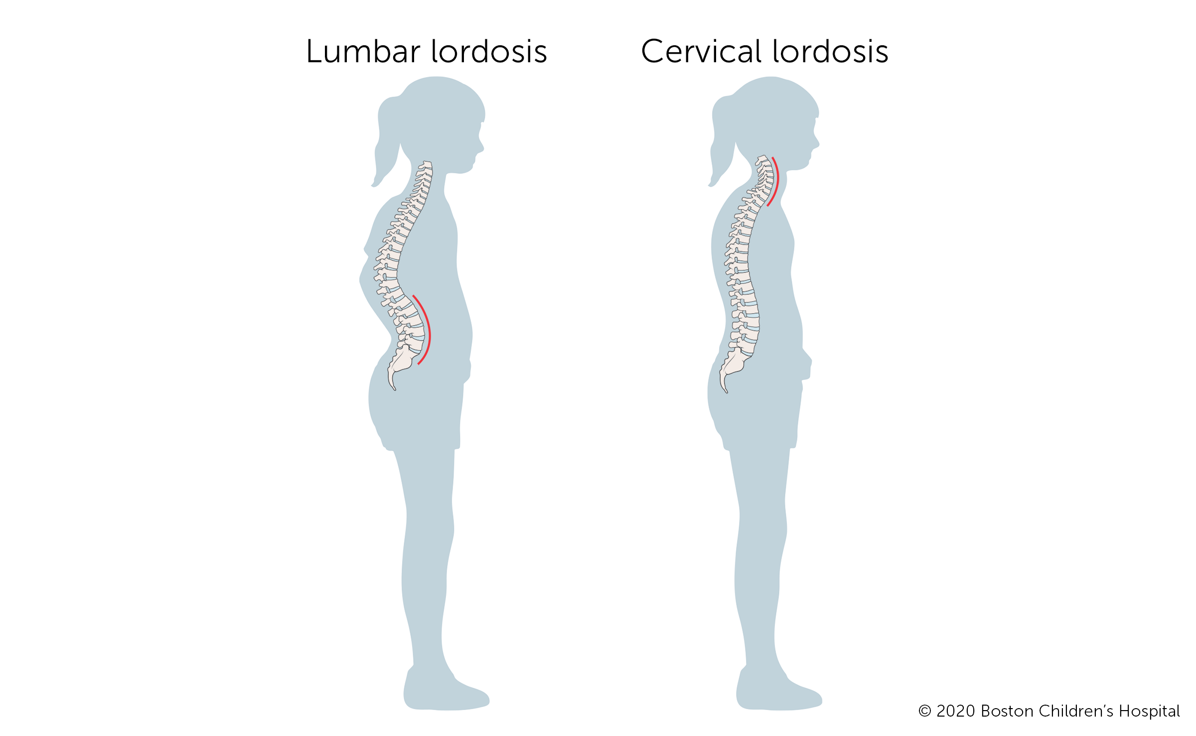
Describe lordosis. (Swayback)
Abnormally increased curvature of cervical and lumbar spine.
Describe kyphosis.
Curvature thoracic.
Hunchback (hyperkyphosis): worsens with age and osteoporosis; more common in women
Heart failure commonly swells which neck vein?
External jugular vein
Define stenosis
Narrowing or stricture of a anatomical tube
Where are the baroreceptors found (bp receptors)?
Located at the bifurcation of the carotid. Sends messages via CN IX (glossopharyngeal) to hypothalamus which sends autonomic response to heart to adjust and correct the potential problem.
What is a carotid body?
A group of cells near carotid bifurcation which are “chemoreceptors,” sensing the concentration of oxygen and carbon dioxide in the blood, also the pH, then sends messages via CN IX (glossopharyngeal) to hypothalamus which sends autonomic message to the medulla to change the respiratory rate appropriately.
Define a Goiter
Swelling of the thyroid gland.
What is the brachial plexus?
Network of nerves; landmark for anterior and posterior scalenes.
If you contract your right sternocleidomastoid muscle how will your head tilt?
Up and left.
Describe a pulled elbow
injury to annular ligament
Define joint capsule
thin, fibrosis sac containing fluid enclosed in a joint.
Define bursa
Closed sac of serous membrane which contains a thin layer of lubricating fluid secreted by the membrane
Define tendon sheath
A layer of membrane around a tendon
Define luxation
Dislocation of a joint.
Define subluxation
joint “out of place” but partially functional.
Partial dislocation
Describe what “double jointed” actually means
Hypermobility
Define flexion
decrease an angle of a joint.
Define extension
Increasing angle of a joint.
Define adduct
Moving toward medial plane
Define abduction
Moving away from the medial plane
What are the three joint types?
Fibrous
Cartilaginous
Synovial
Types of Cartilage
Hyaline
Elastic
Fibrous
Hyaline Cartilage Feature
Hyaline cartilage lines joints. It has little ducts that secrete synovial fluid to lubricate the joint to allow it to move freely.