Wish list - Skull (Completed) - Click on images to see arrows/zoom in
5.0(1)
Card Sorting
1/137
Earn XP
Description and Tags
Listen to this song: https://youtu.be/Yx2T1WwTV1U. All bones needed (BIO LAB) in skull!!!!!!!!!
Study Analytics
Name | Mastery | Learn | Test | Matching | Spaced |
|---|
No study sessions yet.
138 Terms
1
New cards
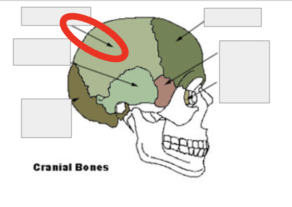
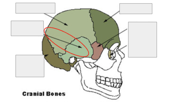
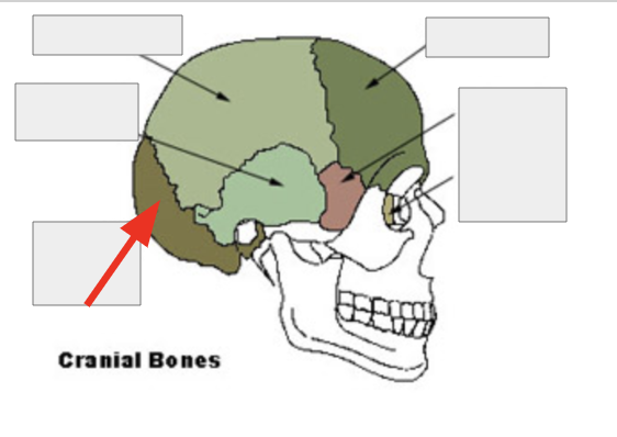
How many Cranial Bones are there
8
2
New cards
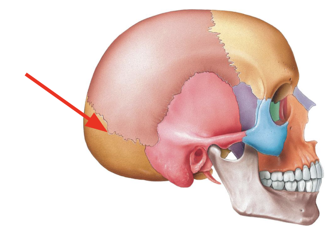
What are the different bones in the CRANIAL region
Parietal (2)
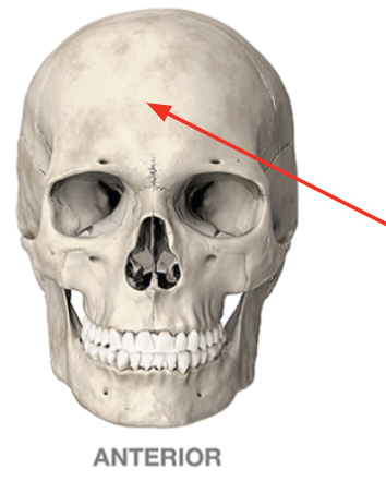
Temporal (2)
Frontal (1)
Occipital (1)
Ethmoid (1)
Sphenoid (1)
Temporal (2)
Frontal (1)
Occipital (1)
Ethmoid (1)
Sphenoid (1)
3
New cards
Parietal
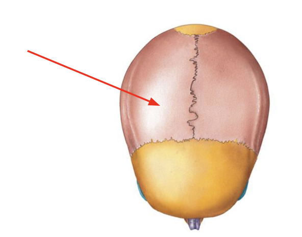
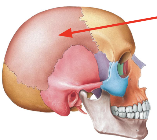
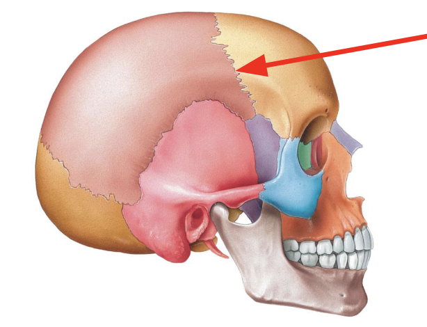
4
New cards
Temporal
5
New cards
Frontal
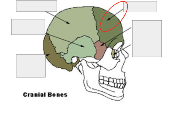
6
New cards
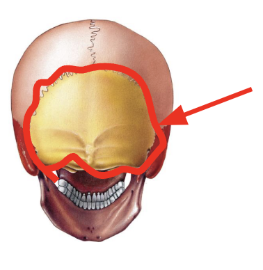
Occipital
7
New cards
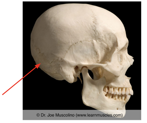
Ethmoid
8
New cards
Sphenoid
9
New cards
Occipital Bone
10
New cards
Occipital Bone
11
New cards
Occipital Bone
12
New cards
Occipital Bone
13
New cards
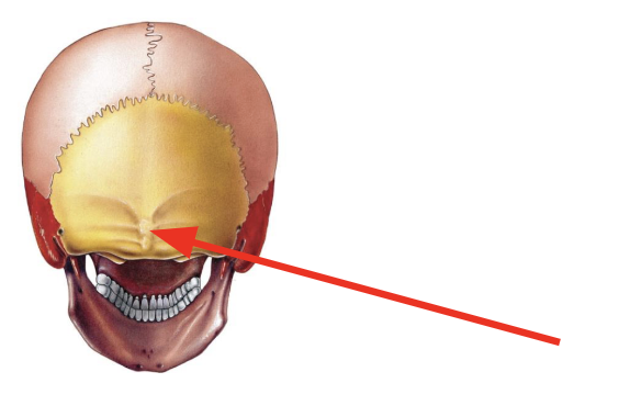
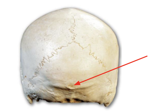
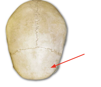
External occipital protuberance
The bump on the posterior side.
14
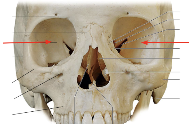
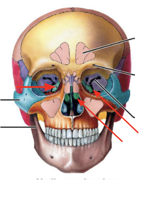
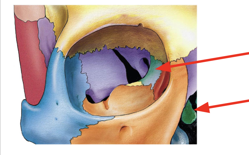
New cards
External Occipital Protuberance
15
New cards
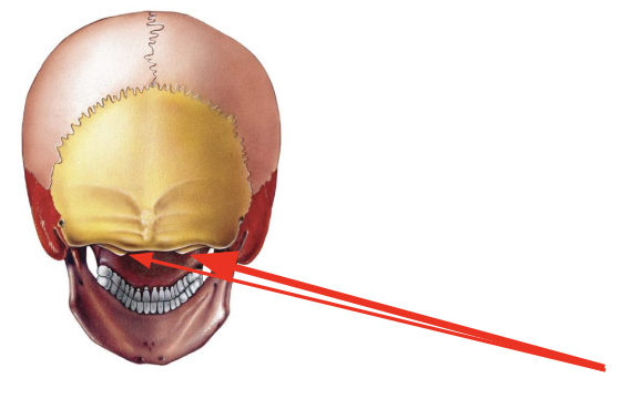
Occipital Condyle__***s***__
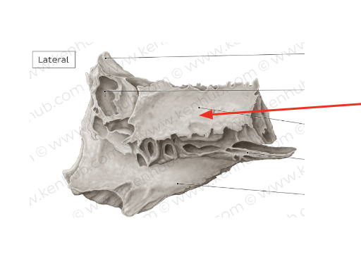
Articulate with the atlas
16
New cards
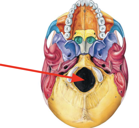
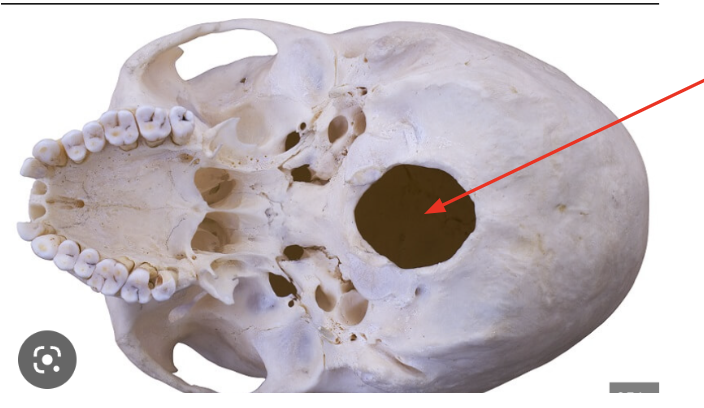
Foramen Magnum
Large hole for the spinal cord to pass through
17
New cards
Foramen Magnum
18
New cards
Occipital condyles
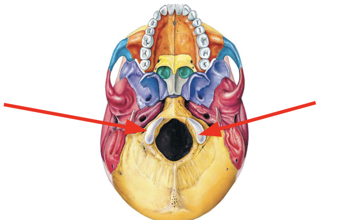
19
New cards
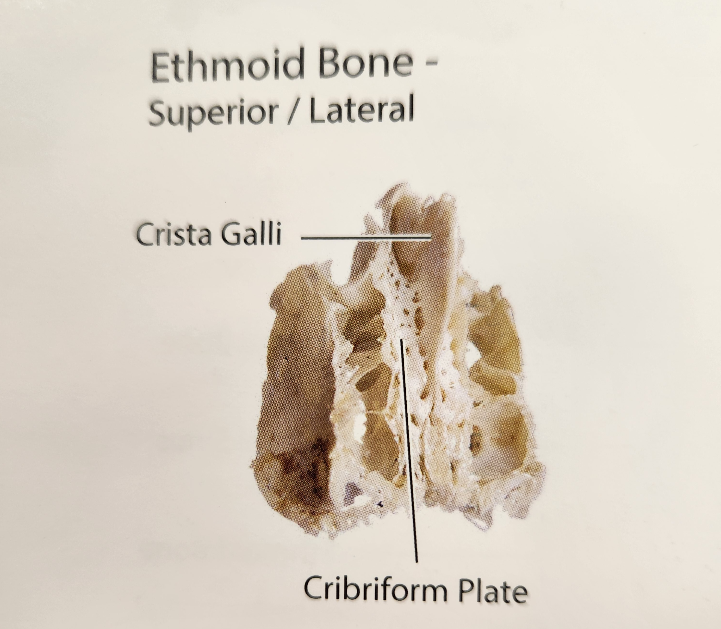
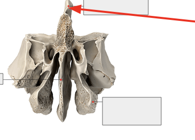
Occipital Condyles
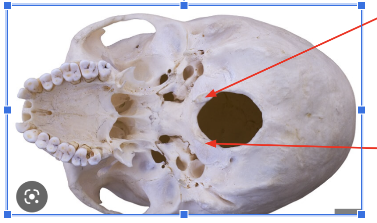
20
New cards
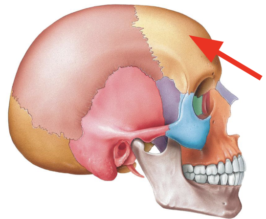
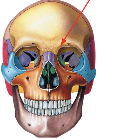
Frontal Bone
Forehead
21
New cards
\
22
New cards
Frontal Bone
23
New cards
Frontal Bone
24
New cards
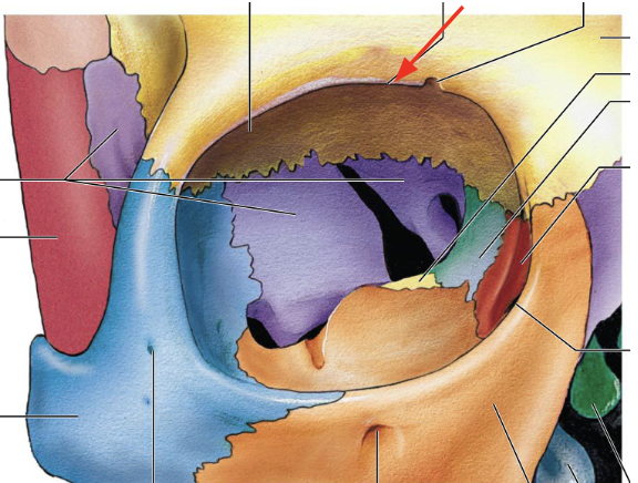
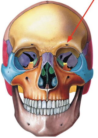
Supraorbital Margin
Ridge of bone above each eye
25
New cards
Supraorbital margin
26
New cards
Supraorbital Foramen
Hole above the supraorbital margin
27
New cards
Supraorbital Foramen
28
New cards
Supraorbital foramen

29
New cards
Right Parietal Bone
30
New cards
Left Parietal Bone
31
New cards
Left Parietal Bone
32
New cards
Right Parietal Bone
33
New cards
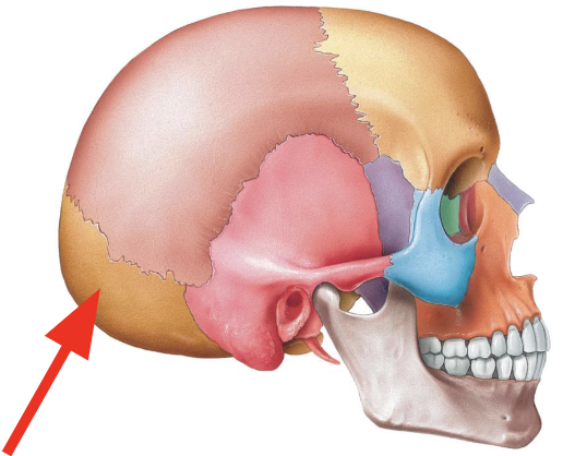
Parietal Bones
Form part of the sidewall and roof of the cranium
34
New cards
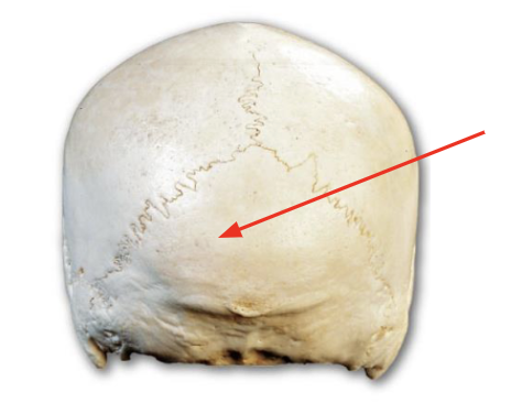
Coronal suture
Junction between frontal bone and 2 parietal bones
35
New cards
Coronal Suture
36
New cards
Coronal Suture
37
New cards
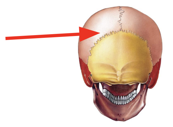
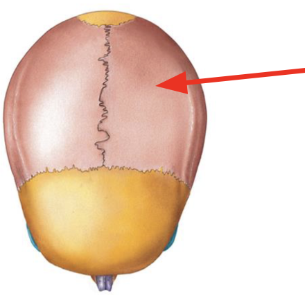
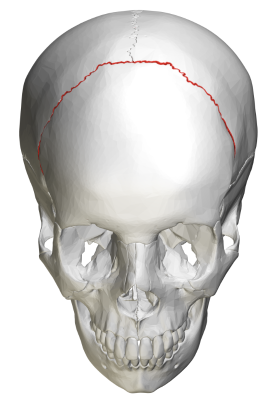
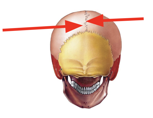
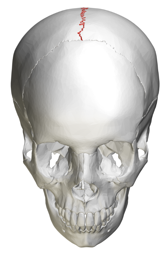
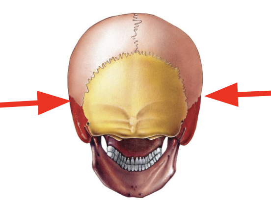
Sagittal Suture
Junction between 2 parietal bones
38
New cards
Sagittal Suture
39
New cards
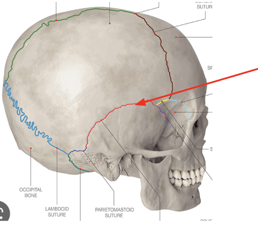
Squamous Suture
Junction between temporal bone and parietal bones
40
New cards
Squamous Suture
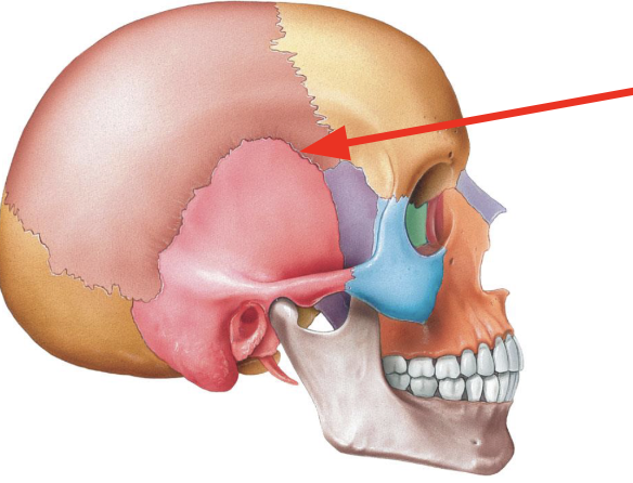
41
New cards
Squamous suture
42
New cards
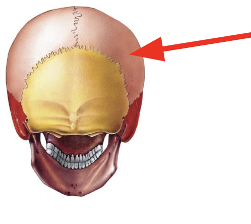
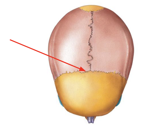
Lambdoidal suture
Junction between 2 parietal bones and the occipital bone
43
New cards
Lambdoidal Suture

44
New cards
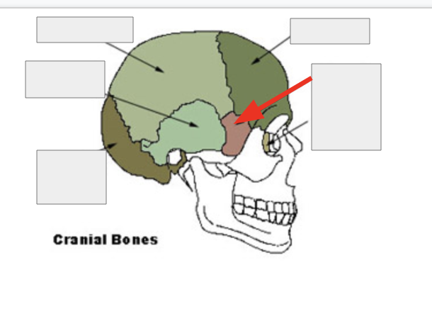
Temporal Bones
Form part of the sidewall of cranium, the temple
45
New cards
Temporal Bone
46
New cards
External auditory meatus
Ear canal
47
New cards
Styloid Process
Elongated, pin-like process on inferior side
48
New cards
Mastoid process
Large bony mass inferior and posterior to ear
49
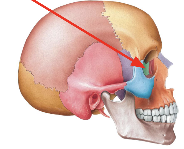
New cards
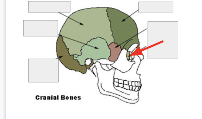
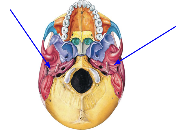
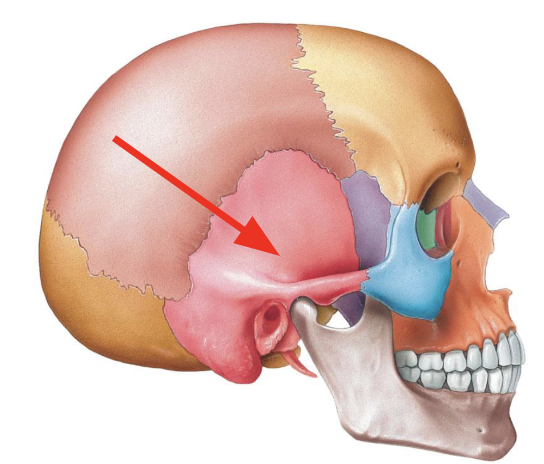
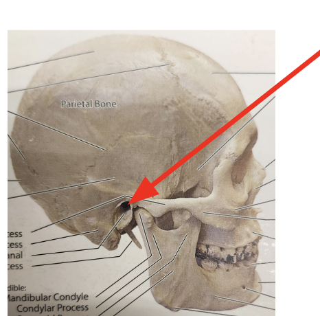
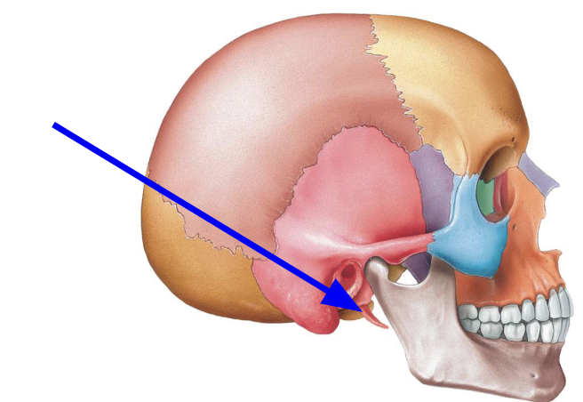
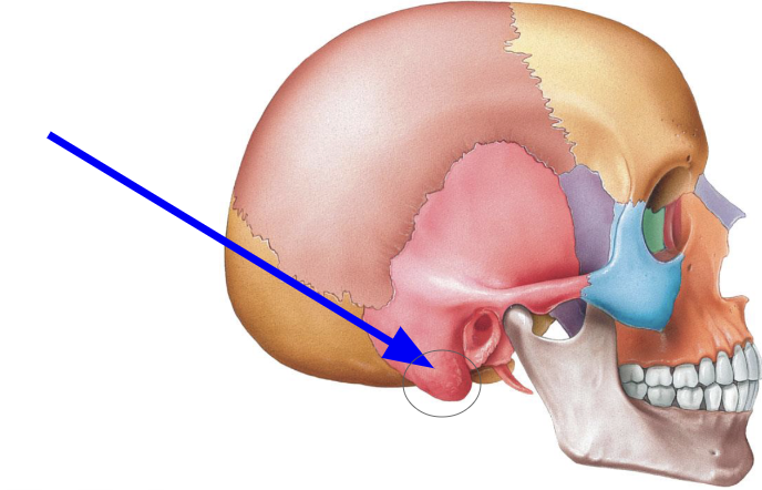
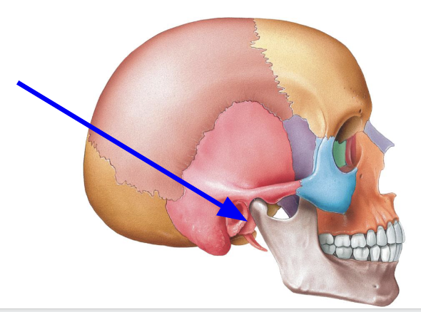
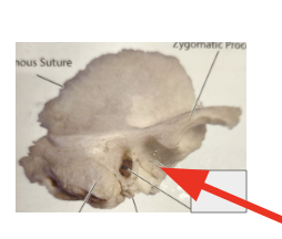
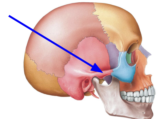
Mandibular fossa
Articulates with the mandibular condyles on mandible
\
\
Note this diagram should be annotated → The mandibular fossa in this picture is hidden, its underneath the condylar process
\
\
Note this diagram should be annotated → The mandibular fossa in this picture is hidden, its underneath the condylar process
50
New cards
Mandibular Fossa
51
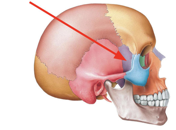
New cards
Zygomatic process
Projection of temporal bone
52
New cards
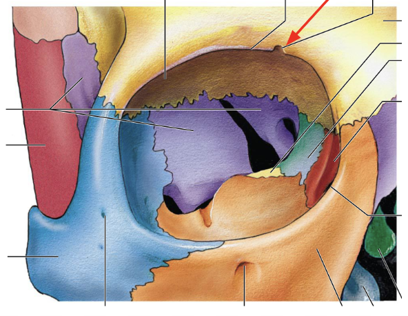
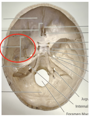
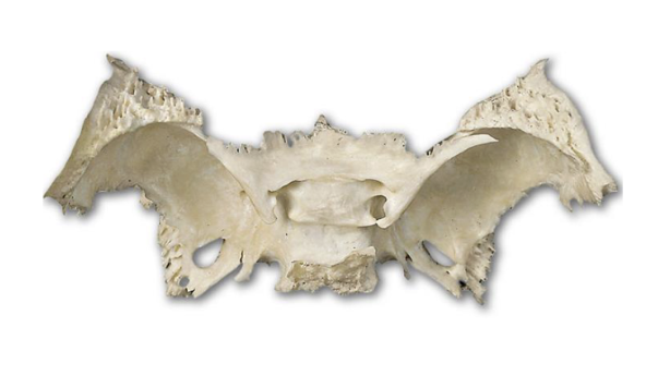
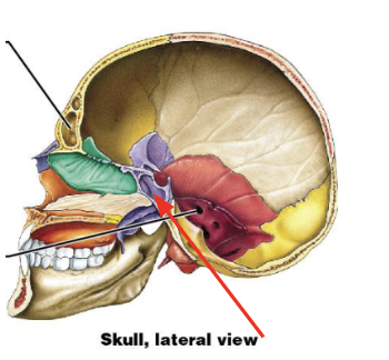
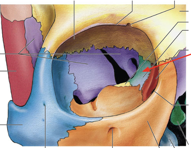
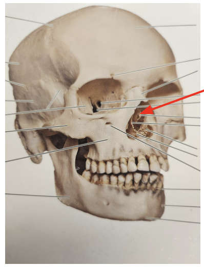
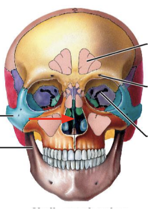
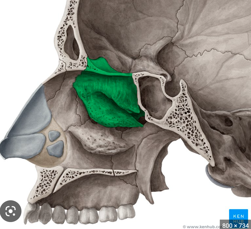
Sphenoid
Irregular bone on floor of cranium, looks like a "bat"
53
New cards
Sphenoid
54
New cards
Sphenoid
55
New cards
Sphenoid
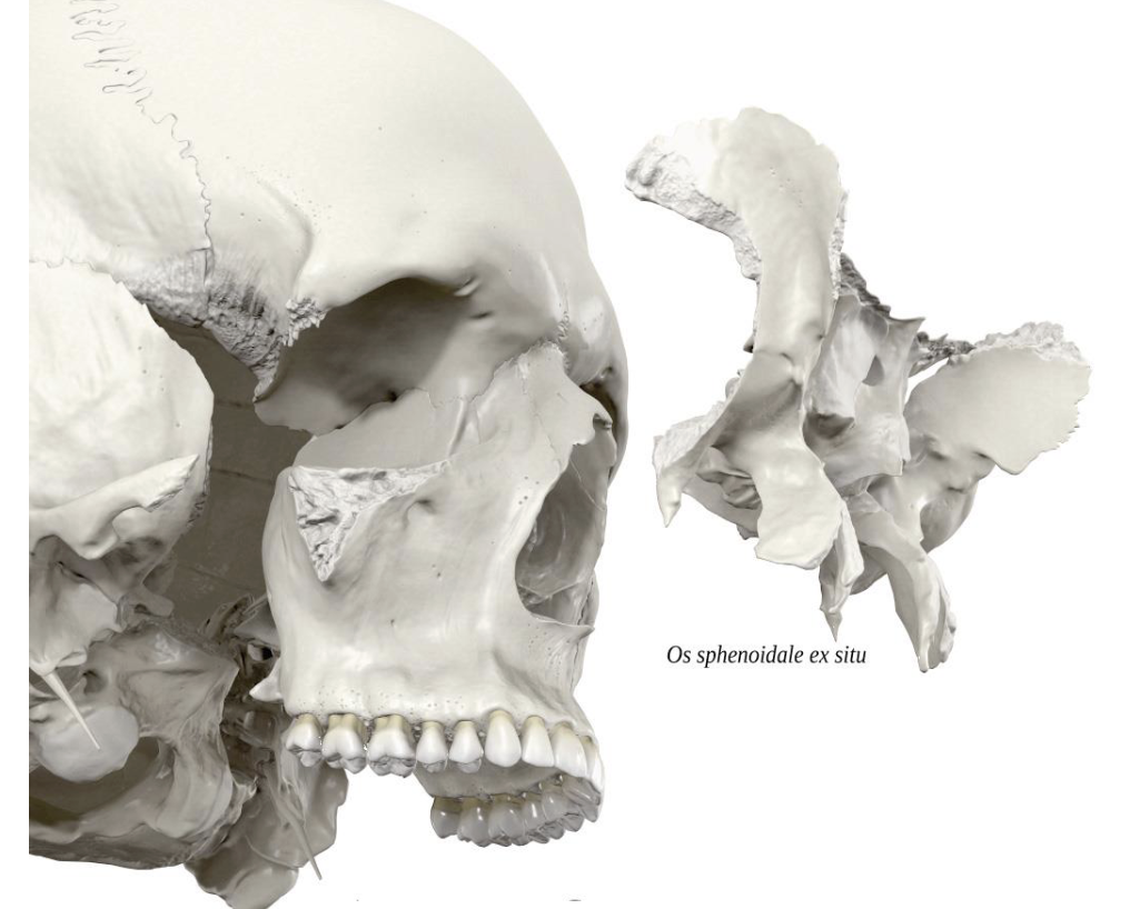
56
New cards
Sphenoid
57
New cards
Sphenoid
58
New cards
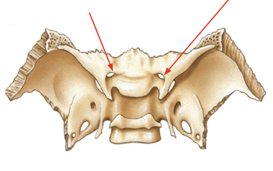
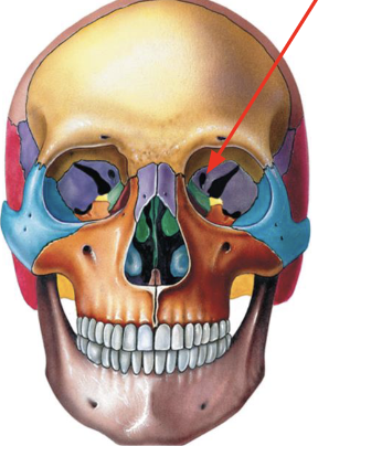
Optic Foramen (Canal)
Hole for the optic nerve to pass through towards each eye
59
New cards
Optic Canal (Foreamen)
60
New cards
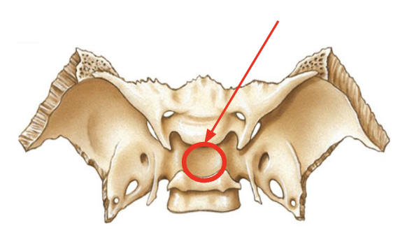
Sella Turcica
Saddle like structure on superior surface (pituitary gland sits here)
61
New cards
Sella Turcia
62
New cards
Greater and Lesser wings
Large and small wing like structures which help form the floor of the cranium
63
New cards
Greater Wing
64
New cards
Lesser Wing
65
New cards
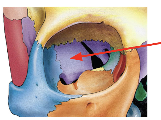
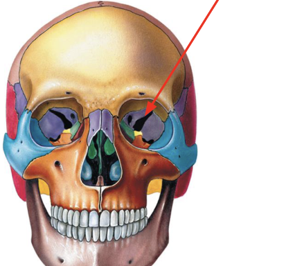
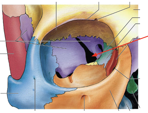
Superior Orbital Fissures
Large slits which can be seen in the eye sockets, it is between the
greater and lesser wings
greater and lesser wings
66
New cards
Superior Orbital Fissure
67
New cards
Superior Orbital Fissure
68
New cards
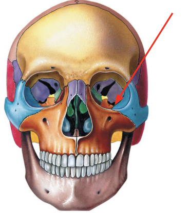
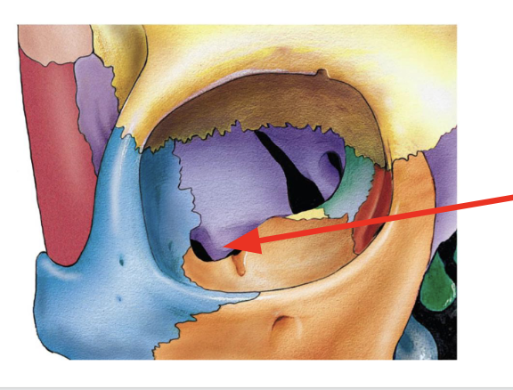
Inferior orbital fissure
Smaller slits, also seen in the eye sockets, just inferior to the
superior orbital fissures.
superior orbital fissures.
69
New cards
Inferior Orbital Fissure
70
New cards
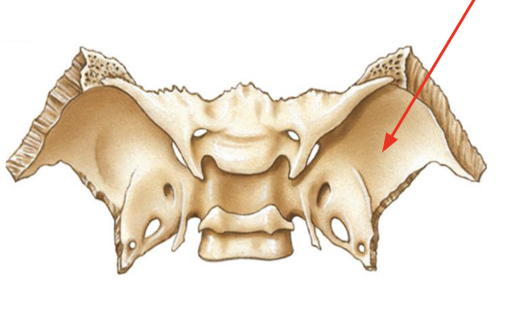
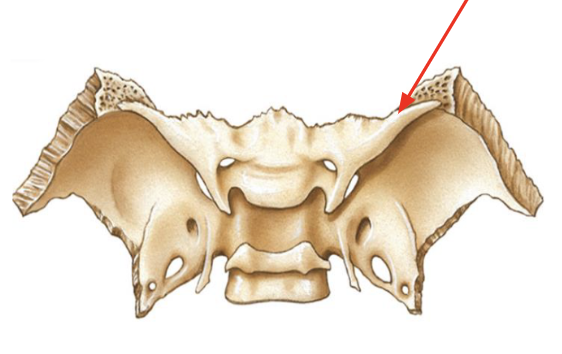
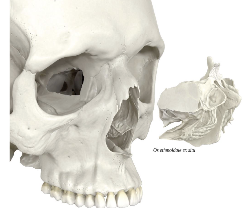
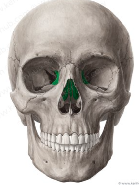
Ethmoid
Irregular bone anterior to sphenoid and between eyes
71
New cards
Ethmoid
72
New cards
Ethmoid
73
New cards
Ethmoid
74
New cards
Ethmoid
75
New cards
Ethmoid
76
New cards
Ethmoid
77
New cards
Ethmoid
78
New cards
Ethmoid
79
New cards
Ethmoid
80
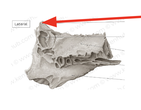
New cards
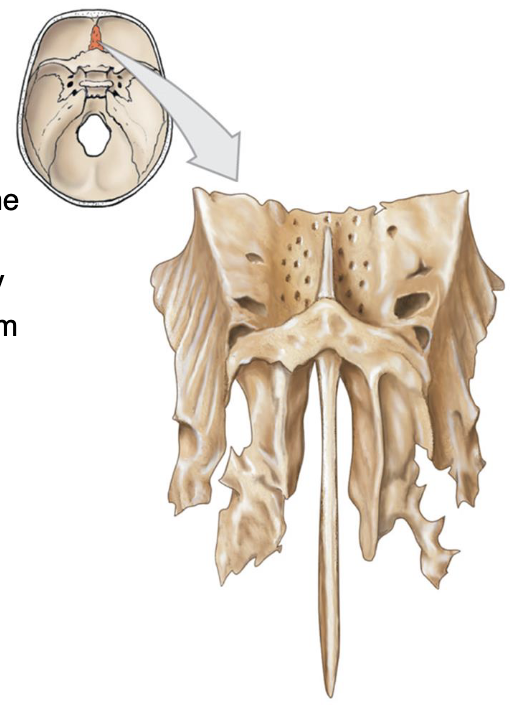
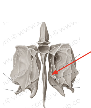
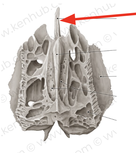
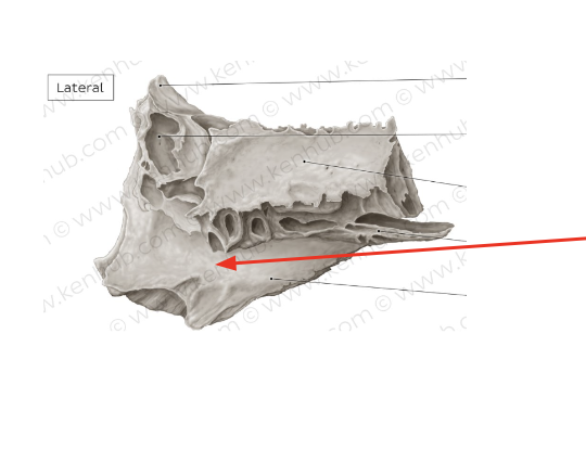
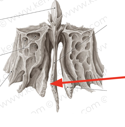
Ethmoid Bone Views

81
New cards
Ethmoid
82
New cards
Ethmoid
83
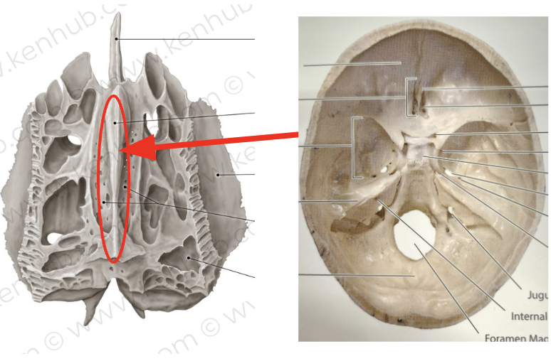
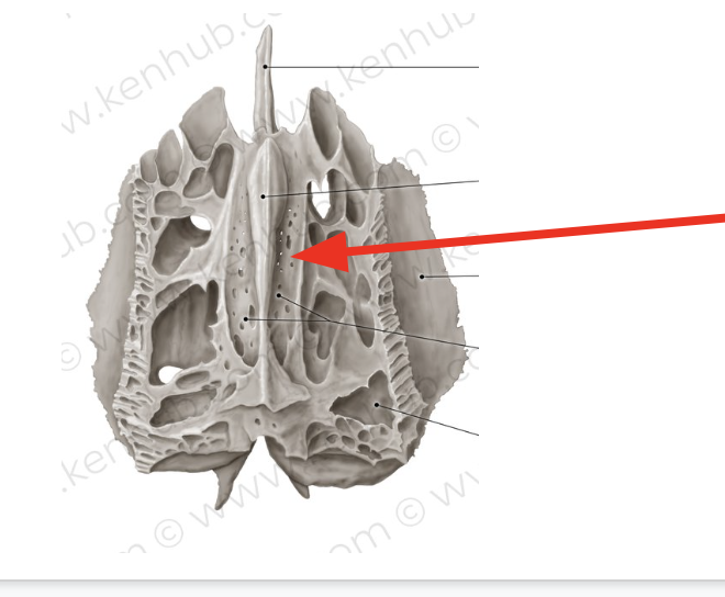
New cards
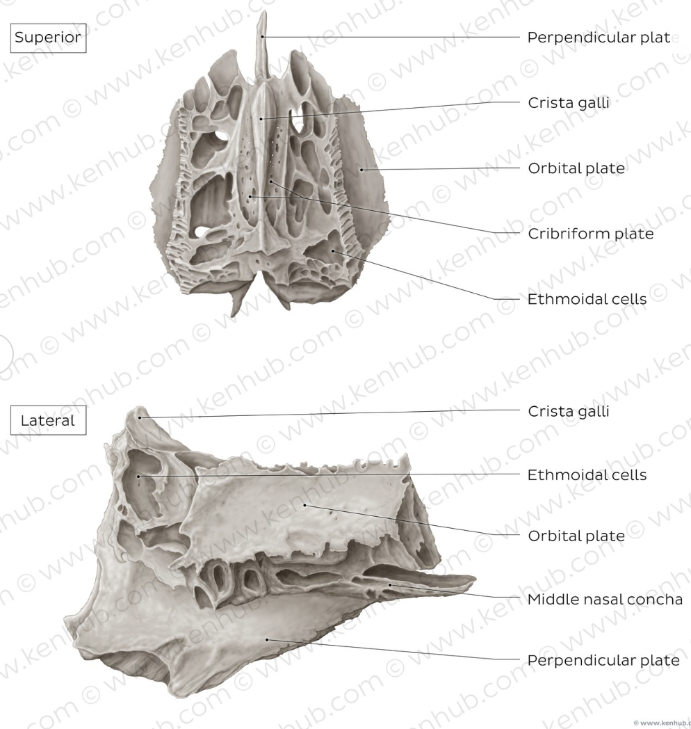
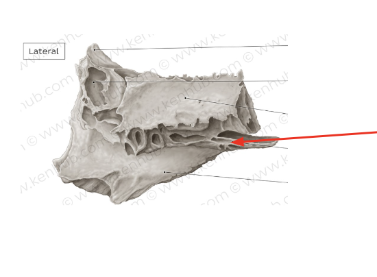
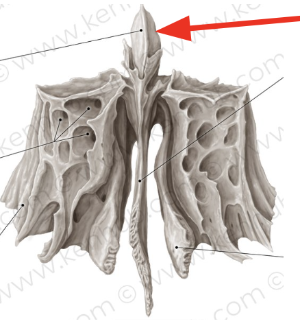
Crista galli
Bony projection on superior surface
84
New cards
Crista Galli
85
New cards
Cribriform plate
Horizontal plate on the superior surface that is **perforated** with
**many small holes** for the olfactory nerve
**many small holes** for the olfactory nerve
86
New cards
Crista Galli
87
New cards
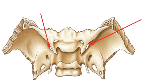
Middle Nasal Concha
\n Not a named bone, but part of the ethmoid bone. Above inferior
nasal concha
nasal concha
88
New cards
Middle Nasal Concha
89
New cards
Middle Nasal Concha
90
New cards
Crista Galli
91
New cards
Superior Nasal Concha
\n Not a named bone, but part of the ethmoid bone. Above middle
nasal concha
nasal concha
92
New cards
Middle Nasal Concha
93
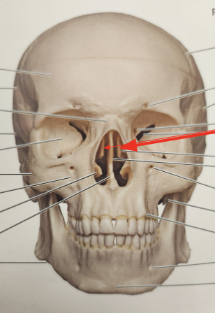
New cards
Perpendicular Plate
Inferior plate that divides the nasal cavity
94
New cards
Perpendicular Plate
95
New cards
Perpendicular Plate
96
New cards
Perpendicular Plate
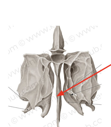
97
New cards
Perpendicular Plate
98
New cards
Perpendicular Plate
99
New cards
Orbital Plate
Smooth plates which form the medial walls of the eye sockets
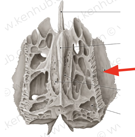
100
New cards
Orbital Plate