Heart and Great Vessels
1/93
There's no tags or description
Looks like no tags are added yet.
Name | Mastery | Learn | Test | Matching | Spaced | Call with Kai |
|---|
No analytics yet
Send a link to your students to track their progress
94 Terms
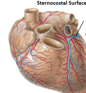
Sternocostal surface of heart
mostly right ventricle; faces sternum and ribs
diaphragmatic surface
mainly left ventricle; resting on the diaphragm
left pulmonary surface
left ventricle facing left lung
right pulmonary surface
right atrium
posterior surface
based formed by left atrium
Innermost layer of pericardium
Serous pericardiumT
Two layers of serous pericardium (superficial to deep)
parietal pericardium, visceral pericardium
Outermost pericardium
Fibrous pericardium
Layers of heart (deep to superficial)
endocardium, myocardium, visceral layer, pericardial cavity, parietal layer, fibrous pericardium
Auricles
Exterior surfaces that sit on top of atria and provide extra force
Pulmonary circuit
carries blood to and from lungs
systemic circuit
carries blood to and from the rest of the bodies
Three veins that return deoxygenated blood to heart
Superior vena cava, inferior vena cava, coronary sinus
Valve in right side of heart
tricuspid valve
Valve in left side of heart
bicuspid valve

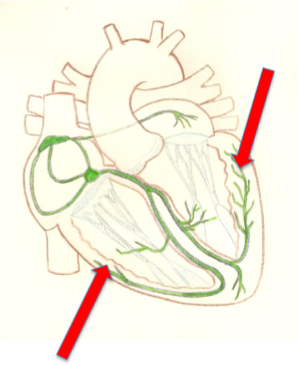
Top Left
Left coronary artery (branching off of aorta)

Top right
Left circumflex artery (branches off left coronary artery)
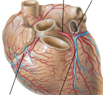
Bottom left
Right coronary artery (branches off of aorta)

Bottom right
Left anterior descending artery (branches off of coronary artery)
What does the right coronary artery supply?
Right atrium, right ventricle, SA and AV nodes, diaphragmatic surface and posterior surface
What does the left anterior descending artery supply?
Left ventricle, left atrium, anterior interventricular septum
Blockage to what vessel is known as the widowmaker?
Left anterior descending artery

Top left
Coronary sinus
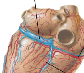
Top right
Right coronary artery

Bottom
Posterior descending artery
Great cardiac vein

Top left
Great cardiac vein

Middle left
coronary sinus
Top right
small cardiac vein

bottom right
middle cardiac vein
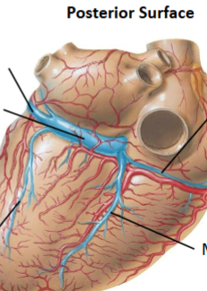
bottom left
posterior cardiac vein
Ostia
small opening in aorta where blood passes through
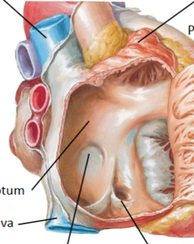
What compartment of the heart is this
Right atrium
Top left
Superior vena cava

2nd left
interatrial septum

2nd bottom
fossa ovalis

Top right
auricle

Bottom left
Inferior vena cava

Top right
Pectinate muscles
Bottom right
opening of coronary sinus
Pectinate muscles
Parallel ridges of cardiac muscle in walls of atria
Interatrial septum
thin tissue separating left and right atria
Fossa oval
Small oval depression in atria that used to be an opening
Foramen ovale
hole in atria at birth
Papillary muscles
cone shaped muscles in ventricles
Trabeculae carneae
irregular muscular ridges that line the two ventricles
Chordae tendineae
Heart strings; tough cords of connective tissue that work the valves
Paradoxical emboli
When the foramen ovale doesn’t properly close and blood clots from leg can enter arterial circulation
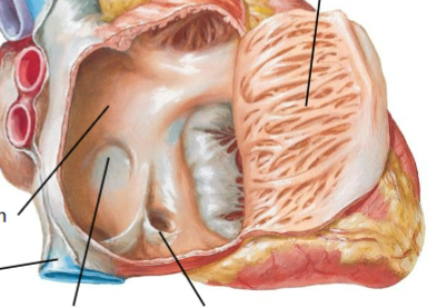
Top right
Pulmonary trunk
2nd from top right
Pulmonary valve


Right center
papillary muscles

Top left
tricuspid valve

left center
chordae tendineae

Bottom left
trabeculae carneae
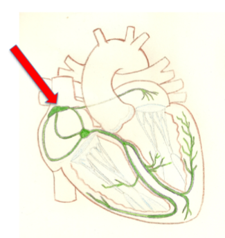
Sinoatrial node
In right atrial wall; sets the rhythm through gap junctions; most rapid conduction rate
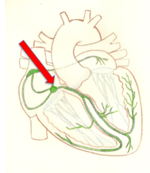
atrioventricular node
in inferior segment of interatrial septum; signals slows down (fewer gap junctions, smaller diameter fibers)
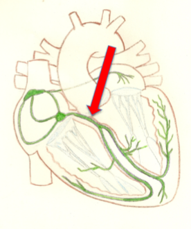
Atrioventricular bundle
in superior portion of interventricular septum; no gap junctions; connects atria to ventricles
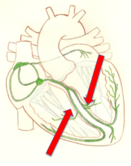
Bundle branches
Along left and right sides of interventricular septum; sends signal to apex of heart
Purkinje fibers
penetrate the apex and then run superiorly in ventricle walls
Five conduction segments
Sinoatrial node
Atrioventricular node
Atrioventricular bundle
Bundle branches
Purkinje fibers
Sinoatrial node
Atrioventricular node
Atrioventricular bundle
Bundle branches
Purkinje fibers
What are the five great vessels?
Superior vena cava, inferior vena cava, pulmonary trunk, pulmonary veins
What does the aortic branch branch off into? (right to left)
Brachiocephalic trunk, left common carotid artery, left subclavian artery

Left
Brachiocephalic trunk
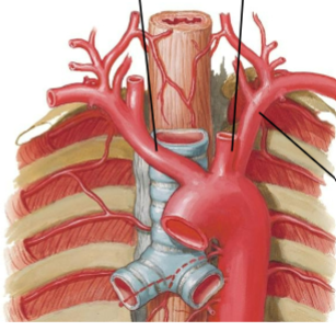
middle
left common carotid artery

right
left subclavian artery
Branches of the brachiocephalic trunk
Right subclavian artery; right common carotid artery

bottom left
right subclavian artery
Top left
right common carotid artery

right
brachiocephalic trunk
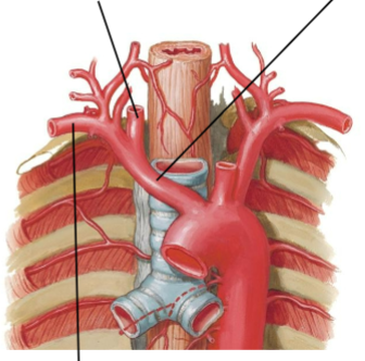
What does the left subclavian artery branch into?
Left vertebral artery and internal thoracic artery

top
left vertebral artery

bottom
internal thoracic artery
What do the common carotid artery branch into?
Internal carotid artery (ascends into skull); external carotid artery (vasuclarizes head and neck)
Left
External carotid artery

top right
internal carotid artery

bottom right
common carotid artery
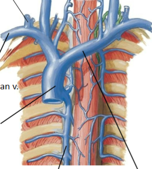
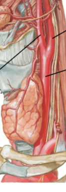
Top left
Right external jugular vein

Top right
left internal jugular vein

2nd of left
right subclavian vein

3rd on left
superior vena cava

bottom left
axygous vein

bottom right
left brachiocephalic vein
What do veins drain through in superior portion of body?
subclavian and jugular veins
What do subclavian and jugular veins form?
Brachiocephalic veins
What do brachiocephalic veins converge to form?
superior vena cava
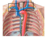
left and right venous angles
Venous angles
Where the lymphatic ducts drain
What does the right lymphatic duct drain?
the right upper extremities
What does the left lymphatic duct drain and what is it called?
It is called the thoracic duct and drains all the body minus the right upper extremities