Practical 2 - Vessels
 Call Kai
 Learn
Learn Practice Test
 Spaced Repetition
 Match
Match Flashcards
 Knowt Play
1/106

There's no tags or description
Looks like no tags are added yet.
Name | Mastery | Learn | Test | Matching | Spaced | Call with Kai | Chat |
|---|
No analytics yet
Send a link to your students to track their progress
107 Terms
tunica interna layers
endothelium
basal lamina
subendothelial connective tissue
internal elastic lamina
tunica media layers
smooth muscle tissues
external elastic lamina
elastic arteries

muscular arteries

arteroiles
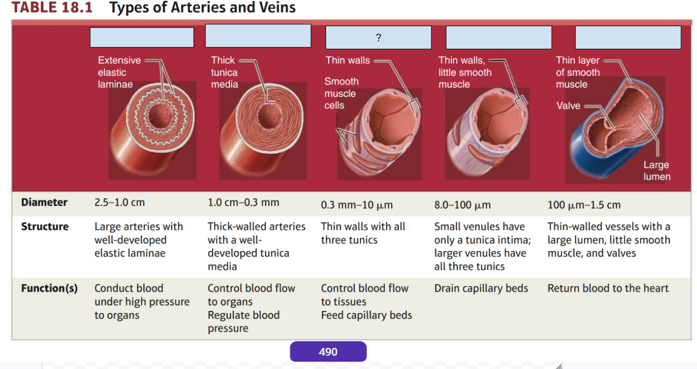
venules
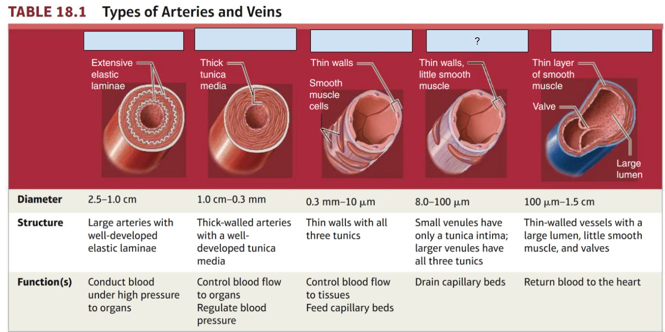
veins

arteries, veins, capillaries differences
Arteries: Have thick, muscular, and elastic walls and a relatively small lumen to withstand high blood pressure.
Veins: Have thinner, less muscular walls and a larger lumen. They often contain valves to prevent backflow.
Capillaries: Have extremely thin walls, only one cell thick, and very small lumens. This allows efficient exchange of oxygen, nutrients, and wastes between blood and tissues.
Arteries
Have thick, muscular, and elastic walls and a relatively small lumen to withstand high blood pressure.
Veins
Have thinner, less muscular walls and a larger lumen. They often contain valves to prevent backflow.
Capillaries
Have extremely thin walls, only one cell thick, and very small lumens. This allows efficient exchange of oxygen, nutrients, and wastes between blood and tissues.
The structural differences help each blood vessel perform its specific function:
Arteries: Their thick, muscular, elastic walls allow them to withstand and maintain the high pressure of blood pumped from the heart.
Veins: Their thinner walls and larger lumens allow them to carry blood back to the heart at lower pressure. Valves prevent blood from flowing backward.
Capillaries: Their walls are only one cell thick, making it easy for oxygen, nutrients, carbon dioxide, and wastes to diffuse between the blood and surrounding tissues.
high pressure
Arteries: Their thick, muscular, elastic walls allow them to withstand and maintain the ___ __________ of blood pumped from the heart.
lower pressure
Veins: Their thinner walls and larger lumens allow them to carry blood back to the heart at ______ _________. Valves prevent blood from flowing backward.
blood and surrounding tissues
Capillaries: Their walls are only one cell thick, making it easy for oxygen, nutrients, carbon dioxide, and wastes to diffuse between the __________ ___ ______________ ______________
capillary

basal lamina

varicose veins
Pathophysiology: Weak or stretched valves cause venous reflux (blood backflow). Gravity pools blood in the leg, raising pressure and ballooning the thin superficial vein walls.
Key Causes: Inactive muscle pumps (standing/sitting for long periods), increased pressure (pregnancy/obesity), and structural weakness (aging/genetics).

occipital artery

Anterior tibial artery
supplies anterior leg and anterior foot
aorta
Branches supply all structures in
the body
Axillary artery
supplies Structures around the axilla
Brachial artery
supplies Arm
Celiac trunk
Structures of the abdominal cavity
common carotid arteries
supplies Head
Neck
Brain
Common hepatic artery
supplies Liver
Stomach
Duodenum
Pancreas
common iliac arteries
supplies Structures of the pelvis and
lower limb
Dorsalis pedis arter
supplies ankle joint and Foot
External carotid artery
supplies neck, face, superficial head
External iliac artery
supplies anterior abdominal wall
femoral artery
supplies thigh
Inferior mesenteric artery
supplies Remainder of the large intestine
internal carotid artery
supplies brain and orbit
internal illiac artery
supplies pelvic structures
left gastric artery
supplies stomach
popliteal artery
supplies knee joint, posterior leg, posterior thigh
posterior tibial artery
supplies posterior leg and plantar foot
radial artery
supplies forearm and hand
renal arteries
supplies kidneys
splenic artery
supplies spleen, stomach, pancreas
subclavian arteries
supplies thorax and upper limb
anterior tibial artery

aorta

axillary artery

brachial artery

Brachiocephalic artery

celiac trunk

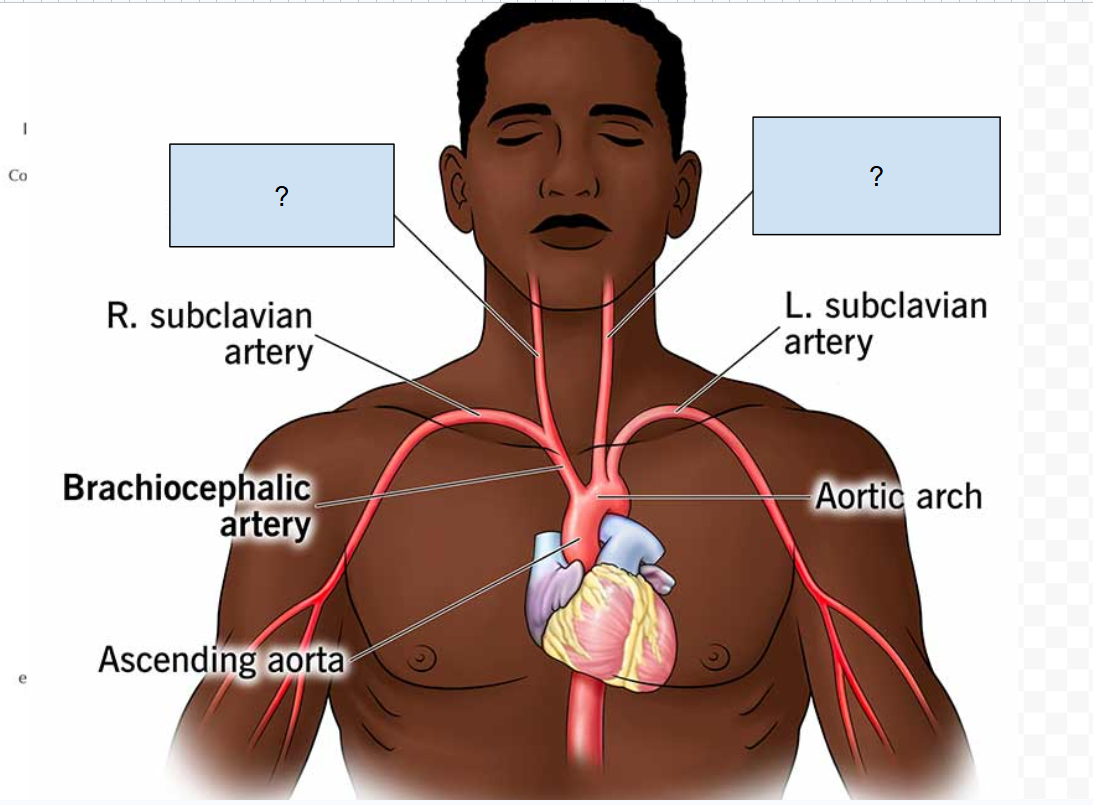
Common carotid arteries
Common hepatic artery

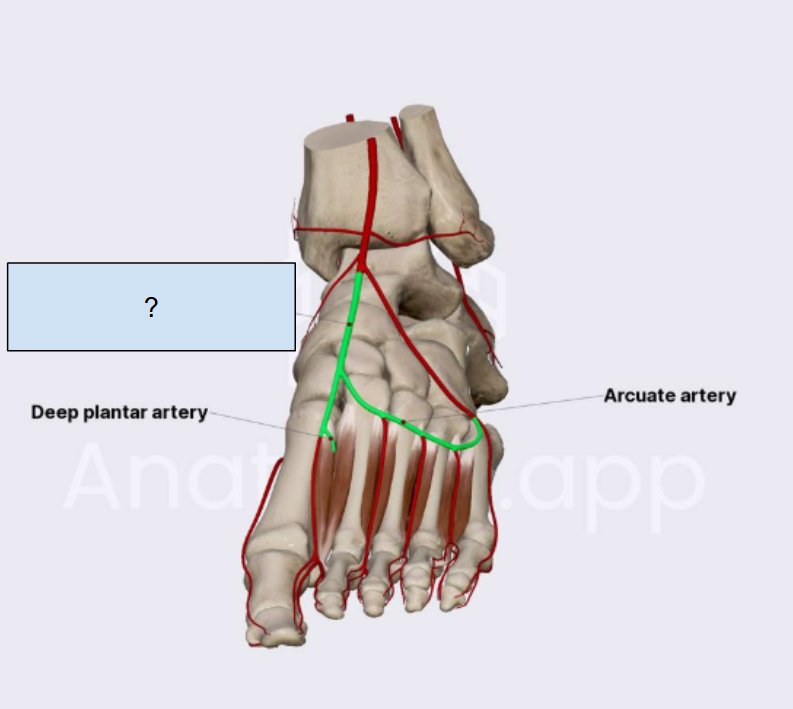
dorsalis pedis artery
external carotid artery

anterior and posterior tibial veins
drains leg and foot
axillary veins
drains arm and axilla
azygos veins
drains Posterior thoracic and abdom-inal wall on the right side
Esophagus
Lungs
Basilic veins
drains from Superficial structures of the
upper limb
brachial veins
drains from arm
brachiocephalic veins
drains from most structures superior to the diaphragm
cephalic veins
drains from superficial structures of the
upper limb
Common iliac veins
drains from Pelvis
Lower limb
Dural sinuses
drains from brain
external iliac veins
drains from Lower limb
external jugular veins
drains the superfacial face and neck
femoral veins
drains from thigh
gastric veins
drains from stomach
great saphenous veins
drains the superficial structures of the
lower limb
Hemiazygos and acces-sory hemiazygos vein
drains the posterior body wall
hepatic portal vein
processes blood from the spleen and digestive organs
hepatic veins
drains the arterial blood from the liver
Venous blood from the hepat-ic portal system
inferior mesenteric vein
drains the remainder of the large intestine
internal illiac veins
drains the pelvis
internal jugular veins
drains the brain, superficial head, neck
popliteal veins
drains the knee joint, leg, thigh
radial and ulnar veins
drains the forearm
splenic vein
drains the spleen
subclavian veins
drains the upper limb and upper thorax
superior mesenteric vein
drains the small intestine and much of the large intestine
vertebral veins
drains the posterior neck
vertebral artery
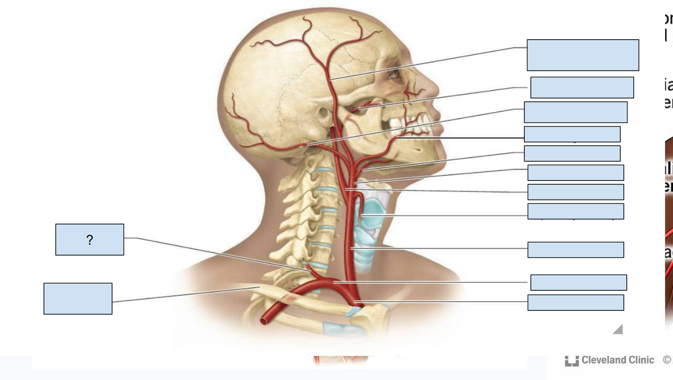
superficial temporal artery
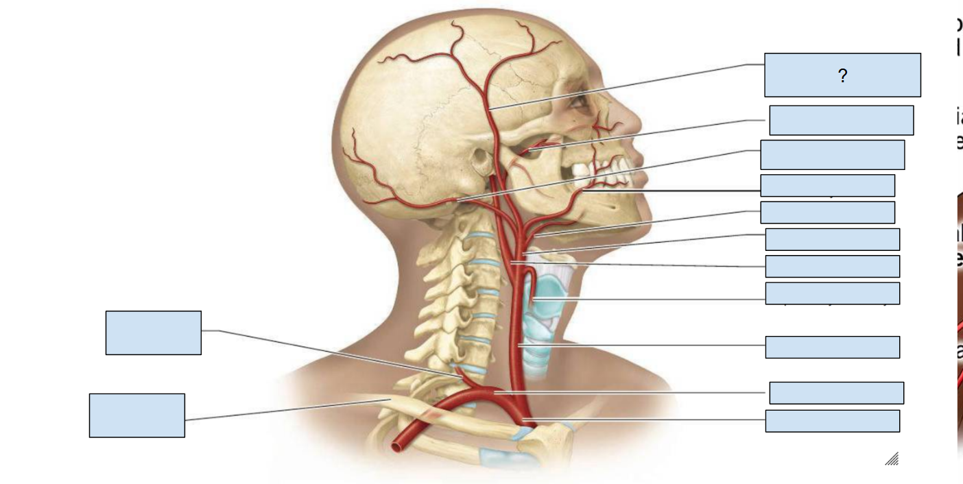
maxillary artery
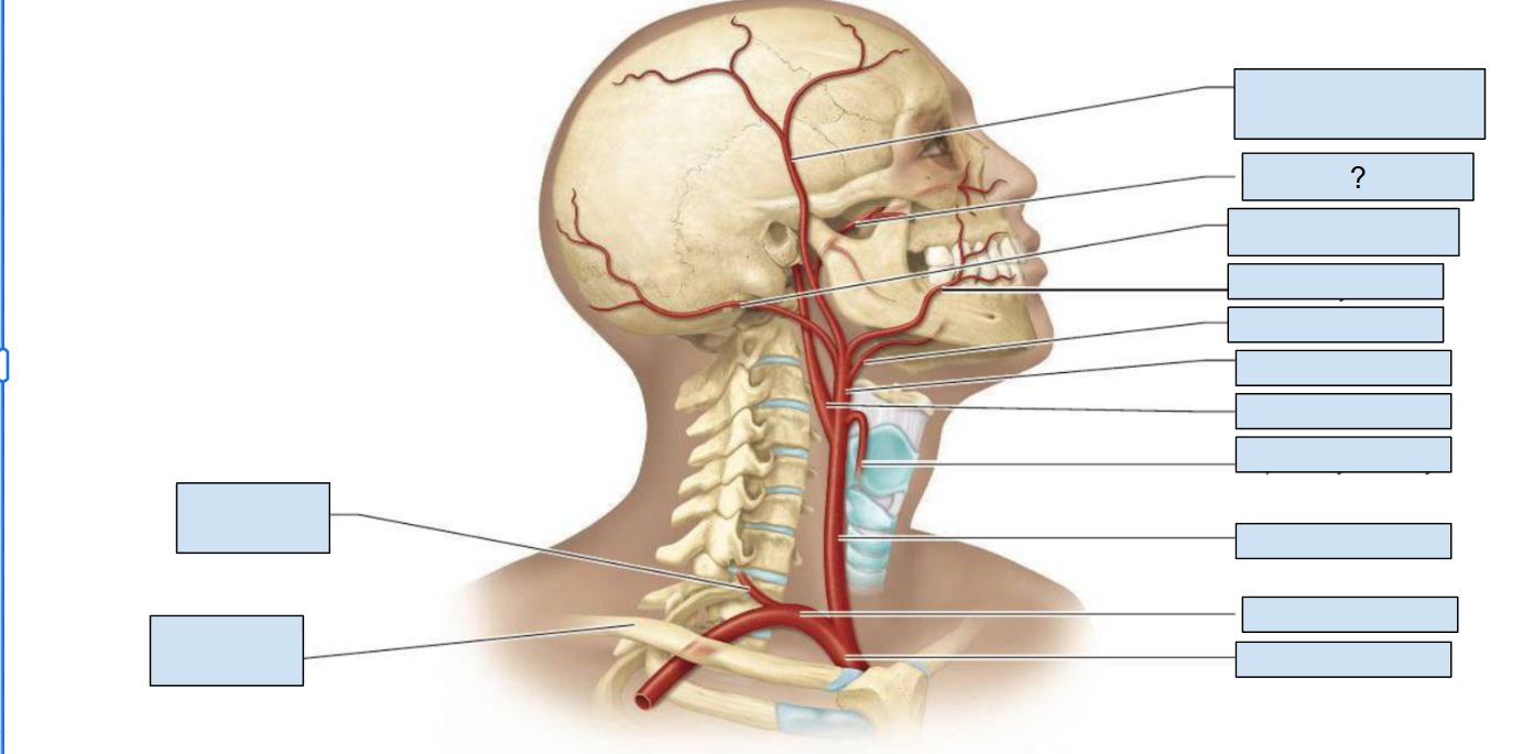
occipital artery

facial artery
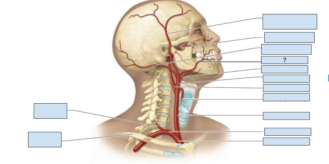
lingual artery

external carotid artery

internal carotid artery

superior thyroid artery
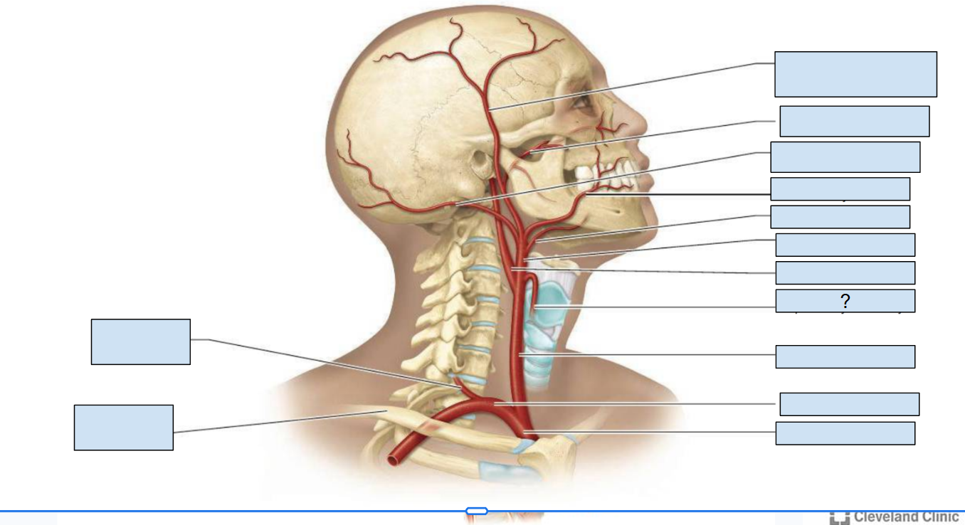
common carotid artery

subclavian artery

brachiocephalic artery

superior mesenteric artery
supplies small intestine and most of the large intestine
ulnar artery
supplies forearm and hand
vertebral arteries
supplies brain
taking a pulse
Place the index and middle fingertips (avoiding the thumb, as it has its own distinct pulse) over a superficial artery where it runs over a bone or firm tissue. Press gently until you feel the rhythmic expansion/pulsation of the vessel. Count the beats for 30 seconds and multiply by 2 (or count for a full 60 seconds if the rhythm is irregular).
radial and carotid
most common areas for checking a pulse
systolic vs diastolic BP
Systolic Blood Pressure (SBP): The peak pressure exerted by blood against the arterial walls during ventricular contraction (systole).
Diastolic Blood Pressure (DBP): The minimum pressure remaining in the arteries during ventricular relaxation (diastole).
systolic blood pressure
The peak pressure exerted by blood against the arterial walls during ventricular contraction (systole).
Diastolic Blood Pressure (DBP)
The minimum pressure remaining in the arteries during ventricular relaxation (diastole)
120/80 mmHg
normal blood pressure
< 120 mmHg
normal systolic BP
< 80 mmHg.
normal diastolic BP