Skull
 Note
Note Call Kai
 Learn
Learn Practice Test
 Spaced Repetition
 Match
Match Flashcards
 Knowt Play
1/83

There's no tags or description
Looks like no tags are added yet.
Name | Mastery | Learn | Test | Matching | Spaced | Call with Kai | Chat |
|---|
No analytics yet
Send a link to your students to track their progress
84 Terms
Skull
The bony structure that forms the head and encases the brain.
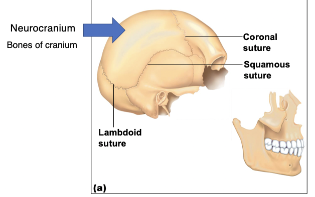
Neurocranium
The part of the skull that surrounds and protects the brain. 8
1.1 ethmoid bone
2.1 frontal bone
3.1 occipital bone
4.2 parietal
5.1 sphenoid
6.2 temporal
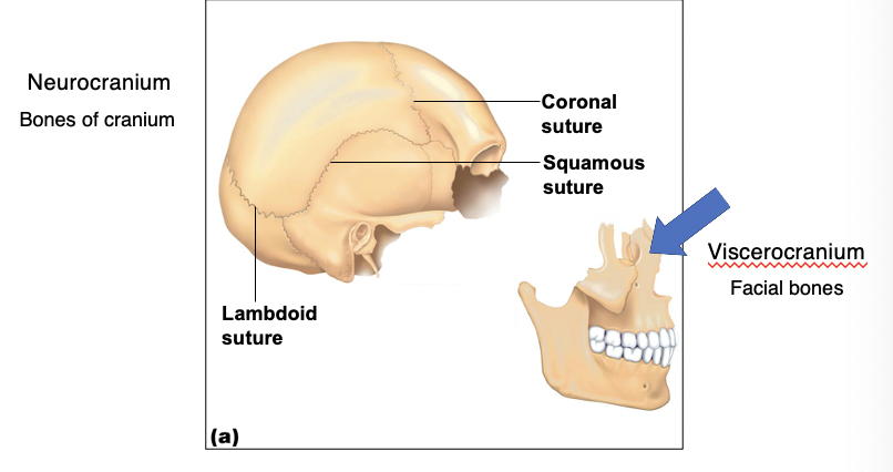
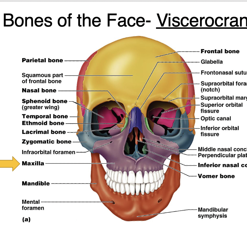
Viscerocranium
The facial skeleton, consisting of bones that form the face. 14
1.Inferior nasal concha (2)
2.Lacrimal bones (2)
3.Mandible
4.Maxilla (2)
5.Nasal bones (2)
6.Palatine bones (2)
7.Vomer
8.Zygomatic bones (2)

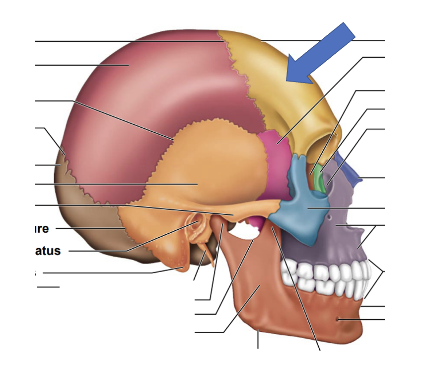
Frontal bone
A single bone that forms the forehead and the upper part of the eye sockets. (neurocranium)
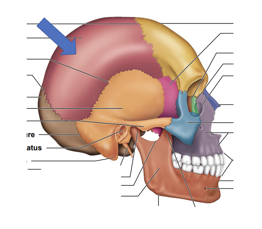
Parietal bones
Two bones that form the sides and roof of the skull. (neurocranium)
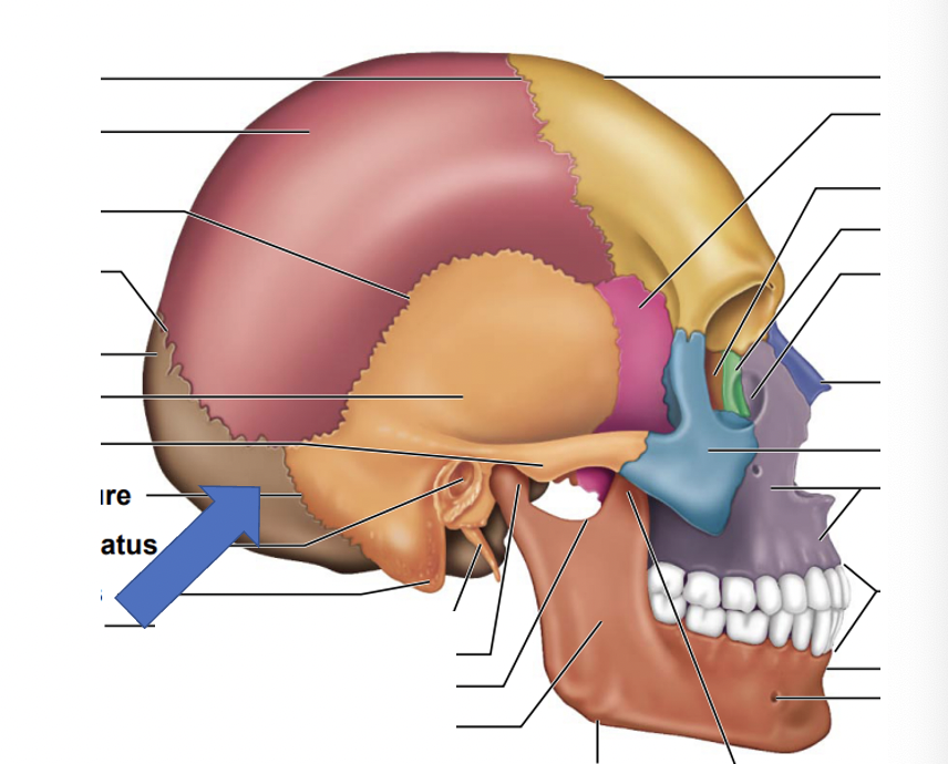
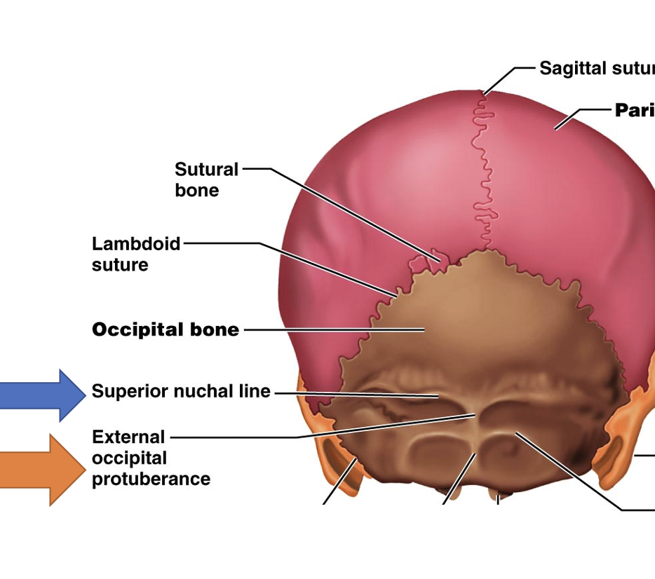
Occipital bone
The bone that forms the back and base of the skull. (neurocranium)
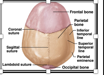
Lambdoid suture
The suture between the occipital and parietal bones, resembling the Greek letter lambda. (neurocranium)
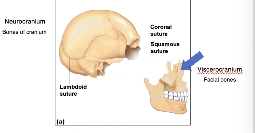
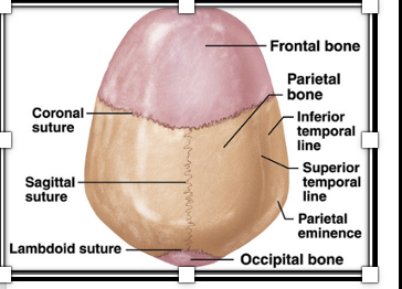
Coronal suture
The suture between the frontal bone and the parietal bones. also known as frontal suture (neurocranium)
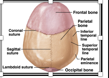
Sagittal suture
The suture between the two parietal bones, running in the sagittal plane.
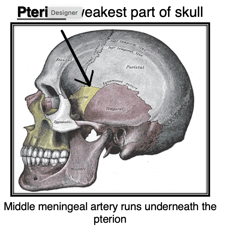
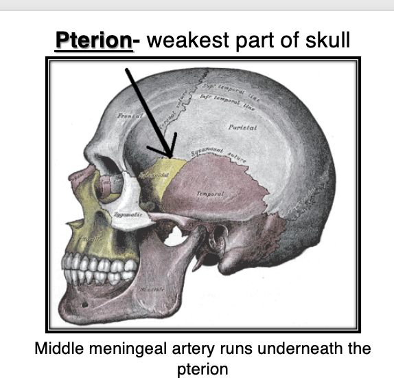
Pterion
A small suture connecting the parietal, frontal, sphenoid, and temporal bones.
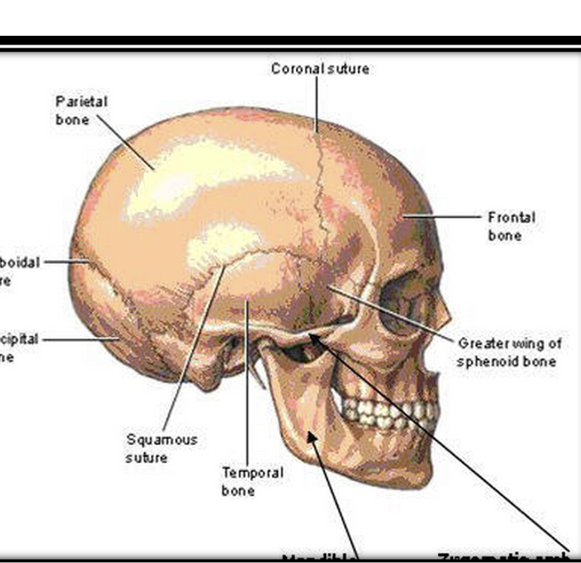
Squamosal suture
The suture connecting the parietal and temporal bones.
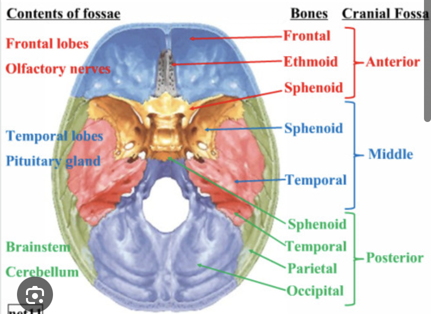
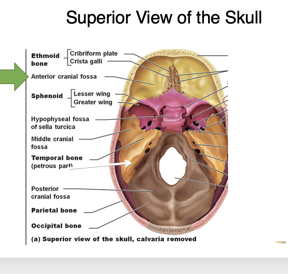
Cranial fossae
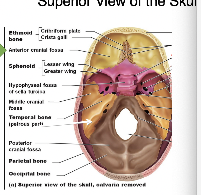
Three large, stair-like depressions in the floor of the cranial cavity.
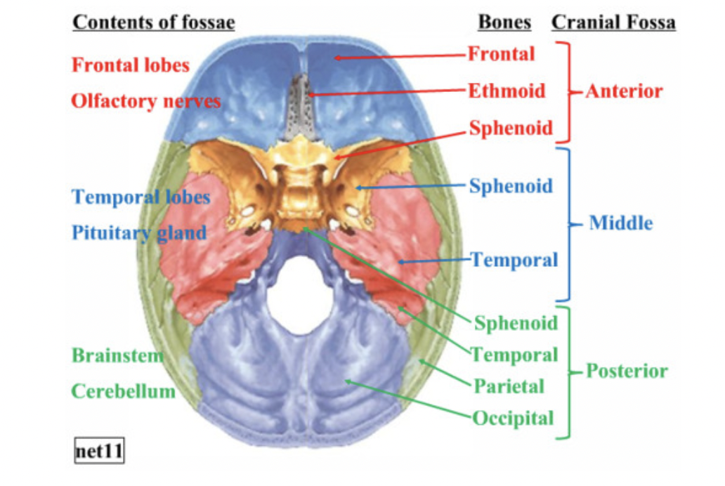
Anterior cranial fossa
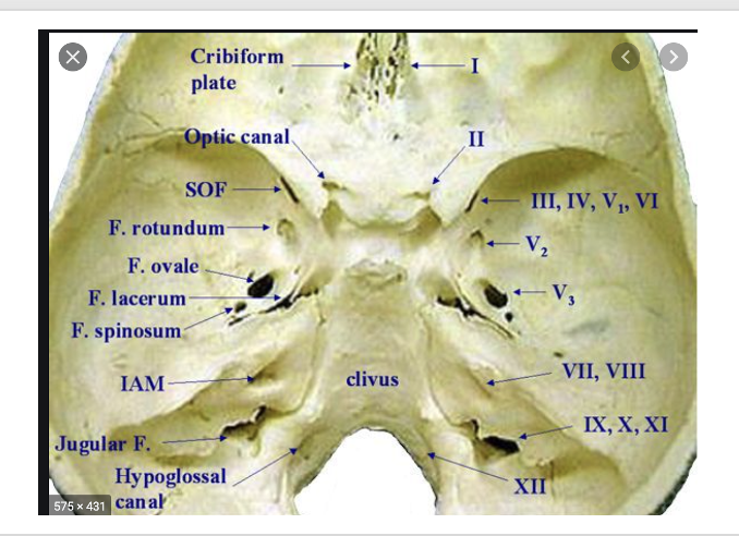
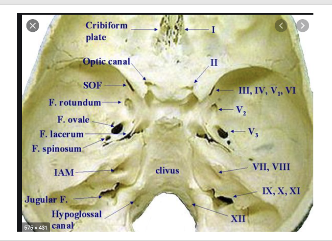
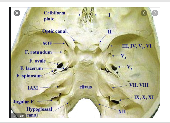
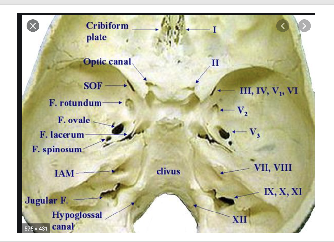
The front depression in the cranial cavity, containing the ethmoid bone, lesser wing of the sphenoid bone and frontal bone
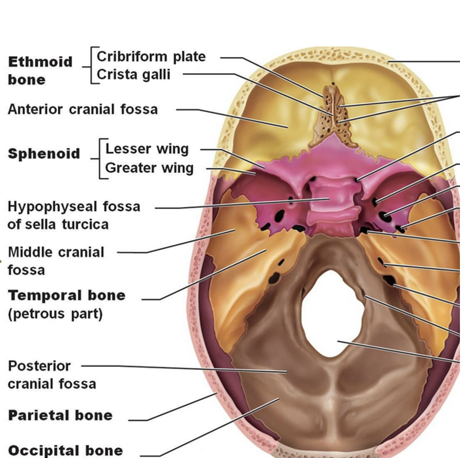
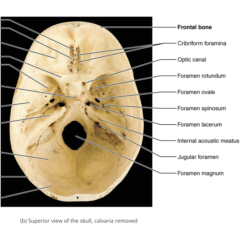
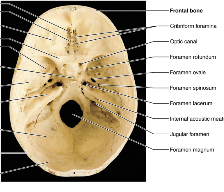
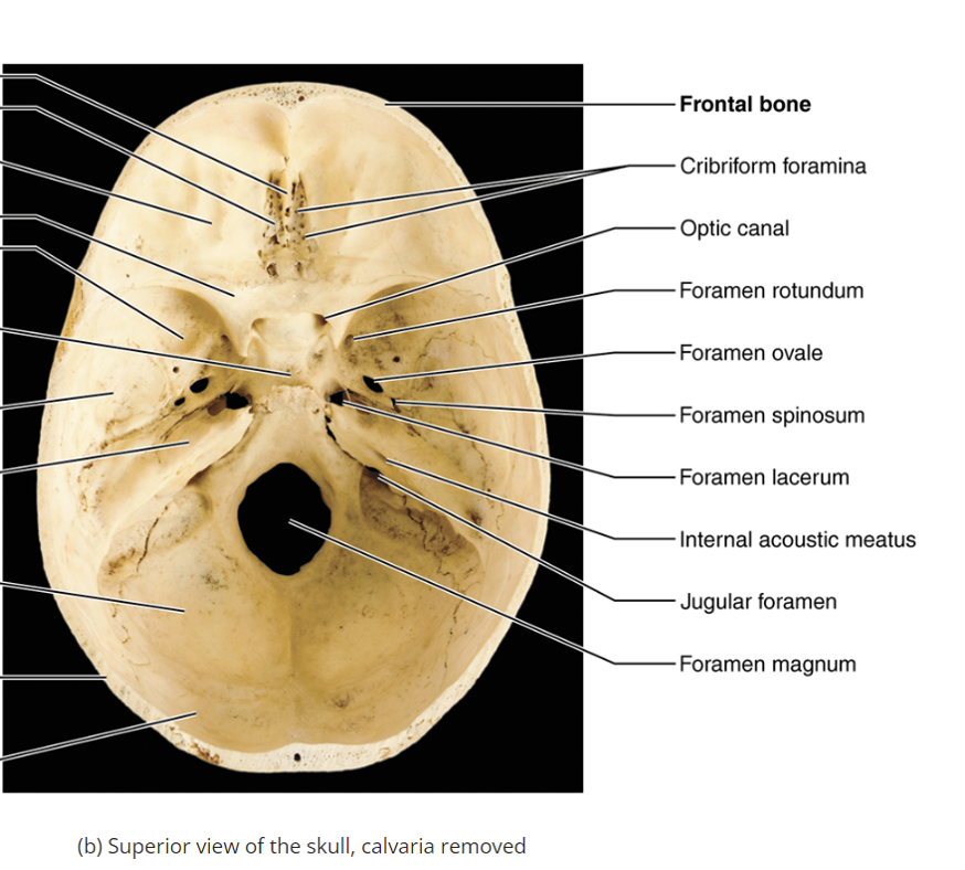
Cribriform foramina
Holes in the ethmoid bone allowing olfactory nerves to pass through/the transversing of the olfactory nerves to the olfactory bulb which brings the sensation of smell to the brain (anterior cranial fossa)
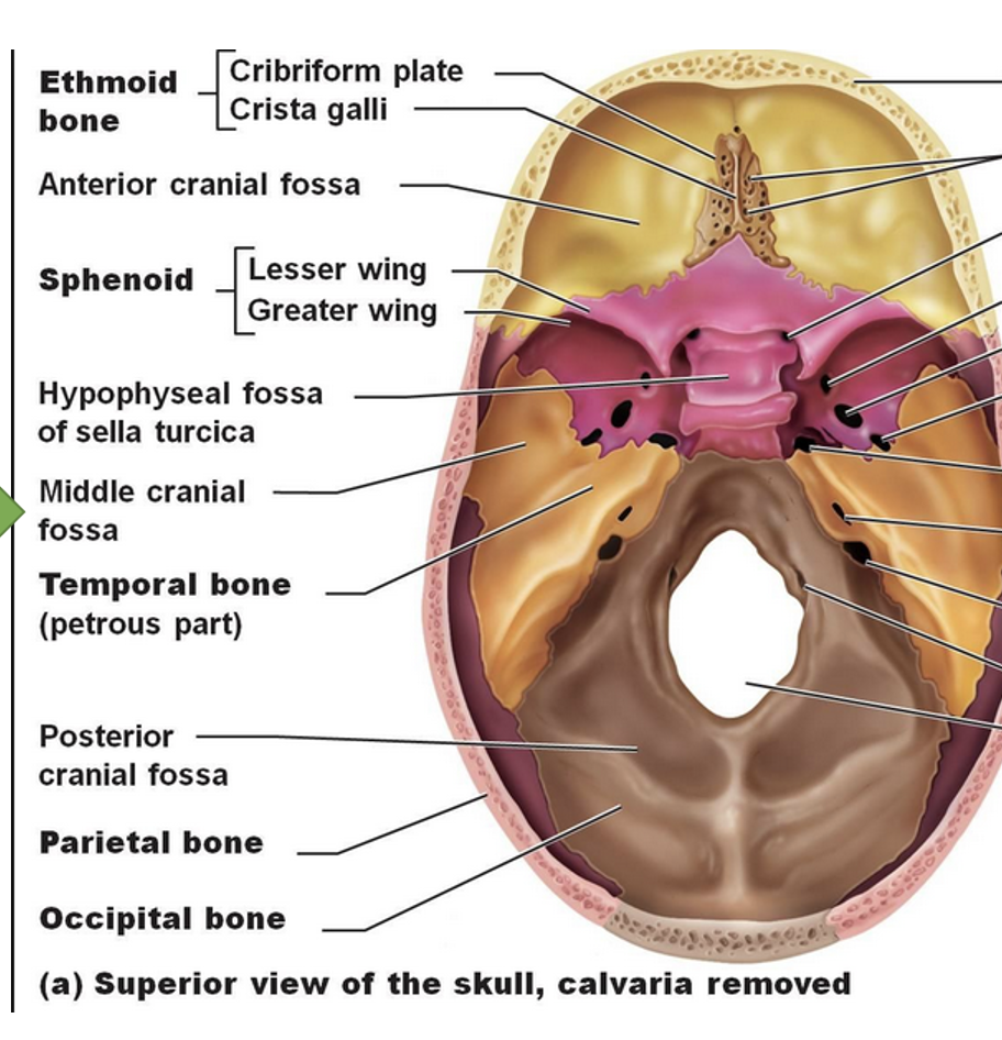
Lesser wing of sphenoid bone
A structure in the anterior cranial fossa that separates the anterior and middle cranial fossae.
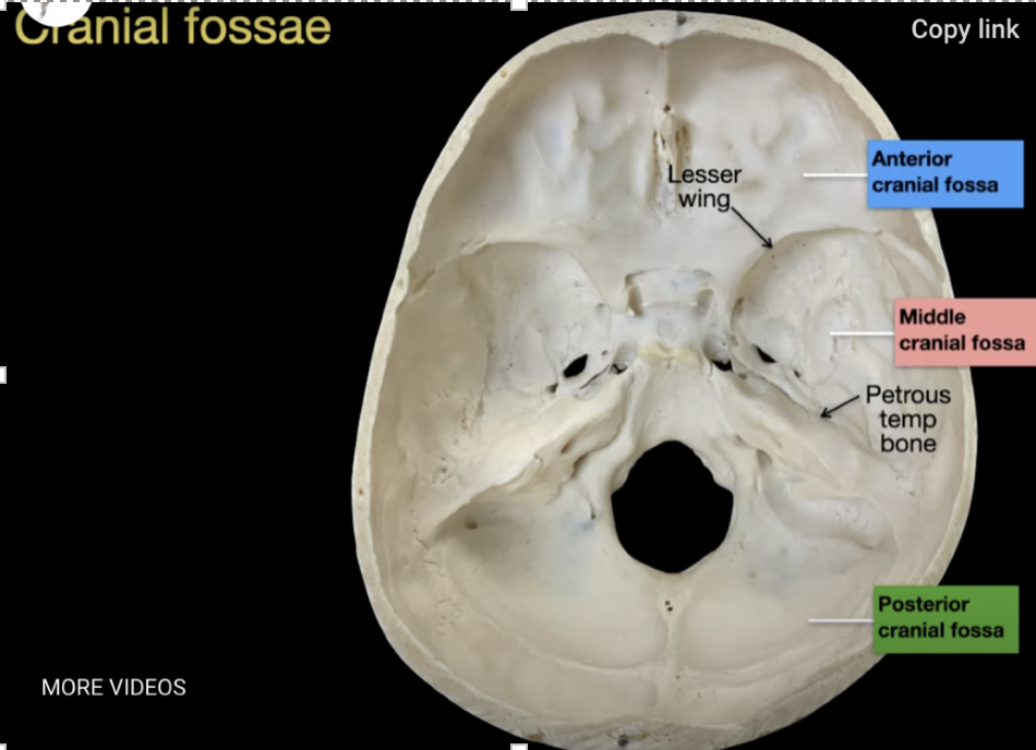
Middle cranial fossa
made up of the sphenoid bone and temporal bone, The central depression in the cranial cavity, containing important structures like the optic canal and superior orbital fissure
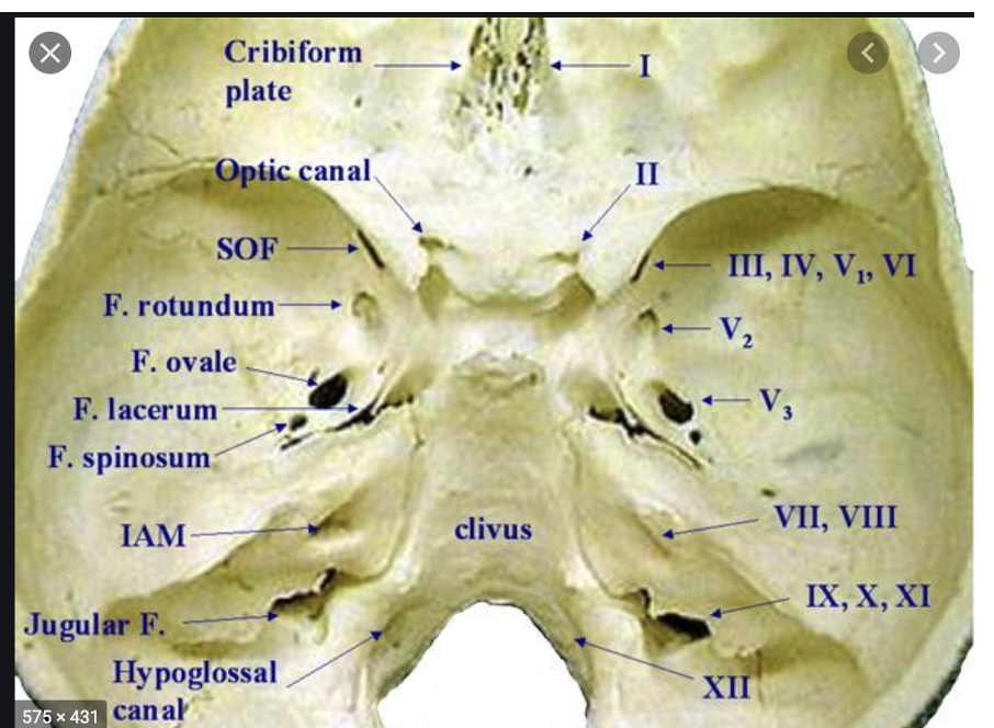
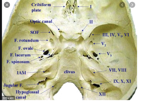
Optic canal
A passage for cranial nerve II (optic nerve which allows us to see) and the ophthalmic artery (branch of internal carotid artery that supplies the orbit and the retina), also known as optic foreman
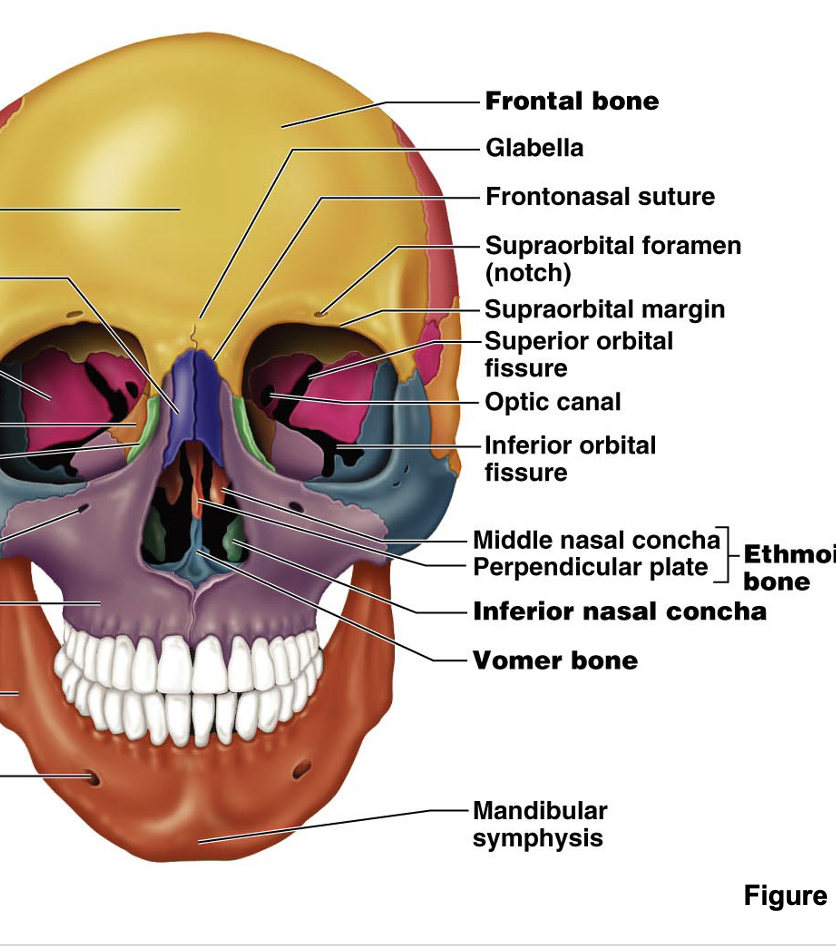
Inferior orbital fissure
inferior Opthalmic vein that goes to the cavernous sinus as well and the pterygoid plexus and facial vein and cranial nerve V-2 infraorbital nerve: the continuation of the V2 branch of the foreman rotundum, and the inferior orbital artery and vein
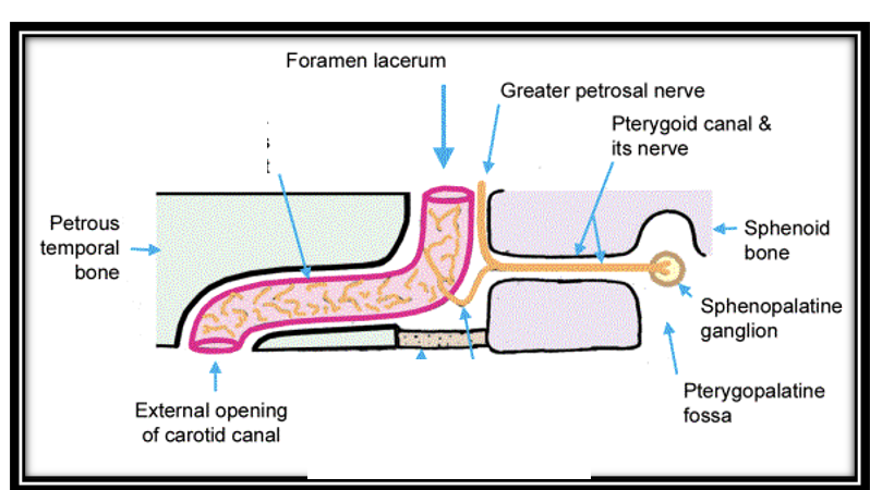
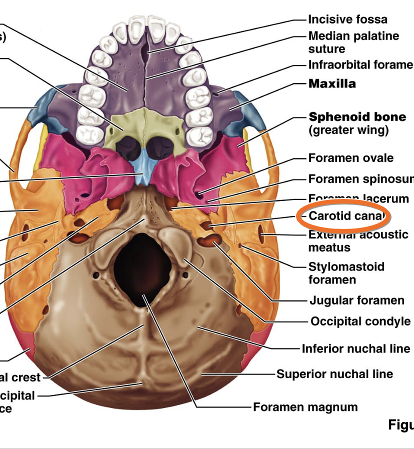
Carotid canal
A passage for the internal carotid artery into the middle cranial fossa. from the bottom of the skull and into the middle cranial fossa (where the carotid canal exits) transmits the internal carotid artery, Finds its way through the foreman Lacerum, Opening is known as the internal carotid artery
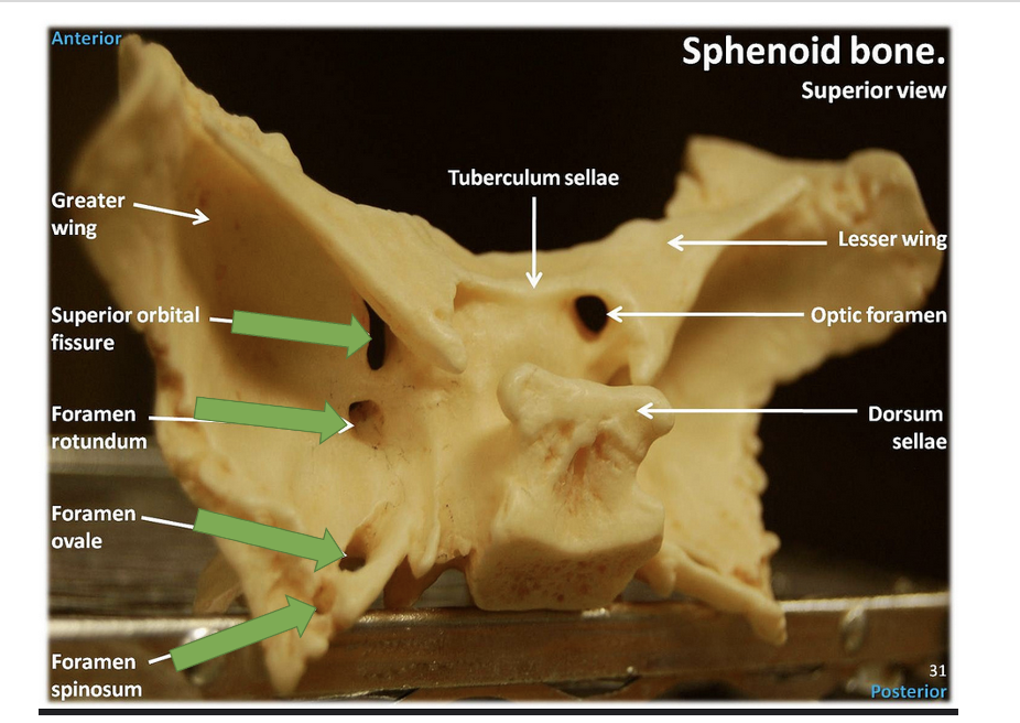
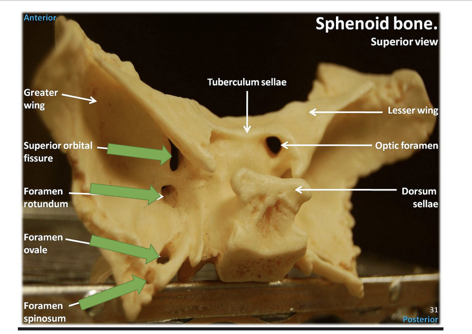
Foramen rotundum
An opening allowing the maxillary branch of cranial nerve V to pass. allows for cranial nerve five and V-2 (maxillary branch) (V-2 goes to the maxilla), trigeminal nerve
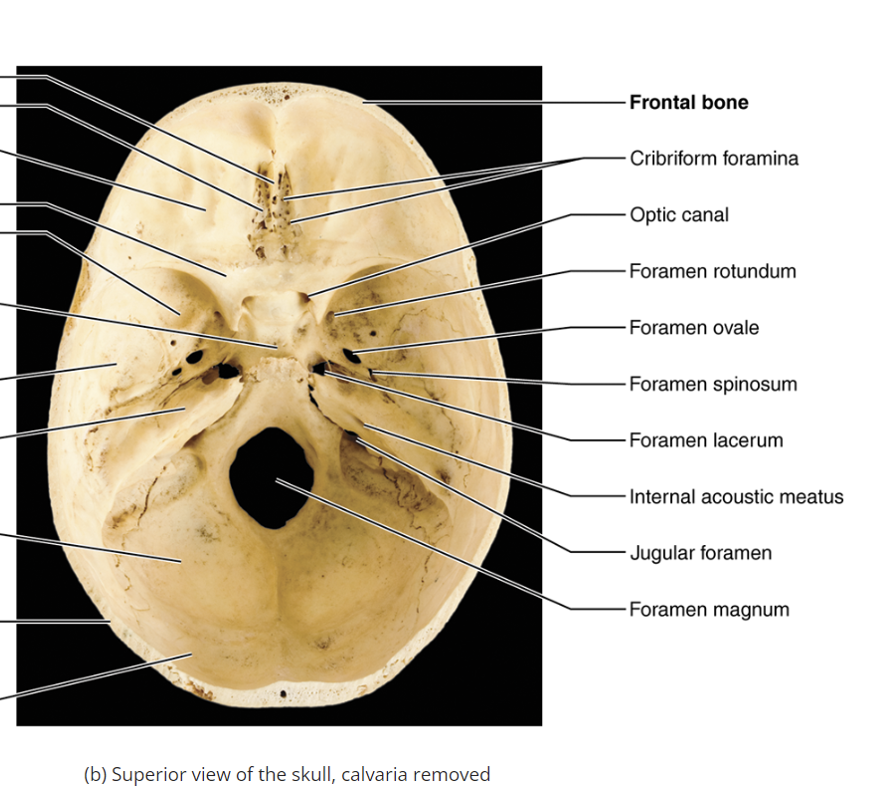
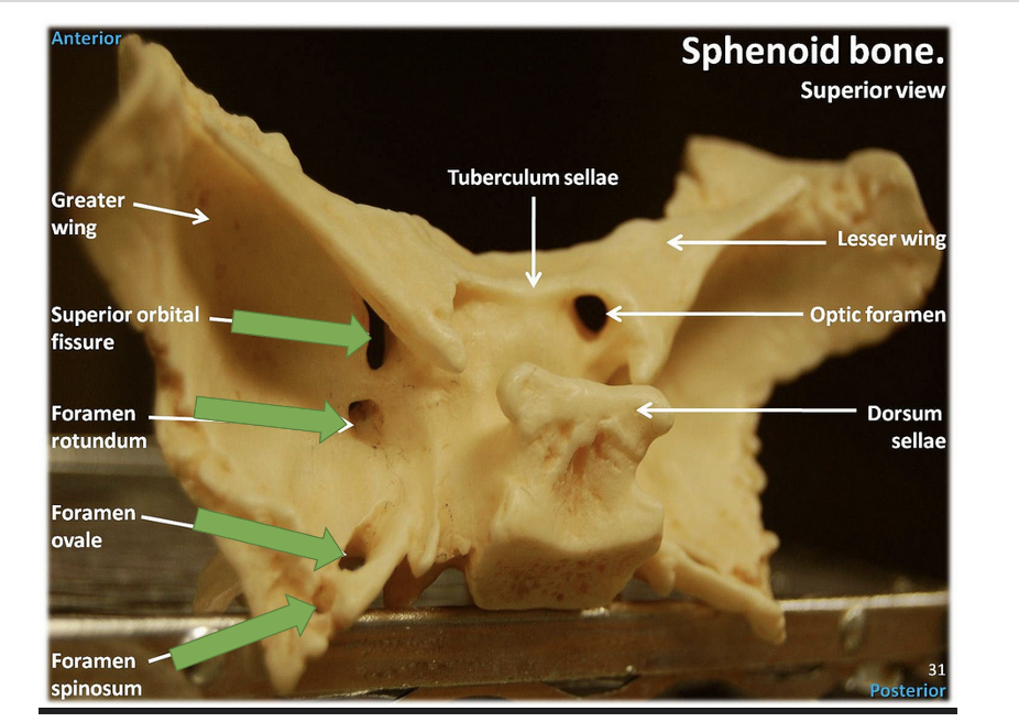
Foramen ovale
An opening for the mandibular branch of cranial nerve V. V-3 branch of trigeminal branch (v-3 goes to mandible)
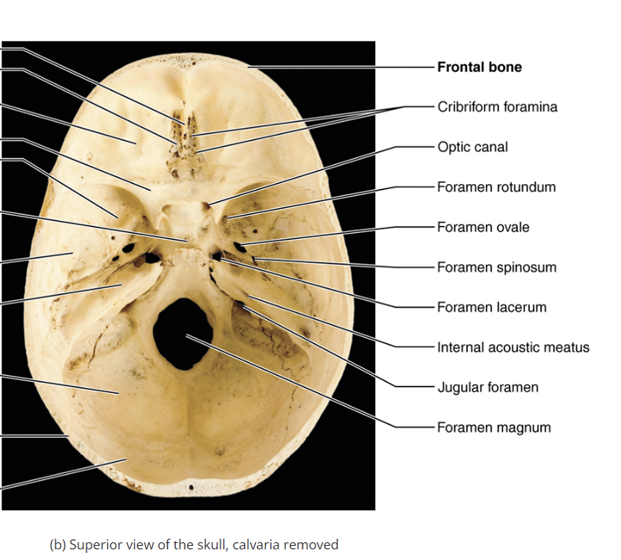
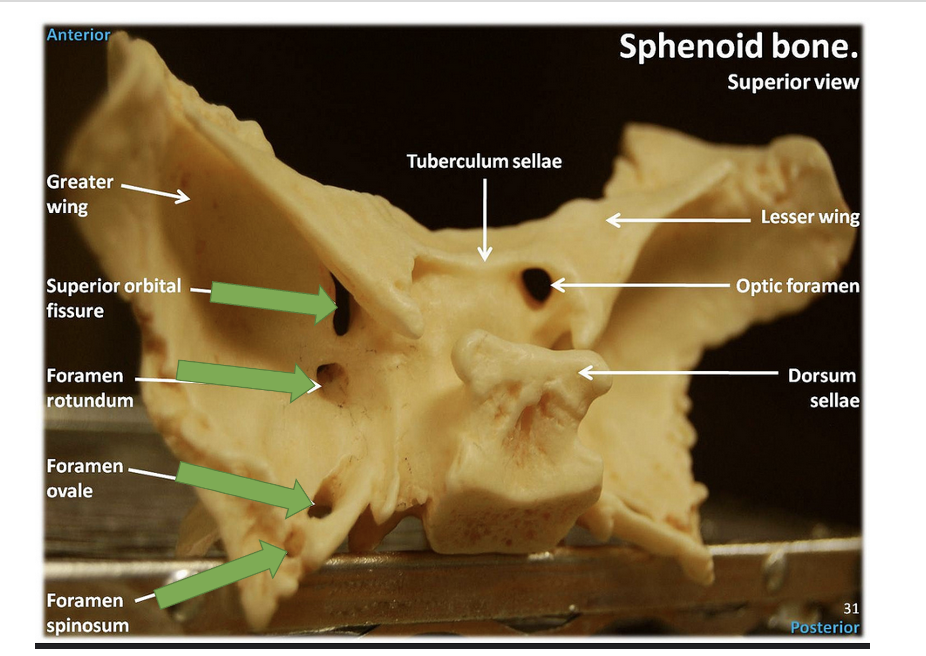
Foramen spinosum
A small opening traversed by the middle meningeal artery that supplies the dura mater, and courses deep to pterion If you crack pterion (very thin portion of bone) could rupture meningeal artery and bleed separating the dura from the skull: epidural hematoma
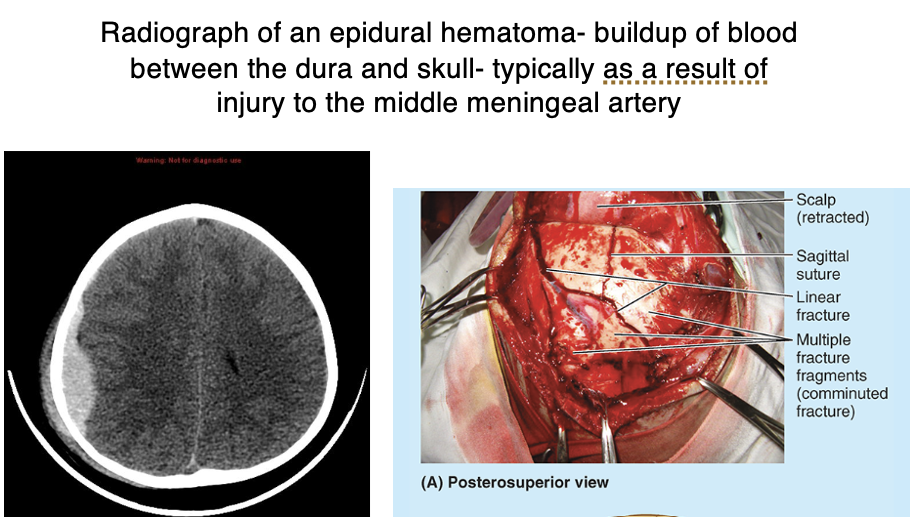
Epidural hematoma
A condition resulting from a rupture of the Pterion → meningeal artery, often due to a fracture at the pterion.
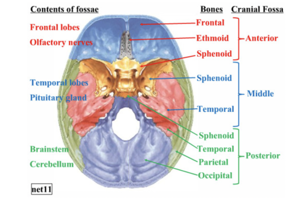
Posterior cranial fossa
The back depression in the cranial cavity, containing the occipital bone and temporal bone. Petris ridge of temporal bone separates the posterior cranial fossa and middle cranial fossa Internal acoustic meatus, Jugular foreman, Hypoglossal canal
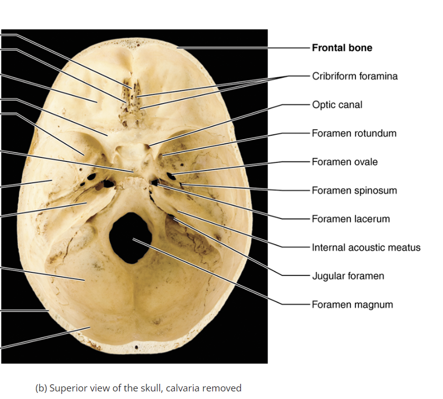
Internal acoustic meatus
passage of cranial nerve that goes into the Petris part of the temporal bone, traverses by cranial nerve VII (facial nerve muscles of facial expression are innervated by the facial nerve) and VIII vestibulocochlear nerve (cranial nerve eight, hearing and balance and innervated by the vestibulocochlear nerve) go into internal ear
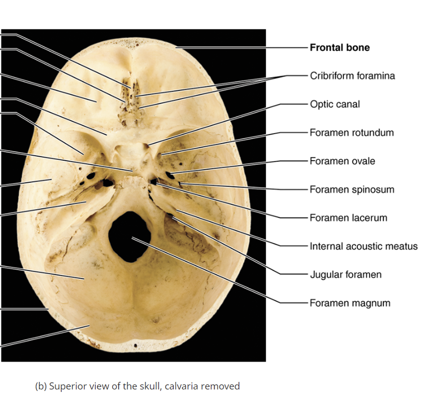
Jugular foramen
An opening for the internal jugular vein and cranial nerves IX, X, and XI. transmits cranial nerve 9 (glossopharyngeal nerve: innervates our tastes to the back of the tongue, pharynx innervates stylopharyngeus muscle ), 10 (vagus nerve that innervates to the heart, the gut, the larynx ), and 11 (accessory nerve: innervates the sternocleidomastoid muscle and the trapezius muscle)
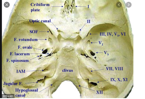
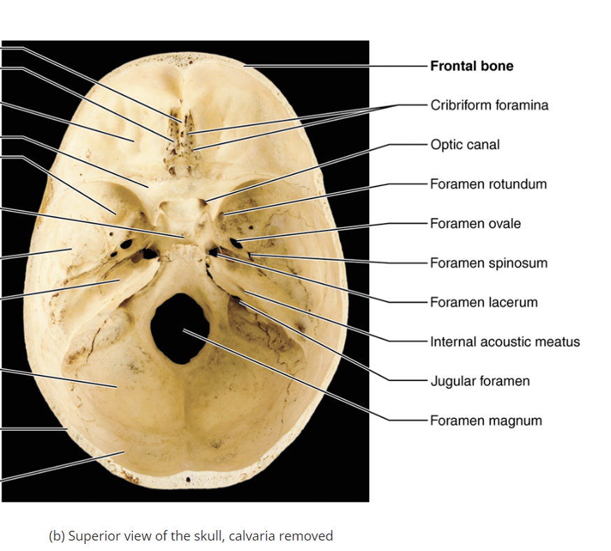
Hypoglossal canal
A passage for cranial nerve XII (hypoglossal nerve: involved in tongue movement.transmits hypoglossal nerve and innervates the muscles of the tongue: when you move your tongue to the left or right when you stick out your tongue
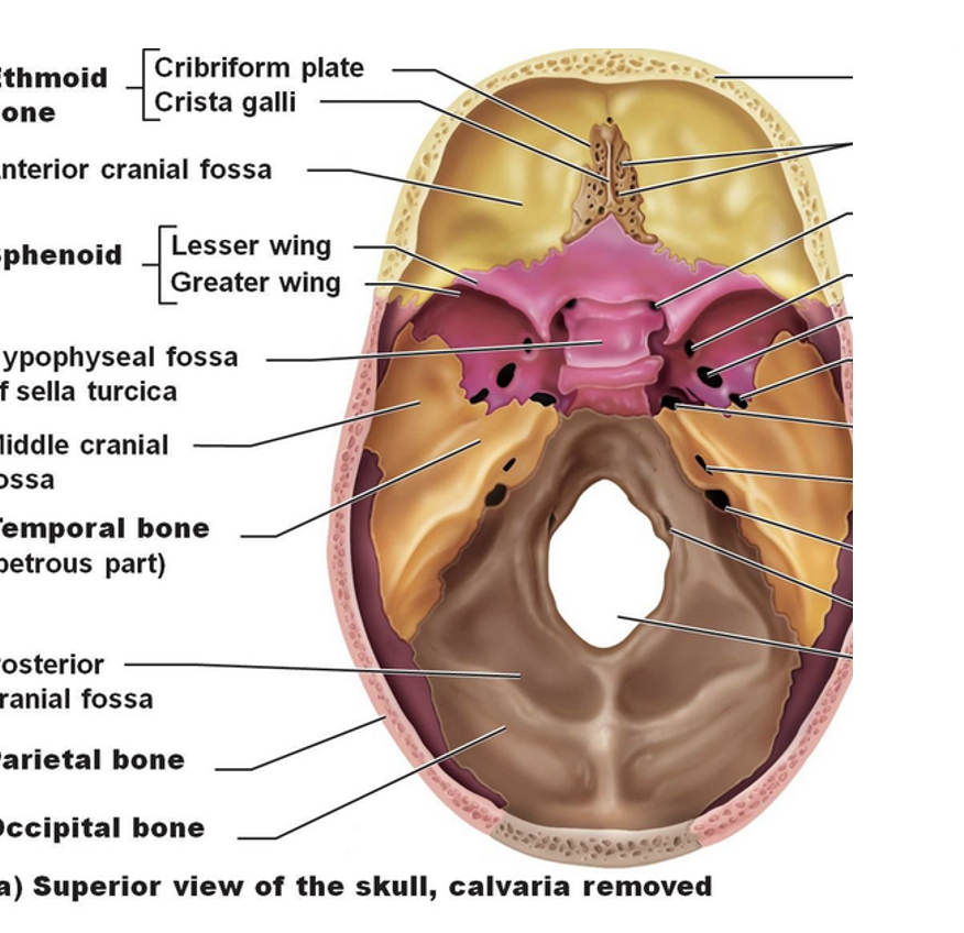
Sella turcica
A depression in the sphenoid bone where the pituitary gland sits.
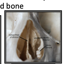
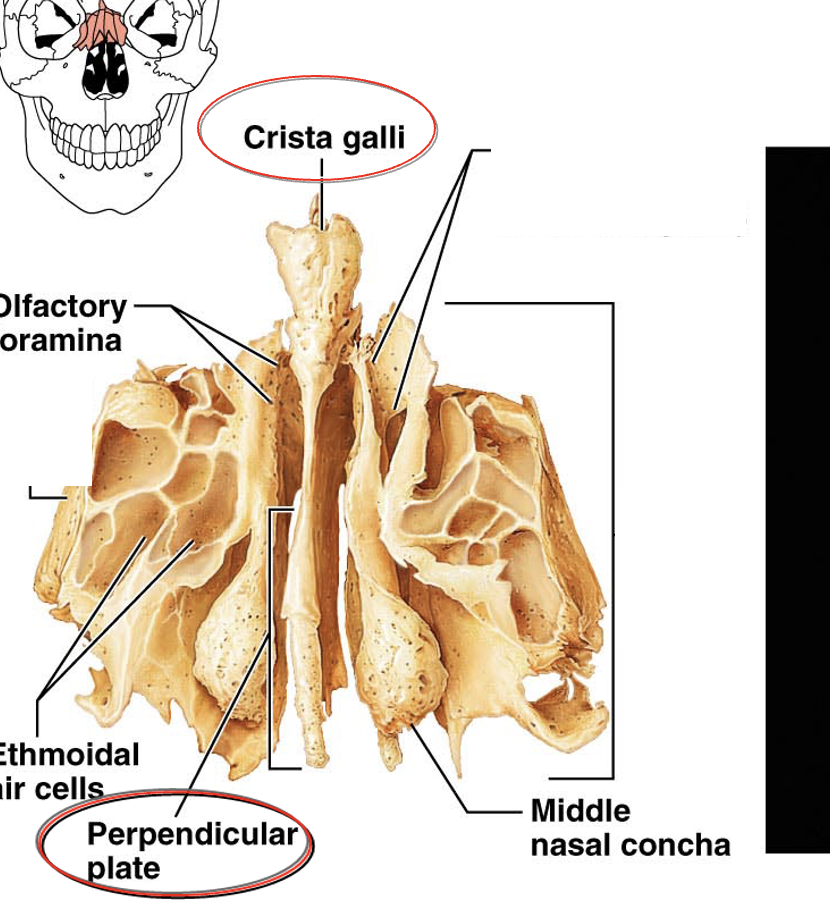
Nasal concha
Structures in the nasal cavity that help circulate air.
Mandible
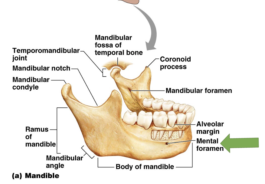
The lower jawbone, consisting of several parts including the body and ramus.
Mental foramen
An opening in the mandible that transmits the mental nerve, transmits the mental nerve, branch of the mandibular branch of the trigeminal
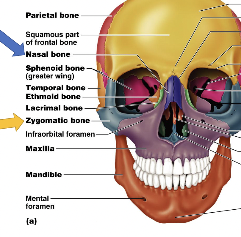
Maxilla
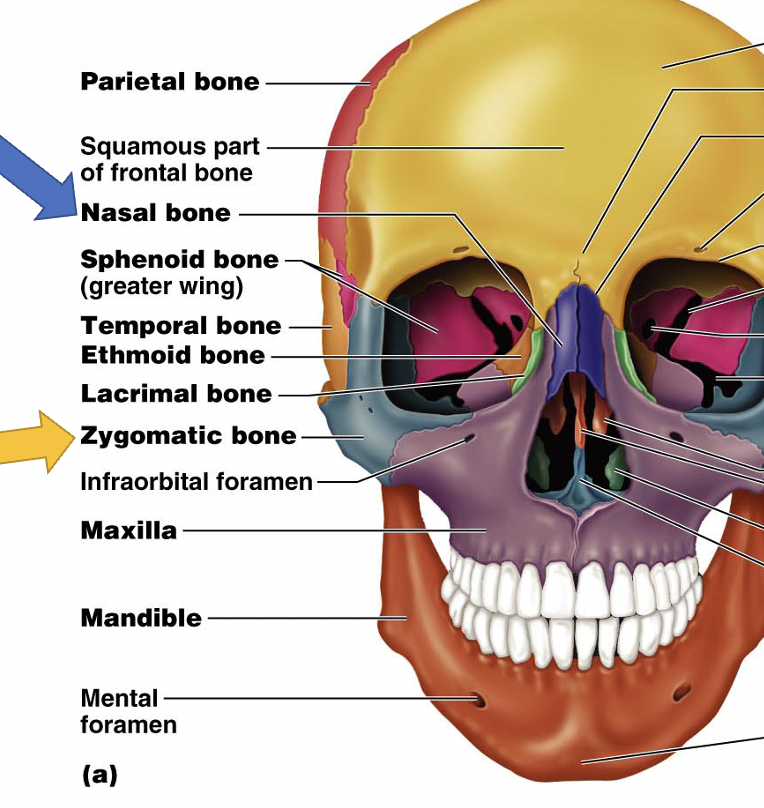
The upper jawbone, part of the bony orbit.
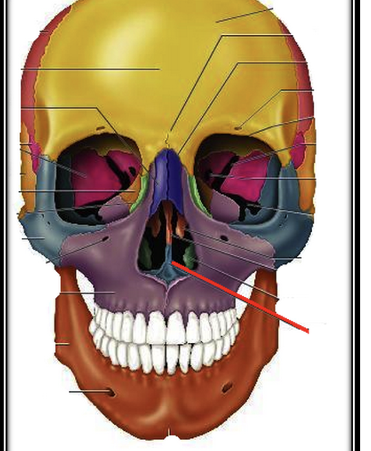
Vomer
A bone forming part of the nasal septum, part of the visceral cranium that makes up the middle part of nasal cavity and meets with the perpendicular plate of the ethmoid bone
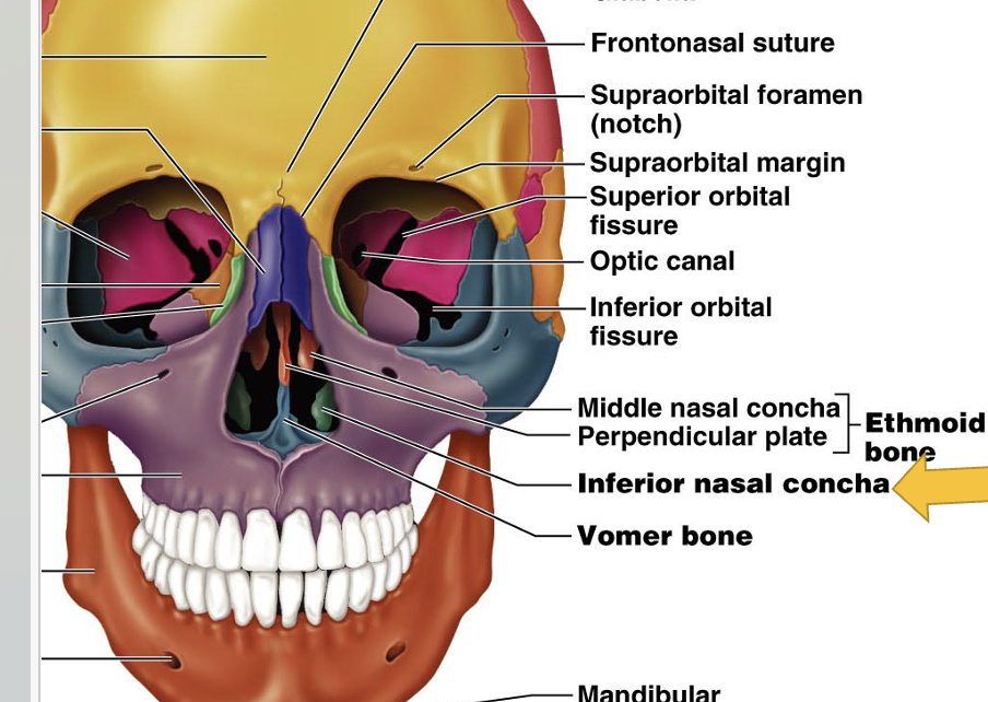
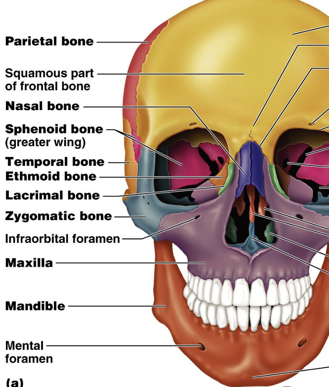
Zygomatic bones
The cheekbones, contributing to the bony orbit. 2 of them
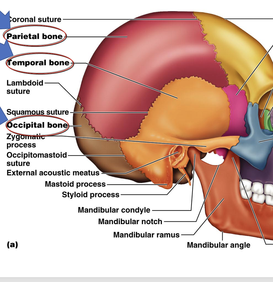
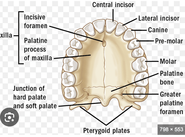
Hard palate
Formed by the maxilla and palatine bones, separating the oral cavity from the nasal cavity. (come together to make the hard palate which is described as the palatine process of the maxilla and horizontal plate of palatine bone
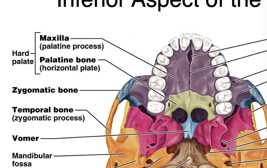
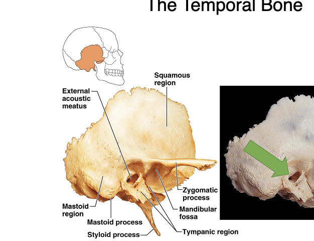
External acoustic meatus
The ear canal within the temporal bone.
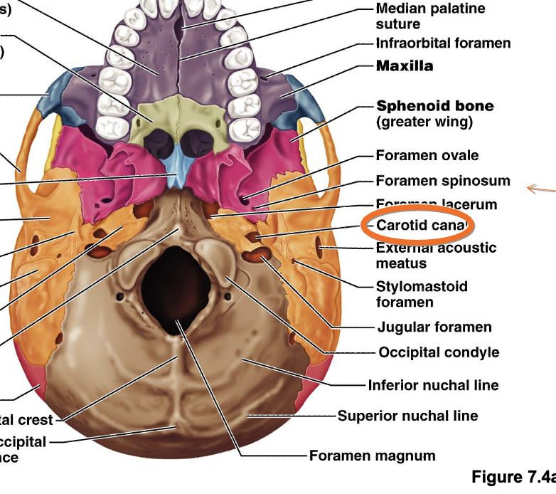
Occipital condyle
The rounded projections on the occipital bone that articulate with the first cervical vertebra.
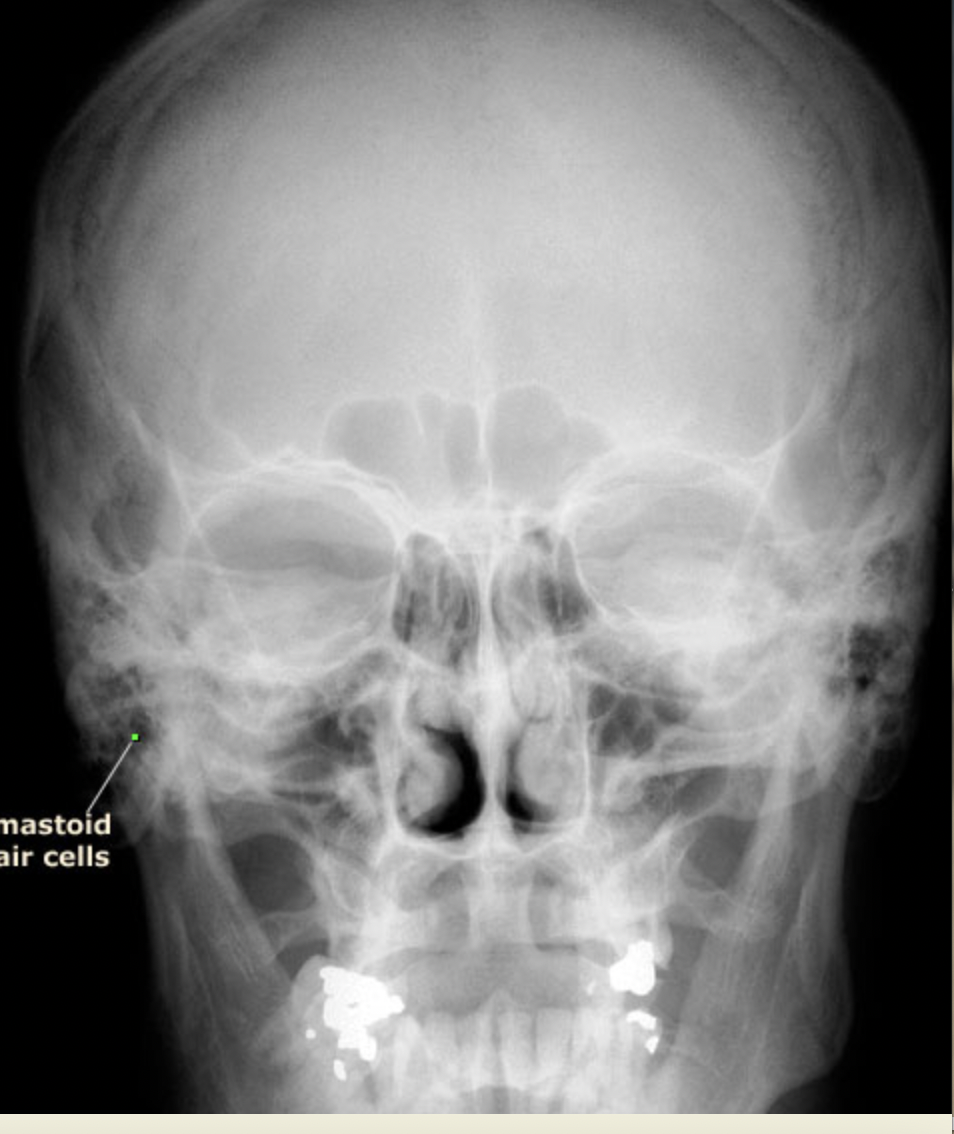
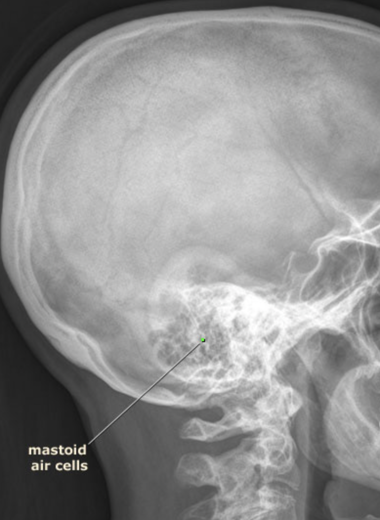
Air cells and sinuses
Structures that lighten the skull and help resonate sound.
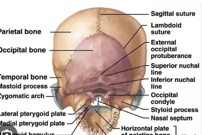
External occipital perturbance
for the attachment of the trapezius muscle
foramen magnum
the spinal cord goes through
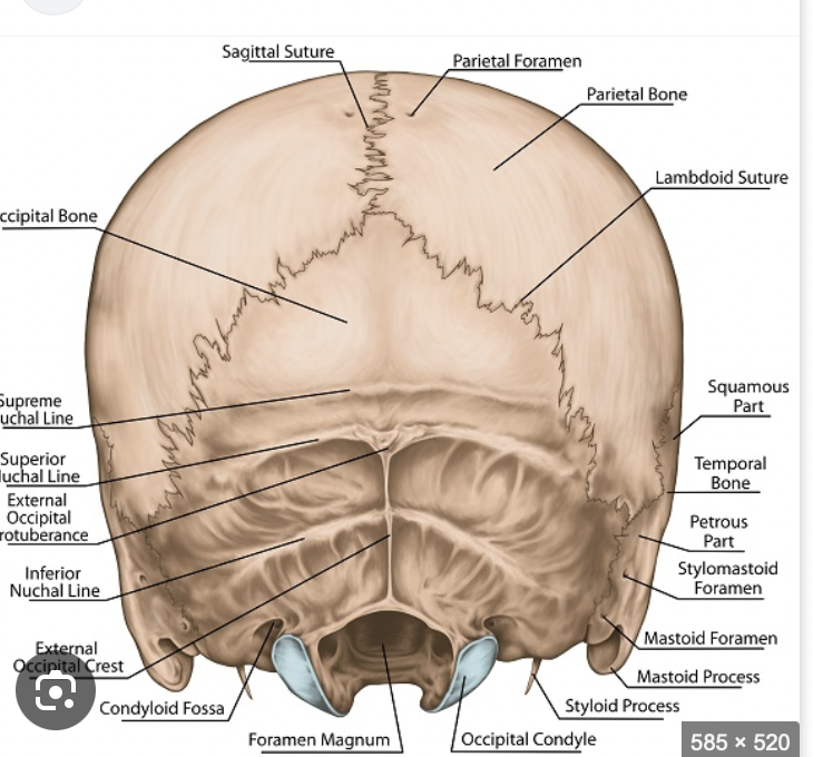
superior nuchal line
nuchal ligament attaches also an inferior nuchal line
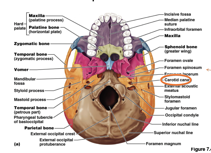
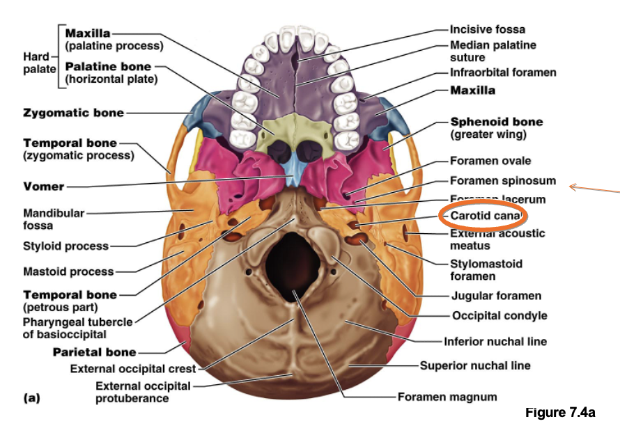
Stylomastoid foreman
between the styloid process and mastoid process
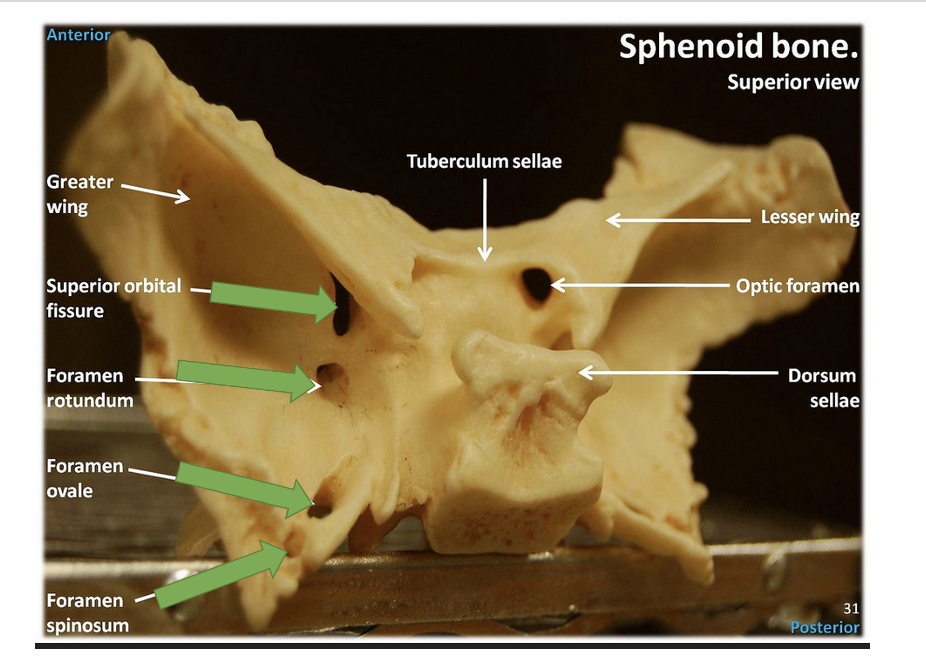
Foreman Spinosum
transmits middle meningeal artery which is important in suppling blood to the calvaria if this is injured one will have an epidural hematoma
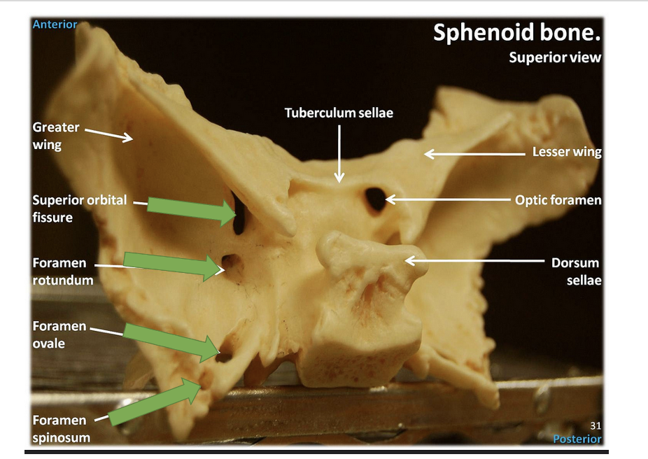
Foreman Ovale
transmits the mandibular division of the trigeminal nerve that involves taking sensation from the lower part of the face involving the lower teeth the lower lip the chin the mandible to the brain
Upper teeth are innervated by the maxillary nerve
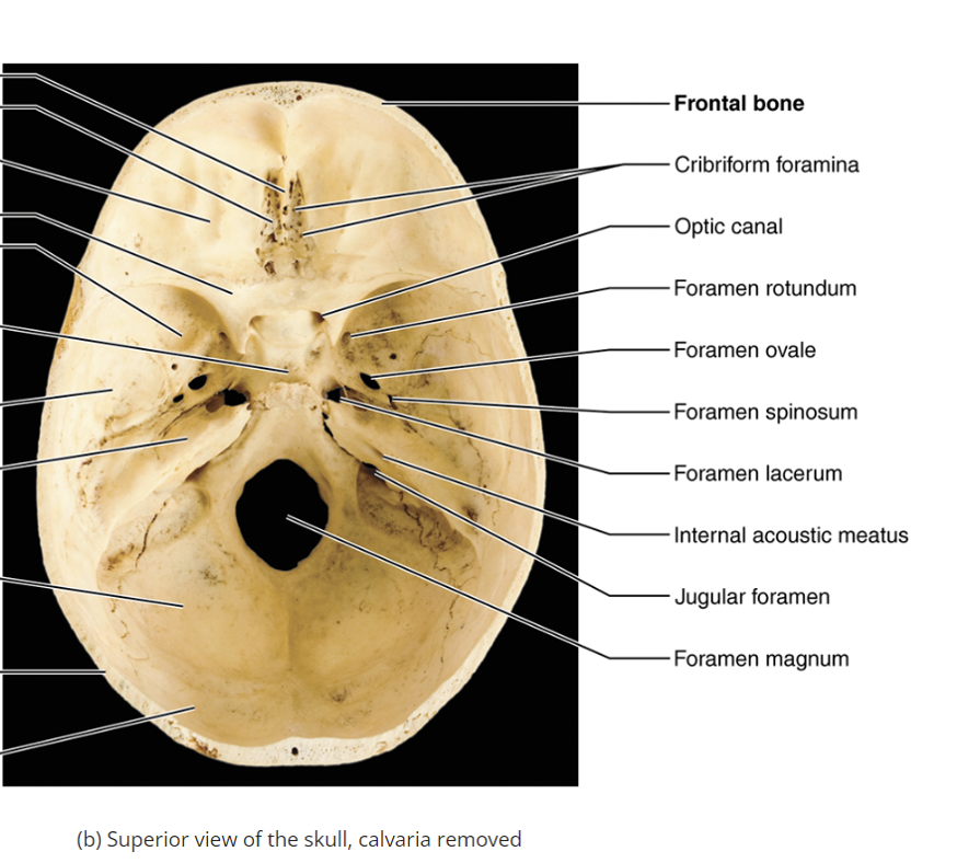
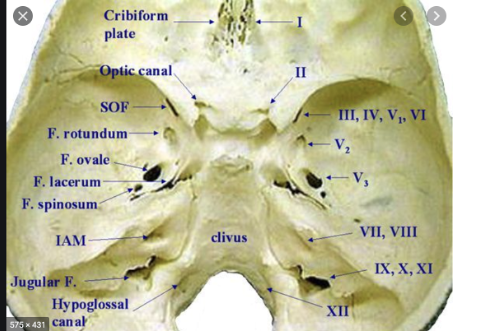
Foreman rotundum
transmits the maxillary nerve (transmits sensation from the lower part of the face meaning maxilla, upper part of the nose, upper lip)
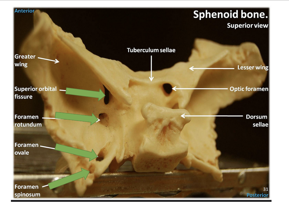
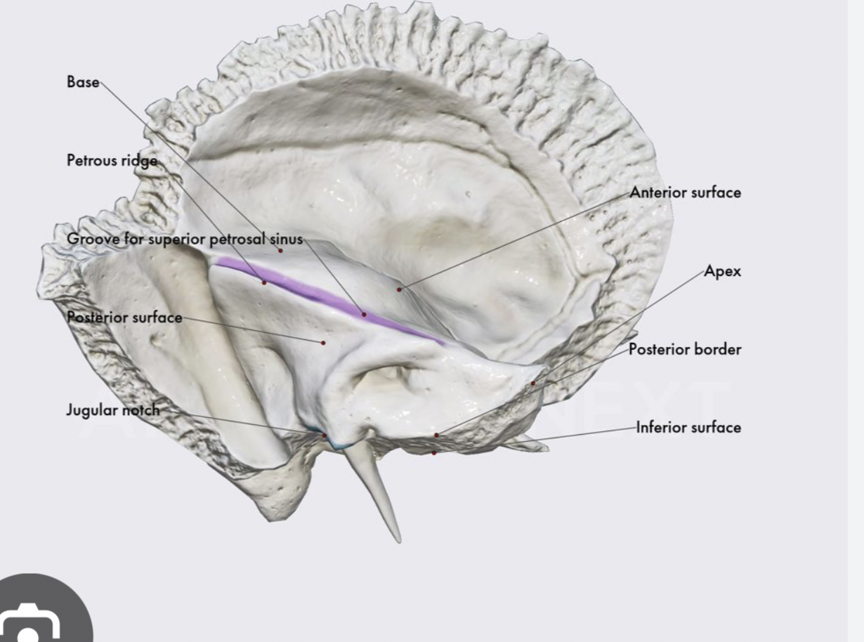
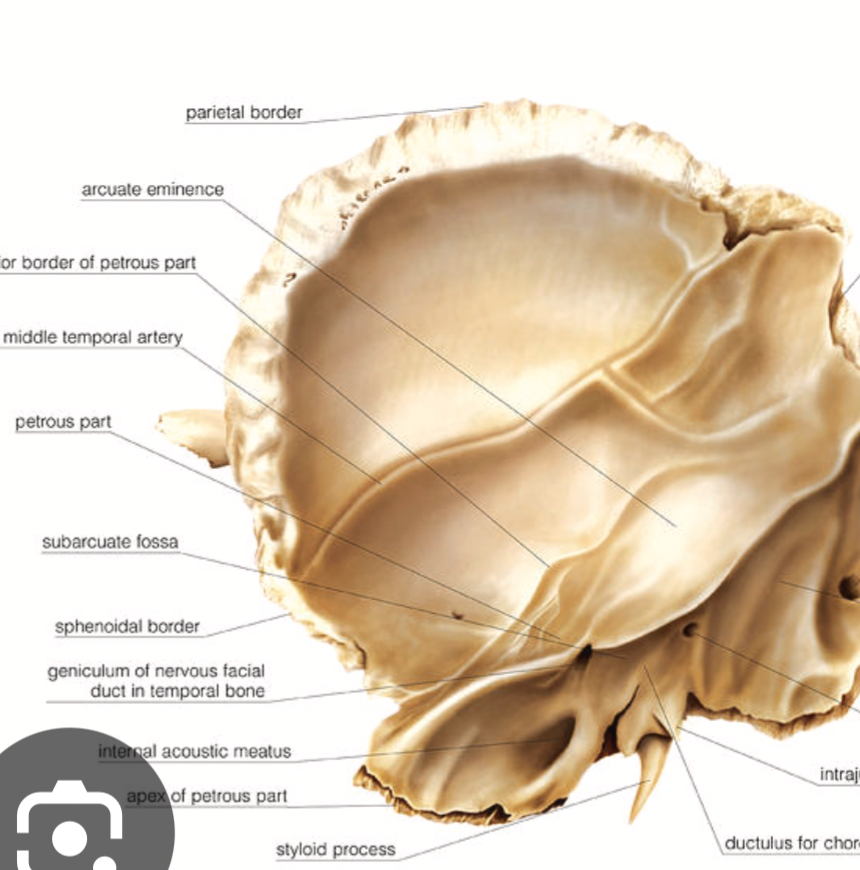
Petrous ridge of temporal bone
separates the posterior cranial fossa and middle cranial fossa
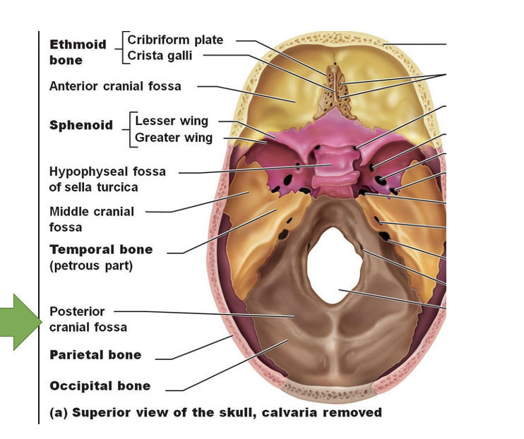
lesser wing of the sphenoid
divides the he middle cranial fossa and the anterior cranial fossa
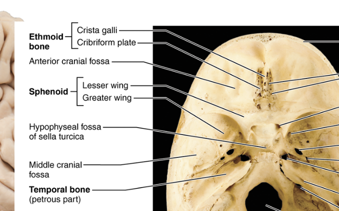
cribriform plate
portion of the ethmoid bone that contains olfactory foramina that allow the olfactory nerve or first cranial nerve to pass through
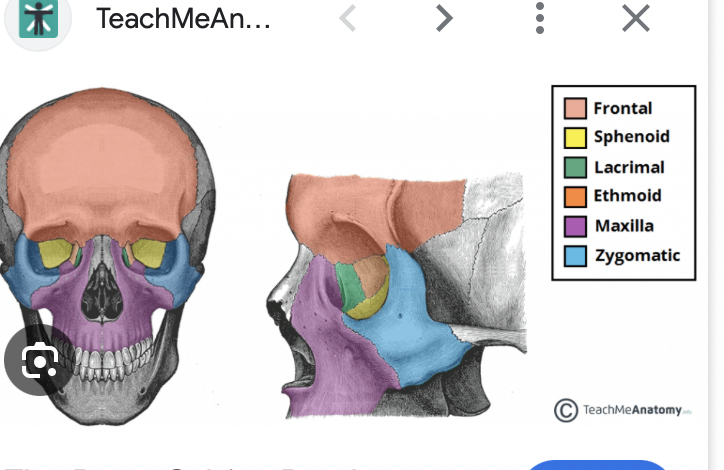
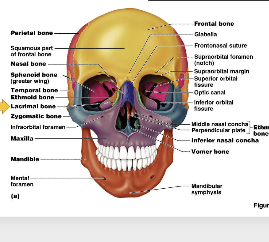
bony orbit
made up of the frontal bone, ethmoid bone, lacrimal bone, the maxillary bone, zygomatic bone, the sphenoid bone, and the palatine bone
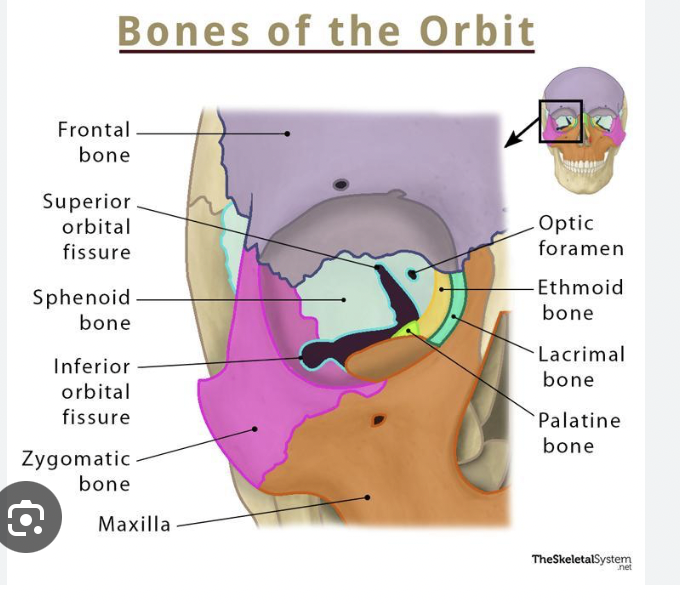
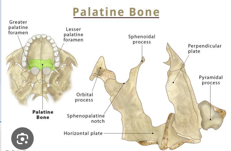
orbital plate of palatine bones
a very small portion of the bony orbit
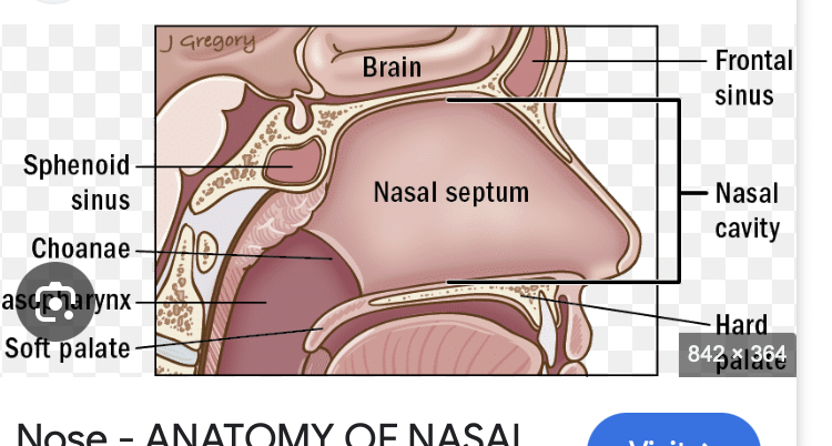
nasal septum
made up of two bones: the perpendicular plate of ethmoid and the Velma
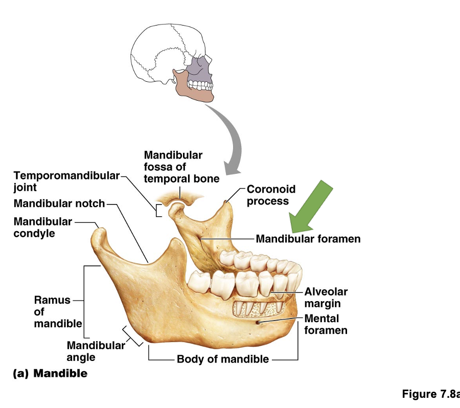
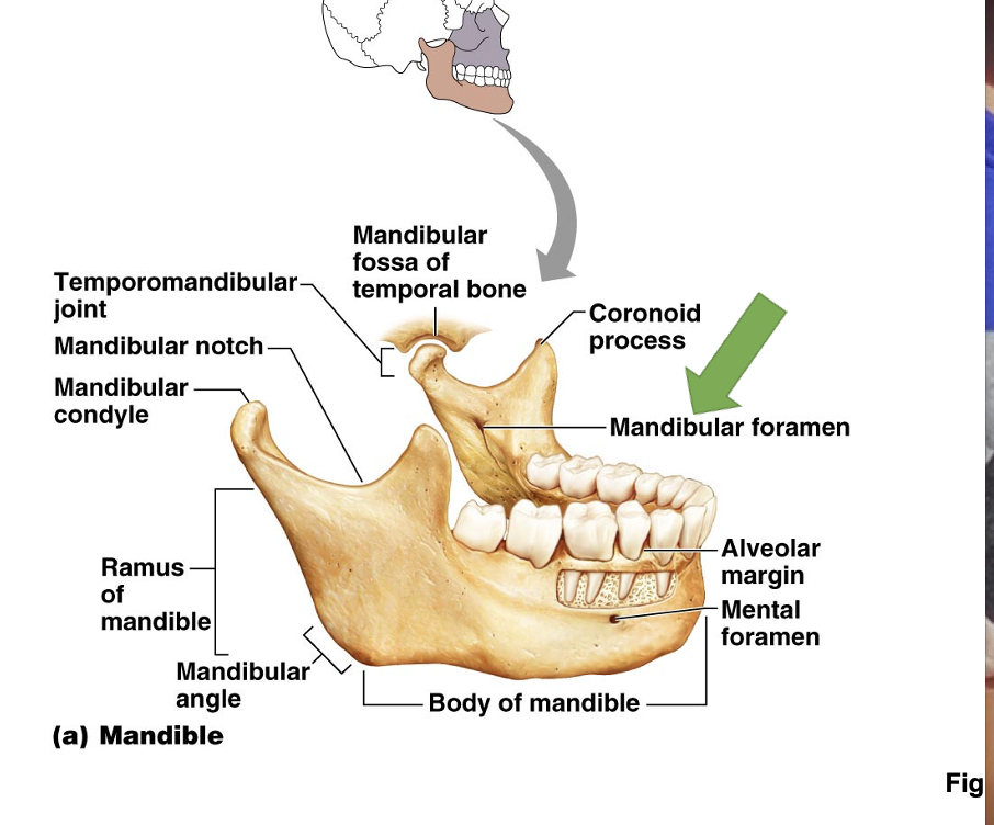
mandibular foreman
on the inside of mandible), it transmits the inferior alveolar nerve, a branch of the mandibular division of trigeminal
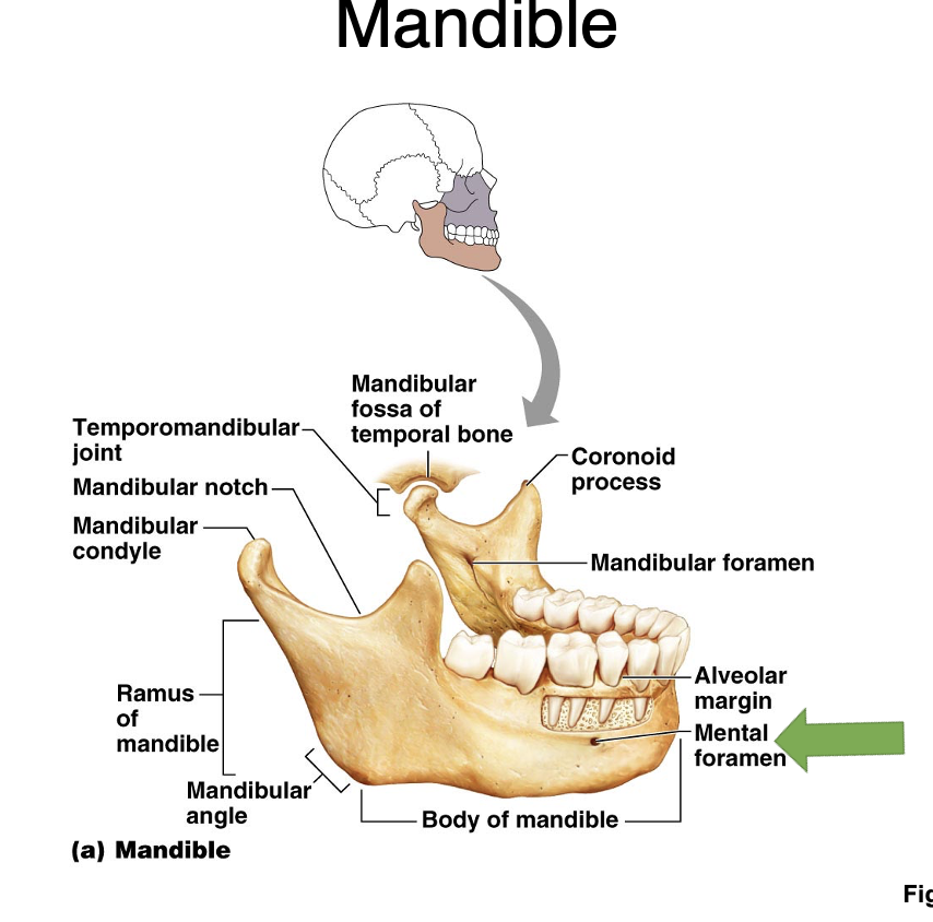
mandible
two foramina (mandibular foreman and mental foreman), contains body of mandible, angle of mandible, the ramus of the mandible, condyle of the mandible, coronoid process (part of viscerocranium)
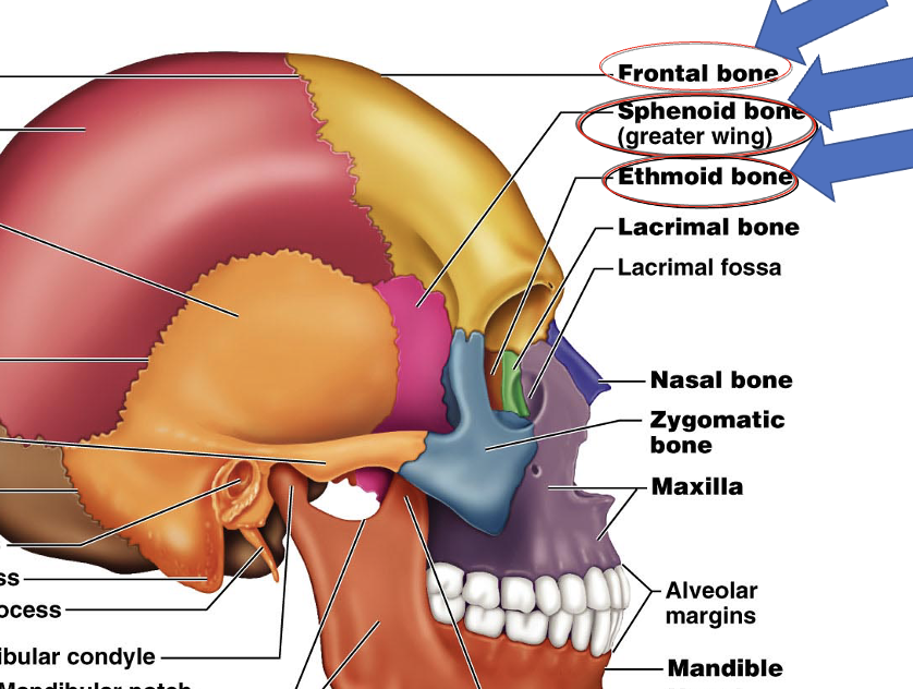
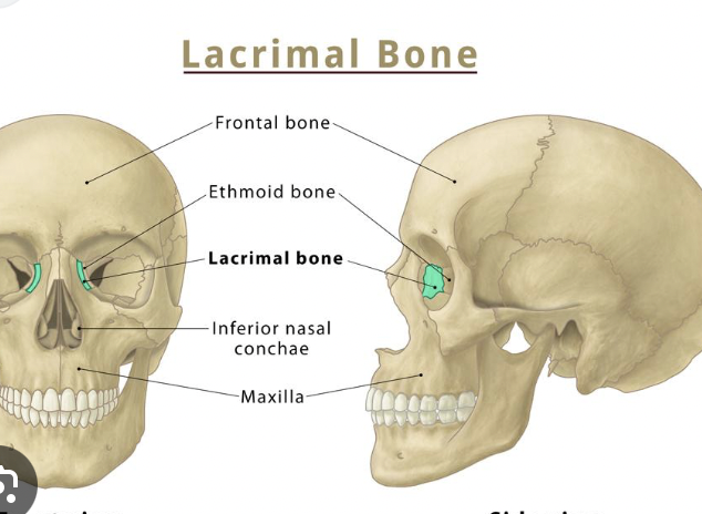
lacrimal bone
a visceral cranium bone that it part of the bony orbit
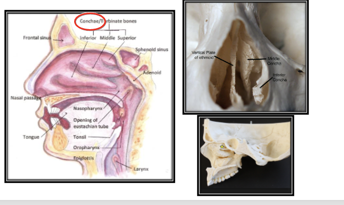
nasal cavity
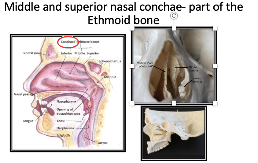
Within is the superior and middle nasal concha (part of the ethmoid bone) there is a small bone known as the inferior nasal concha (part of the viscera cranium)
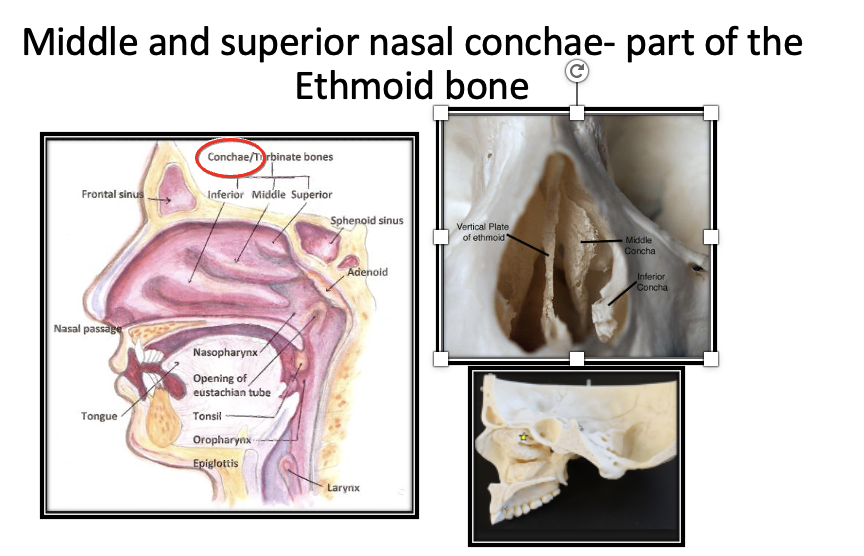
Cauchy
shells in nasal cavity that allow for circulation of air as you breathe in
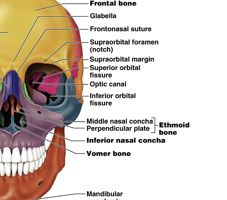
ethmoid bone
perpendicular plate and superior nasal concha
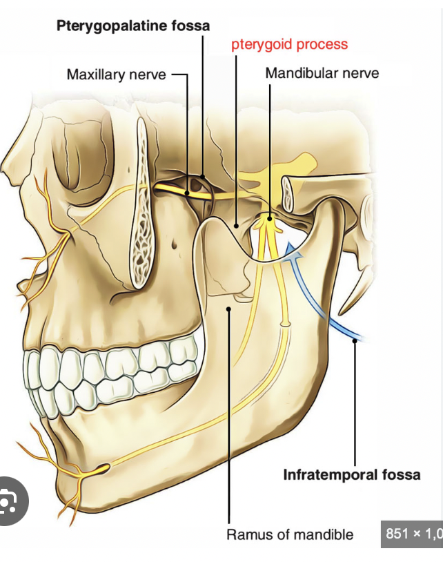
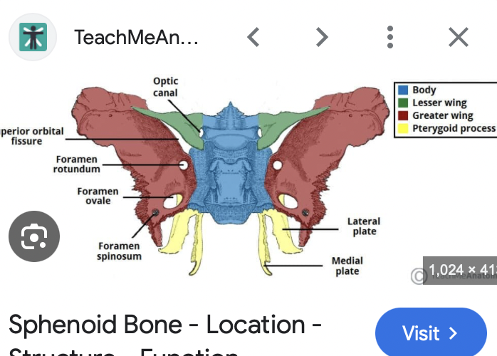
pterygoid process
two processes together are called the pterygoid plate (lateral and medial) Attachments for muscles of mastication (chew)
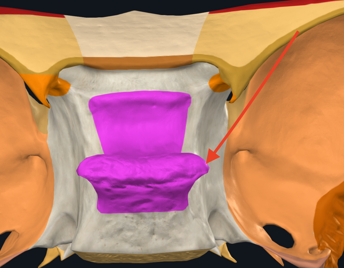
posterior projection processes
Two bony projection off of Sella Turcica
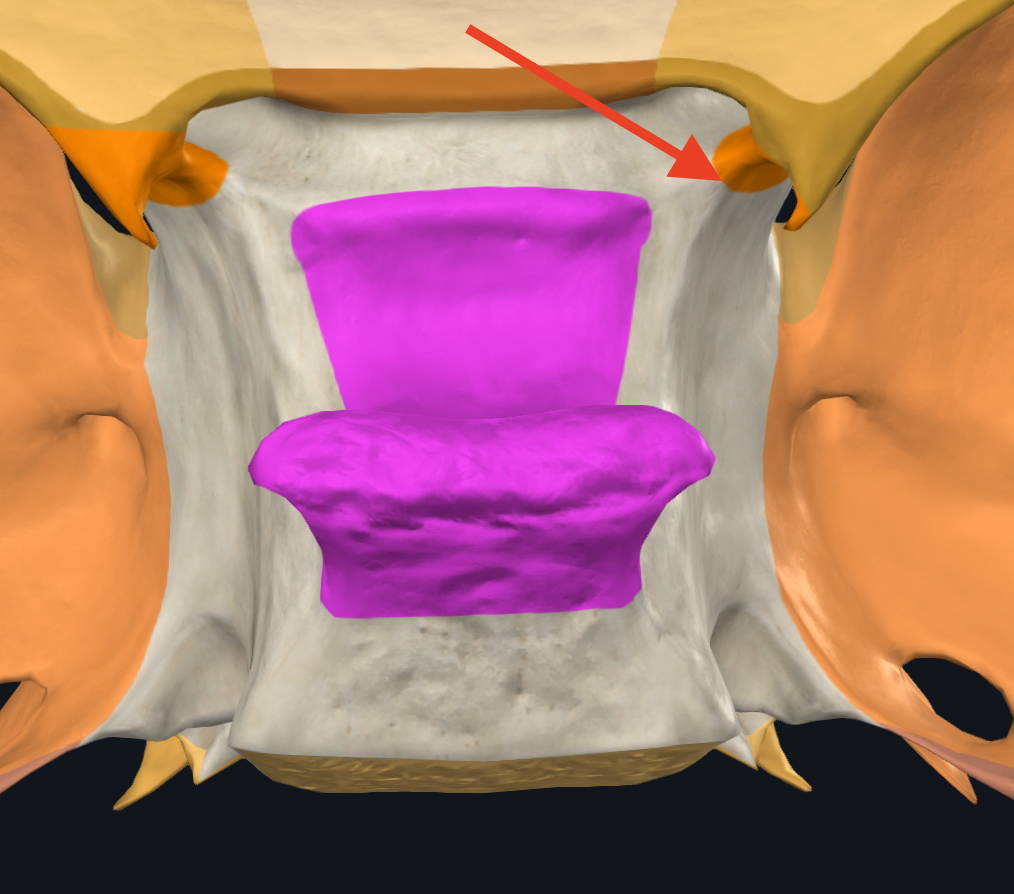
anterior clinoid processes
Two bony projections off of lesser wings
sella turcica
portion in the middle of sphenoid bone where the hypophyseal gland or pituitary gland sits
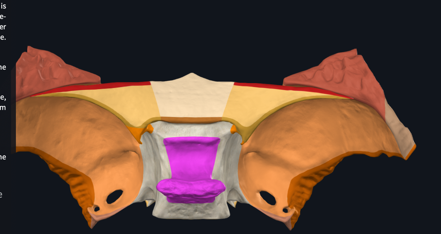
Sphenoid bone
forms the posterior portion of anterior cranial fossa and much of the middle cranial fossa
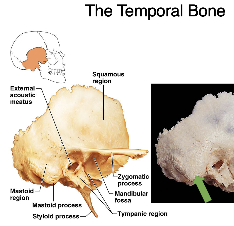
Squamosal of temporal bone
thin portion of temporal bone
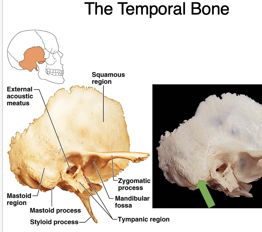
temporal bone
External auditory meatus
Petris ridge of temporal bone: hard and more prominent part of temporal bone, in middle cranial fossa
Mastoid process
Styloid process: pointy
Internal acoustic meatus
Squamosal of temporal bone
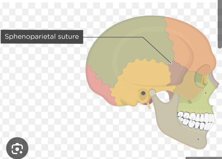
Sphenoparietal suture
between sphenoid and parietal
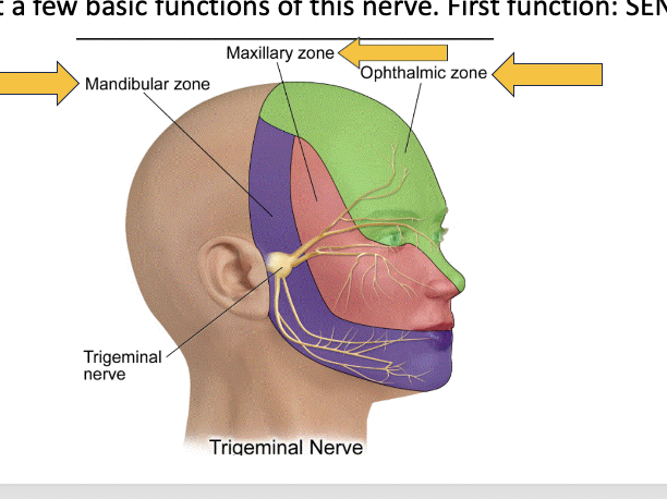
CN V
trigeminal nerve mandibula zone, maxillary zone, ophthalmic zone, arises from the pons
V-1 ophthalmic, sensation to upper part of the face such as cornea mucosa of the upper part of the nasal cavity upper eyelid, Inferior orbital fissure
V2- maxillary- sensation to the middle part of the face such as inferior eyelid, upper teeth, upper lip, inferior part of the nasal cavity, and anterior cheek, Foramen rotundum, allows for cranial nerve five and V-2 (maxillary branch) (V-2 goes to the maxilla)
V3- mandibular- sensation to the lower part of the face such as lower teeth, lower lip, anterior 2/3 of the tongue, tympanic membrane. In addition, this nerve has somatic motor functions: it innervates all muscles of mastication (chewing muscles). foreman Ovale V-3 branch of trigeminal branch (v-3 goes to mandible)
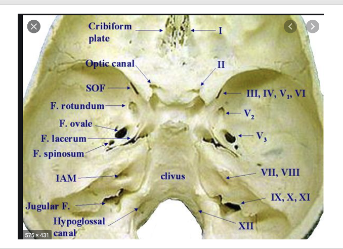
CN II
optic nerve- special sensory (sight). The receptors are in the retina and the axons project to the occipital lobe of the brain, through optic canal
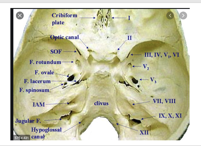
CN I
olfactory nerve- special sensory (sense of smell). The receptors are within the nasal mucosa. The axons travel through the tiny foramina of the cribriform plate. These axons travel to the frontal and temporal lobes of the brain, through cribriform plate
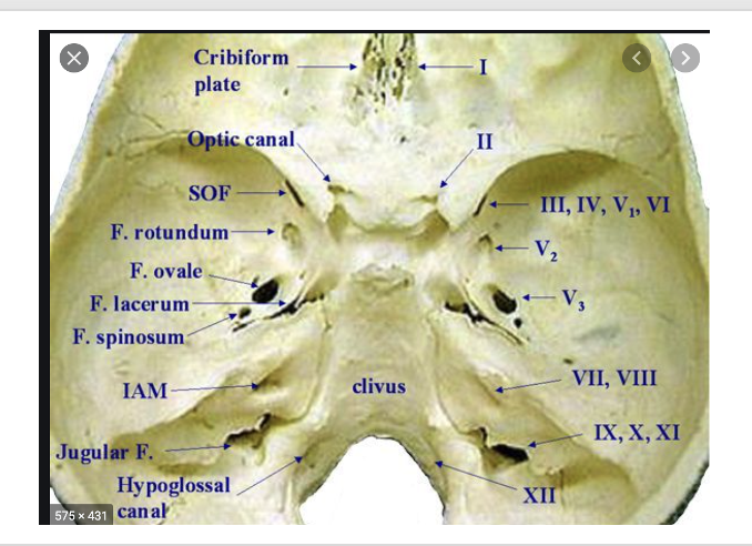
CN III
oculomotor nerve- This nerve originates from the midbrain. It innervates muscles of the eye (we will learn about these later). It also has parasympathetic functions: causes constriction of the pupil and is responsible for lens accommodation. This nerve has visceral motor and somatic motor functions. arise from the midbrain, superior occipital fissure
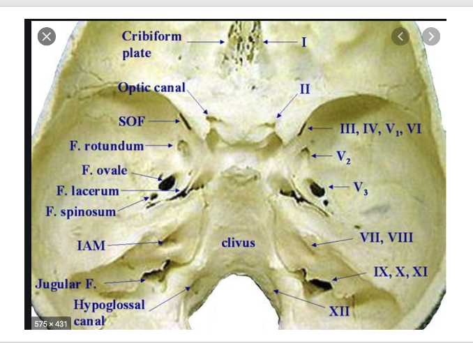
CN IV
trochlear nerve- This nerve innervates one muscle of the eye. Somatic motor, arise from the midbrain, superior occipital fissure
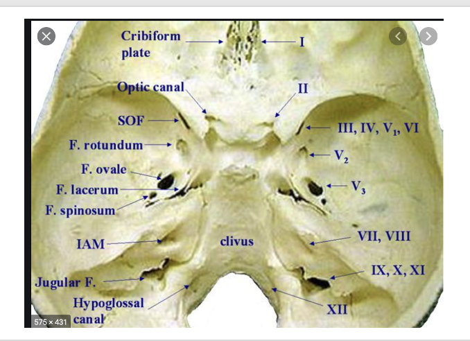
CN VI
abducens nerve- innervates one muscle of the eye- somatic motor, arise from pontomedullary Junction, superior occipital fissure
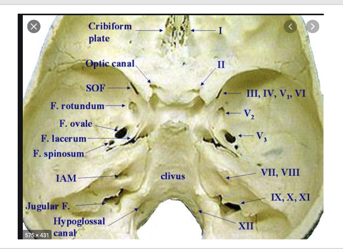
CN VII
facial nerve- somatic motor to muscles of facial expression (muscles that make you smile, raise your eyebrows, wrinkle your nose etc). This nerve also innervates the lacrimal gland (makes tears) and glands in the palate and nose (parasympathetic function). It also has a branch that provides taste to the anterior 2/3 of the tongue. arise from pontomedullary Junction, internal acoustic meatus
CN VIII
vestibulocochlear nerve- special sensory- hearing and balance via the cochlea and the semicircular canals in the inner ear arise from pontomedullary Junction, internal acoustic meatus
CN IX
glossopharyngeal nerve- innervates one muscle (somatic sensory to stylopharyngeus). Also innervates the back of the tongue and pharynx. This nerve also innervates the parotid gland (visceral motor). arise from the medulla, through jugular foreman
CN X
vagus nerve- innervates muscles of the larynx and the vocal cords (somatic motor- more on this later). It has parasympathetic functions innervating the heart, bronchi and lungs and most of the gastrointestinal system, through jugular foreman
CN XI
accessory nerve- innervates two muscles- somatic motor- sternocleidomastoid and trapezius, jugular foreman
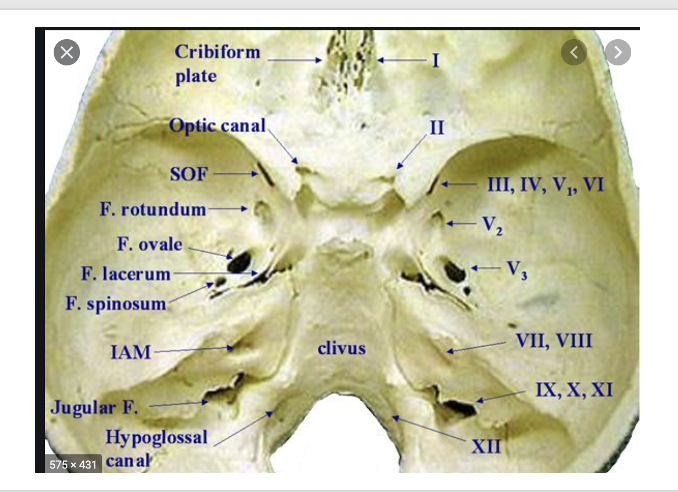
CN XII
hypoglossal nerve- innervates muscles of the tongue,arise from the medulla, through hypoglossal canal
Somatic motor
innervating muscles
Somatic (general) sensory
general senses such as touch pain temperature and pressure
Visceral motor
parasympathetics function of cranial nerves
Visceral sensory
sensations of internal organs such as stretch visceral pain
Special sensory
taste, vision, hearing, balance
peripheral nerves
can be either spinal nerves (these nerves innervate the muscles, bones, joins, tendons and skin of the body except the head) or cranial nerves that innervate structures in the head.
Nasal Septum
made up of the perpendicular plate of the ethmoid bone and the vomer (an independent bone)