1. Diseases of the eyelids, conjunctive and third eyelid.
1/93
There's no tags or description
Looks like no tags are added yet.
Name | Mastery | Learn | Test | Matching | Spaced |
|---|
No study sessions yet.
94 Terms
What are the anatomical structures of the eyelid?
Skin, muscle layer, stoma and tarsus, palpebral conjunctiva

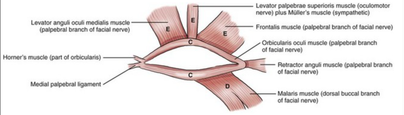
Which muscles control eyelid movement and what are their functions?
M. orbicularis oculi (eye closure – CN VII)
Horner muscle (pumping tears)
M. retractor anguli oculi lateralis (stabilises lateral canthus)
M. levator anguli oculi medialis (medial levitator of upper eyelid)
What are some diseases affecting the periocular area (around the eyelids)?
Demodex
Sarcoptes
Dermatomycoses
Autoimmune skin diseases
Seborrhoea
Allergic/atopic dermatitis
Solar dermatitis
Hypothyroidism
What are examples of congenital diseases of the eyelids?
Eyelid agenesis/atresia/coloboma
Dermoid
Growth disorders of the cilia
Distichiasis
Ectopic cilia
Trichiasis
Madarosis
Entropion
Ectropion
What is eyelid agenesis/atresia/coloboma?
Partial or complete underdevelopment of the eyelid margin and/or lid itself

How is eyelid agenesis/atresia/coloboma treated?
If the margin of eyelid is developed w/o meibomian glands, no surgical therapy is necessary.
If there are ectopic hairs in the area → remove.
If cornea is only slightly irritated: administer topical lubricants on a daily basis until work.
If lesions are larger, a substitute eyelid should be created by blepharoplasty
What is a dermoid?
A congenital, benign neoplasia (choristoma) of ectopic, abnormally developed skin tissue in or at the eyelid margin, frequently associated with some dysplastic deformities of the adjacent conjunctiva. Can occur everywhere.

What is distichiasis?
Hair(s) growing from the free lid margin, often from meibomian gland openings on upper or lower eyelids
What are the clinical signs of distichiasis?
Corneal irritation (rubbing hairs), increased lacrimation, blepharospasm, and potentially corneal ulceration

How is distichiasis treated?
Manual epilation every 4-5 weeks (temporary)
Permanent removal/destruction/redirection of hair follicles (electrocautery, cryotherapy, laser therapy, eyelid splitting)
What are ectopic cilia?
Cilia emerge through the palpebral conjunctiva and impinge directly on the cornea. Predisposed in animals with distichiasis

What are the clinical signs of ectopic cilia?
Severe corneal irritation, increased lacrimation, blepharospasm, and potentially corneal ulceration
How are ectopic cilia treated?
Evert lid, conjunctiva removed by electric knife, hair follicle is visualised and destroyed
What is trichiasis?
Misdirection of normally located eyelashes, often in the medial canthus or nasal fold

What are the causes of trichiasis?
Congenital (abnormal follicle development)
Acquired (blepharitis, scarring, trauma/surgery, entropion, ocular surface disorders, systemic conditions)
What are some specific types of trichiasis?
Nasal fold trichiasis
Upper eyelid trichiasis (dorsolateral)
Trichiasis combined with entropion
Brachycephalic ocular syndrome (caruncular hairs irritating the cornea)
What are the clinical signs of trichiasis?
Ocular irritation, redness, tearing, corneal abrasions/ulcers/scarring, and blepharospasm (or complete eyelid closure if severe)
What is entropion?
Partial or complete inward rolling of the eyelid margin
What are the causes of entropion?
Congenital (breed-specific: Chow Chow, Bulldog, Poodle, Shar Pei)
Acquired (spasm after trauma)
What are the clinical signs of entropion?
Hyperlacrimation, blepharospasm, conjunctivitis, keratitis, and mucopurulent discharge
How is entropion treated?
Conservative (ocular lubricants)
Surgical (temporary or permanent [Hotz-Celsus] tacking sutures)
![<ol><li><p>Conservative (ocular lubricants)</p></li><li><p>Surgical (temporary or permanent [Hotz-Celsus] tacking sutures)</p></li></ol><p></p>](https://knowt-user-attachments.s3.amazonaws.com/6f1cd012-9863-4163-86ad-7c7fb066e34f.png)
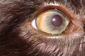
What is ectropion?
Outward drooping of the lower eyelid

What are the causes of ectropion?
Congenital (breed-specific: hunting dogs/heavy-skinned dogs)
Acquired (VII nerve paralysis)
How does ectropion lead to pathologies?
Conjunctiva is exposed to environmental irritants, drying, 2nd bacterial infections
What are the clinical signs of ectropion?
Keratitis, conjunctivitis, and hyperlacrimation
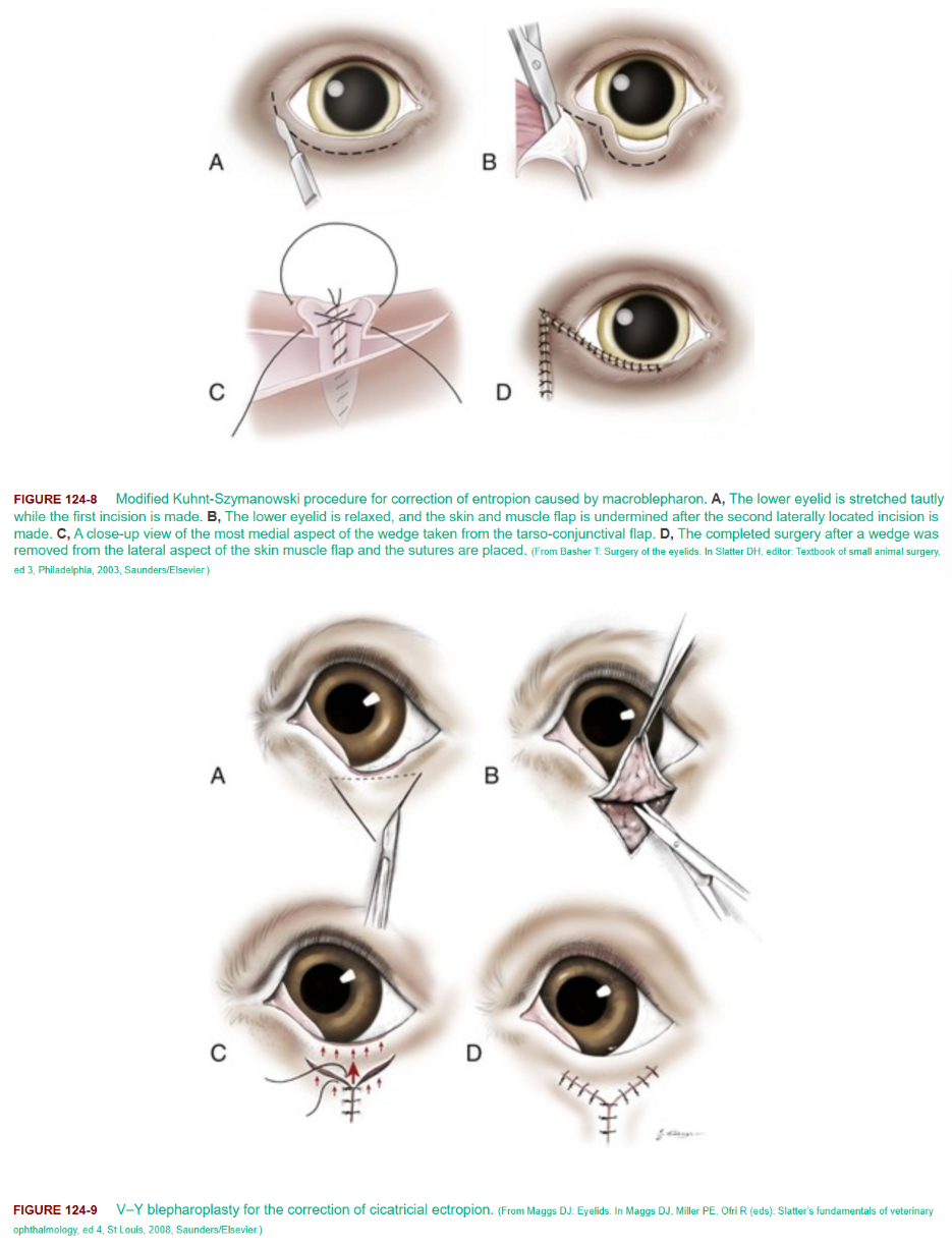
How is ectropion treated?
Conservative (daily eye cleaning)
Surgical (V-Y-blepharoplasty, modified Kuhnt, or wedge resection)
What are examples of acquired eyelid disorders?
Blepharitis
Lid trauma/laceration
Hordeolum
Chalazion
Eyelid neoplasia
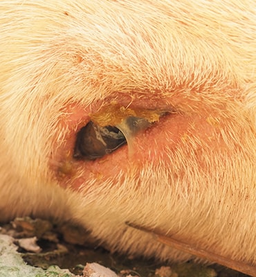
What is blepharitis?
Inflammation of the eyelid skin, deep eyelids, or free margin

Blepharospasm, hyperaemia, swelling, exudation, alopecia, pruritus, and lacrimation
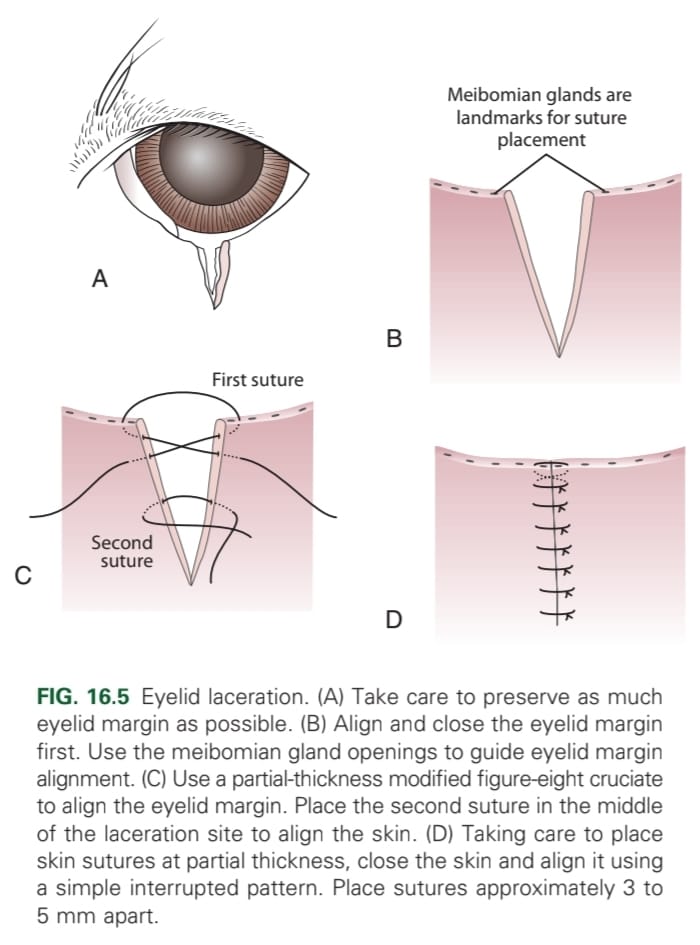
How should eyelid trauma/lacerations be managed?
Repaired ASAP, preserving as much tissue as possible, to protect the cornea
How are eyelid lacerations treated?
Use partial thickness modified figure-8 cruciate at the margin then in the middle of the laceration to align the skin. Fill in with simple interrupted sutures 3-5 mm apart.
What is a hordeolum?
Inflammation of eyelid glands (Zeis, Moll, or Meibomian)

What is a common causative agent of hordeolum?
Staphylococcus aureus. Typically due to poor eyelid hygiene, chronic blepharitis, meibomian gland dysfunction, or ocular surface inflammation
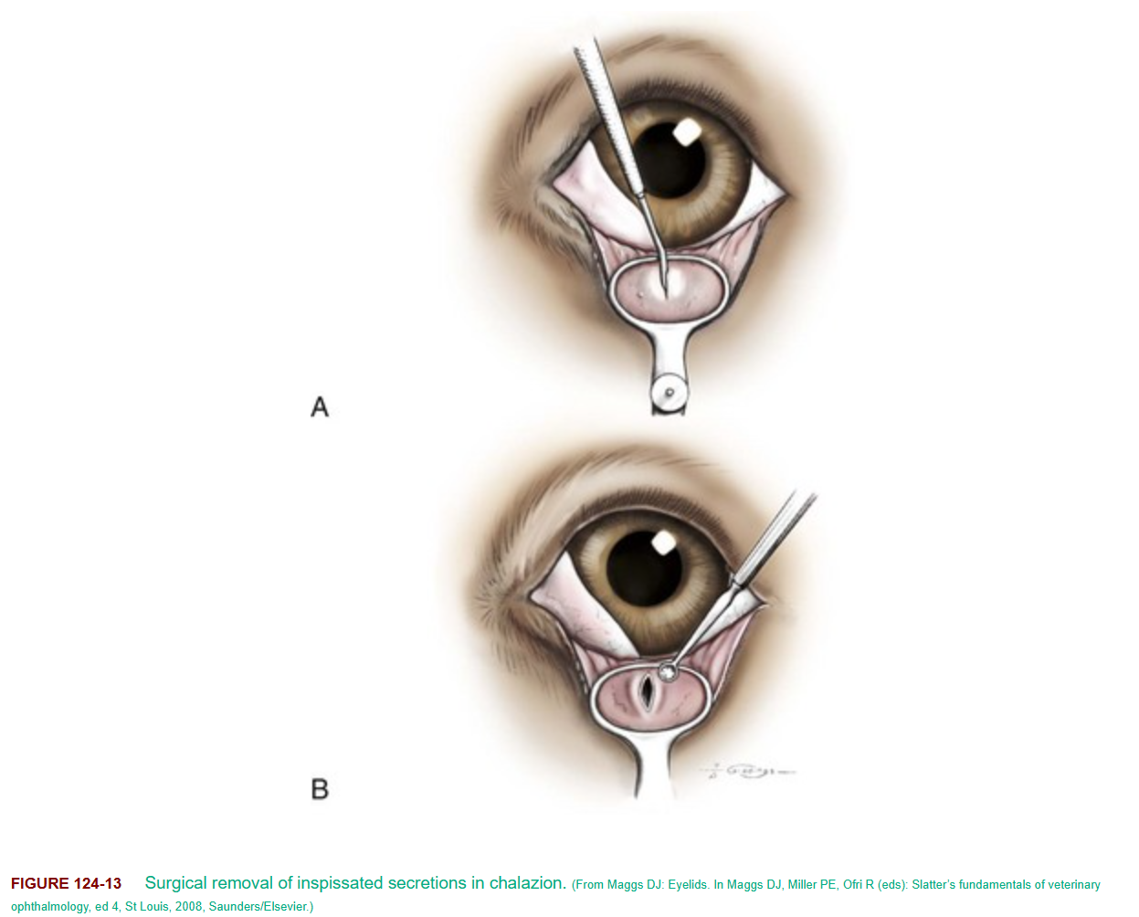
What is a chalazion?
A small bump in the eyelid caused by a blocked oil gland.
What types of eyelid neoplasia can occur?
Adenoma, papilloma, adenocarcinoma, benign melanoma, malignant melanoma, haemangioma, haemangiosarcoma, and squamous cell carcinoma.
Histology is essential for diagnosis and prognosis
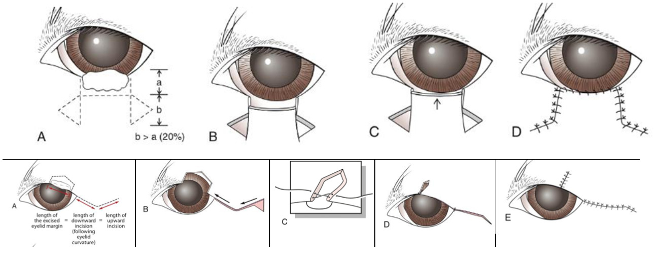
What is the treatment for eyelid neoplasia?
If < 30% of eyelid margin: V/ house-shaped mass removal align margin with figure-8 modified cruciate suture, then close remaining skin partial thickness in simple interrupted pattern 2-4mm apart
If > 30% of lower eyelid margin: H-plasty pedicle advancement flap. Suture conjunctiva and skin at the new lid margin with a simple continuous pattern and try to align so no growing hairs will touch the eye. Place interrupted sutures to oppose the remaining incision.
If > 30% of upper eyelid margin: Semi-circular flap. Remove mass, make a lateral semicircular incision (length of curve x2 the excised portion, where peak of curve is middle of incision). At the lateral end of the incision, excise a triangle area, advance the skin flap and close with figure-8 suture. Oppose the skin of new lateral canthus with the peak using figure-8 suture. Close skin with simple interrupted.
What are examples of diseases of the conjunctiva?
Conjunctivitis
Chlamydial conjunctivitis
Feline viral rhinotracheitis (FVR)
Feline calicivirus infection
Canine distemper
Kennel cough/ Canine Infectious Respiratory Disease Complex (CIRDC)
What are the causes of conjunctivitis?
Infectious (bacteria like Chlamydia, viruses like distemper, FHV, FCV, parasites like Thelazia)
Allergic
Mechanical/physical irritation
Keratoconjunctivitis sicca (KCS)
How is conjunctivitis treated?
Topical/oral antibiotics (e.g., tetracycline or doxycycline for Chlamydia)
Topical antivirals (for FHV)
Chlamydophila felis and C. pneumoniae
Which age group of cats is most commonly affected by chlamydial conjunctivitis?
Kittens between 2-6 months old. Kittens under 8 weeks are protected by maternal antibodies
What are the clinical signs of FVR?
Mild to severe acute rhinitis and conjunctivitis, pyrexia, depression, ocular discharge and pain, and anorexia.
Severe signs, including fatal pneumonia, can occur in young kittens.
Ulcerative dendritic keratitis is also associated with FHV-1
What type of disease is canine distemper?
A contagious multisystemic disease of canids. Known as hardpad disease.
What are the clinical signs of canine distemper related to the eyes?
Conjunctivitis and corneal ulcers (ocular form)
What are the clinical signs of kennel cough?
Harsh, dry coughing and gagging, often with ocular discharge. More severe signs (fever, anorexia, vomiting, purulent nasal/ocular discharge, depression, productive cough) may indicate additional infections like distemper or bronchopneumonia, especially in puppies
What is an example of a disease of the third eyelid/nictating membrane?
Cherry eye
What is the surgical name for cherry eye?
Hypertrophy and prolapse of the third eyelid gland
What are the clinical signs of cherry eye?
A red, swollen mass on the lower eyelid, mucoid discharge, and conjunctival inflammation

What is the difference between pre corneal tear film and actual tears?
Pre-corneal has fat and mucin included
What’s the function of tear film?
Reduce evaporation from eye
What are examples of disorders of the eyelashes?
Ectopic cilia, distichiasis, trichiasis
What are infectious diseases affecting the eye?
Chlamydia, calicivirus, herpesvirus
How can chlamydia be treated?
Systemic ATB (long) but also topical ATB drops (doxycycline, tetracycline)
What happens to the cartilage in cherry eye?
There is a laxity in attachment of the gland to the cartilage of the third eyelid and the periorbita, allowing for prolapse.
The cartilage can become bent or curved abnormally, prevents third eyelid from lying flat against eye, which can exacerbate condition & make surgical treatment harder.
What is meibomitis?
Inflammation of the meibomian glands
Where are the meibomian glands?
Inside the eyelids