Cellular Concepts PPT
 Call Kai
 Learn
Learn Practice Test
 Spaced Repetition
 Match
Match Flashcards
 Knowt Play
1/61

There's no tags or description
Looks like no tags are added yet.
Name | Mastery | Learn | Test | Matching | Spaced | Call with Kai | Chat |
|---|
No analytics yet
Send a link to your students to track their progress
62 Terms
Cell functions
-growth
-energy production
-metabolism
-DNA replication
-protein synthesis
Sodium-Potassium Pump
-uses ATP (active transport!)
-Na+ = extracellular
-K+ = intracellular
Na-K+ pump rate
pump 3 Na+ out
bring 2 K+ in
Na-K+ pump roles
1) establish resting membrane potential
2) maintains fluid volume
Mitochondrion “cell powerhouse”
-converts nutrients → ATP
-contain own DNA, can replicate when more ATP is needed
-different cell types have various # of mitochondria based on metabolic needs
DNA mutations in mitochondria can occur from…
free radicals
Aerobic metabolism
oxygen → mitochondria → 34 ATP
3 types of cells that have different # of mitochondria based on metabolic needs
1) cardiac
2) skeletal muscle
3) bones
2 types of cellular metabolic processes
1) Anaerobic metabolism (glycolysis)
2) Anaerobic metabolism (krebs cycle)
Anaerobic Metabolism (i.e. Glycolysis)
= no oxygen
produces 2 ATP + lactic acid
Aerobic Metabolism (i.e. Kreb’s cycle)
= requires oxygen
produces 34-36 ATP per glucose
Disease Etiology
The study of the causes or origins of diseases, including genetic, environmental, and infectious factors that contribute to health conditions.
Etiology
-original cause of cell alteration or disease
ex) infection, trauma
Characteristic changes with specific etiologic agents
ex) atrophy of skeletal muscle cells with paralysis, HPV may lead to cervical dysplasia
In response to an etiologic agent, the cell may…
-develop adaptive, compensatory changes
-develop maladaptive changes
Types of Cellular Adaptations
-atrophy
-hypertrophy
-hyperplasia
-metaplasia
-dysplasia
Left Ventricular Hypertrophy
thickened muscle wall of LV, due to heart disease or hypertension or overworking
Gingival hyperplasia
enlargement of gum tissue, often due to medications or chronic inflammation.
Benign Prostatic Hyperplasia
A non-cancerous enlargement of the prostate gland, commonly occurring in older men, which can lead to urinary symptoms
-urine may go back up inside of exiting out the urethra
Metaplasia: Barrett’s Esophagus
condition where normal squamous cells of the esophagus are replaced by columnar tissue, from chronic acid exposure from GERD
-duodenal bile reflux + gastric acid reflux
Metaplasia v.s. Dysplasia - conditions of the normal ciliated epithelium
metaplasia: chronic injury/irritation
dysplasia: persistent severe injury or irritiation
Neoplasia
-new growth
-disorganized, uncoordinated
-tumor
Neoplasia tumors
can be benign or malignant
*assess for differentiation
well differentiated cells = good (benign)
poorly-differentiated cells = bad (malignant)
Benign Neoplasms
-cells resemble normal cells
-well differentiated
-do not metastasize
-well defined borders
Malignant Neoplasm
-cells appear different from healthy cells
-poorly differentiated
-increased likelihood of metastasis
-poorly defined borders
Cellular injury pathways
*VIEW IMG - Refers to the various mechanisms through which cells can sustain damage due to stressors, leading to functional impairment or cell death. These pathways include necrosis, apoptosis, and autophagy.
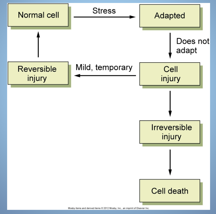
Changes that occur in cell injury
-dysfunction of Na+/K+ pump
-loss of plasma membrane integrity
-mitochondria dysfunction
-defects in protein synthesis ability
-intracellular accumulations
-cell swelling
-gene damage
Dysfunction of Na+/K+ pump
cause: lack of ATP
-too much Na+ in cells → cell swelling
-loss of electrochemical gradient
Cellular Injury Mechanisms
-hypoxic injury
-free radical injury (oxidative stress)
-physical agents of injury
-chem injury
-infectious injury
-immunologic injury
-gene defects
-nutritional imbalance
Cellular Hypoxia
cell hypoxia → anaerobic metabolism → 2 ATP + pyruvate → LACTIC acid
More on Cellular Injury
-single, unpaired electron that create instability and reactivity w/ adjacent molecules
-react w/ plasma mem. and organelles (including nucleus/DNA) → oxidative degradation
-free radicals can be removed by the cell, but it may be overwhelming and cause oxidative stress
Cellular Injury: Physical injuries
-trauma (laceration, fall, gun shot wound)
-burns (sunburn, electrical)
-frostbite
Cellular Injury: Chemical injuries
-hypernatremia (cell dehydration)
-hyperglycemia → damage endothelial cells that line arteries
-medications (nephron/neurotoxic), pollutants, poisons
Cellular Injury: Infectious agents
-bacteria, virus, fungi, parasites
i.e.) HPV can cause cervical dysplasia
i.e.) H. Pylori causes gastric and duodenal ulcers
Cellular Injury: Immunological agents
-autoimmune, ex) RA
-chronic inflammation
Cellular Injury: Genetic Defects
damage and mutation to DNA → RNA → ribosomes → proteins
Cellular Injury: Nutritional imbalances
-starvation: not enough protein, carb, lipid, vitamins, minerals
-inadequate protein: = low albumin = fluid shift from vascular space into interstitial space (i.e. peritoneal cavity)
-obesity: fat accumulation, stress on heart, increased metabolic needs
What can happen with inadequate protein intake?
this leads to low albumin, so fluid will shift from the vascular space → interstitial spaces such as the peritoneal cavity
Endothelial Cells
-line arterial BV
-”largest organ” → responsive and secretory tissue
-key in angiogenesis
-have Nitric Oxide (NO) → vasodilator
-have Endothelin → vasoconstrict
Endothelial Cells in Angiogenesis
-vascular endothelial growth factor (VEGF)
-stimulates collateral BV branches
Nitric Oxide (NO) is a ….
vasodilator
Endothelin is a …
vasoconstrictor
Endothelial Cell Injury
initiates arteriosclerosis and cell changes of CV disease
Significant Injurious agents that cause Endothelial Cell Injury
-HTN
-DM/hyperglycemia
-Free radicals
-Persistent secretion of Angiotensin II
-LDL cholesterol
Inflammatory Response to cell injury
-caused by a variety of things
-local manifestations (signs / symptoms) → redness, heat, swelling, pain, loss of function
Things that cause an inflammatory response to cell injury
infection, mechanical damage, ischemia, nutrient deprivation, temp. extremes, radiation, etc.
Inflammatory Response - Vascular
1) blood vessel dilation
2) increased vascular permeability + leakage
3) WBC adherence
3 Stages of Acute Inflammatory Rxn
1) Vasodilation — histamine, bradykinin, prostaglandin
2) Vascular Permeability
3) Cellular Migration — WBC, platelets
5 Markers of Inflammation
1) CRP - c reactive protein
2) ESR - erythrocyte sedimentation rate
3) Leukocytosis
4) Calcitonin
5) Lactic Acid (marker of sepsis!)
Sepsis is defined as…
Systemic Inflammatory Response Syndrome (SIRS) that is due to infection
Leukocytosis = high WBC
how many cells is this??
approx. 5,000-10,000 cells/cm
Necrosis
cell death due to overwhelming stressors or injury
-irreversible → lysosomes break open, initiating autolysis
-inflammatory rxn results
Apoptosis
programmed cell death in an organized process
-no inflammation
ex) WBCs undergo apoptosis after their participation in an inflammatory rxn
Ischemia
lack of blood
Necrosis (short answer)
cell death
Infarction
tissue death caused by prolonged ischemia (lack of blood)
Prolonged ischemia leads to…
Ischemic necrosis
CKmb
a cardiac enzyme released during myocardial infarction (MI)
troponin
a protein complex found in cardiac muscle that is released into the bloodstream during myocardial injury or MI
Any tissues that sustains prolonged ischemia is susceptible to… ???
infarction
Individual cells have different tolerances to ischemia
Brain: 6 min
Heart: 20 min
Skeletal muscle: several hours
2 things that may come up during MI and/or heart infection
1) CKmb = cardiac enzyme
2) troponin = cardiac protein