Module 7: Occipital Lobes
1/50
There's no tags or description
Looks like no tags are added yet.
Name | Mastery | Learn | Test | Matching | Spaced | Call with Kai |
|---|
No analytics yet
Send a link to your students to track their progress
51 Terms
Occipital Lobes
Brain region primarily responsible for visual processing
- only lobe dedicated to one sensory system
- visual perception begins in this lobe but continues into parietal, prefrontal etc.
Primary Visual Cortex (V1)
Initial processing area for visual information and builds the image
- columns in cells in V1 that respond to colour are called Blobs
Secondary Visual Cortex (V2)
Processes visual information after V1
- has increased levels of complexity
V2 is made up of stripes (thin and thick) and pale zones, each having different functions
What is V1 made up of and what do they do? What type of cells are in V2?
V1: Blobs and Interblobs
- V1 has columns of cells called blobs
- Blobs process colour and will relay this information to thin stripes (which process colour) in V2
- Interblobs feed information into pale zones (which process form) in V2
----------
V2: Stripes
Thin - receive colour info. and processes it
- sends info. to V4 (Ventral stream) Temporal
Thick - Processes motion
- sends info. to V5 (Dorsal Stream) Parietal
Pale Zones - process form
What is the pathway from V1 (Blobs) onward?
Blobs --> Thin Stripes --> V4 (Ventral Stream)
What is the pathway from V1 (Interblobs) onward?
Interblobs --> Pale Zones --> V4 (Ventral Stream)
Interblobs --> Thick Stripes --> V5 (Dorsal Stream)
Dorsal Stream (Where?)
Pathway for visual guidance of movements
- plans motor action
- location and spatial awareness
Ventral Stream (What?)
Pathway for object perception
- processes form and recognizes objects
- identification
What happens to a monkey with dorsal stream damage? (Drawer Task)
- a monkey with dorsal stream damage has difficulty in understanding the "where," which is location and spatial awareness
A monkey with this type of damage would not be able to identify where the apples are based on the cylinder marking their location
HOWEVER, they would be able to identify where the apples are based on recognizing the form/shape of the drawer (ventral task)
What happens to a monkey with ventral stream damage? (Drawer Task)
- a monkey with ventral stream damage has difficulty in understanding the "what," which is the identification of objects
A monkey with this damage would have trouble distinguishing b/w the two trays based on the percept of what it looks like (object recognition)
HOWEVER, they would be able to identify where the apples are based on recognizing the location of the cylinder next to the drawer that has the apples (dorsal task)
What is double dissociation? How does this relate to the monkey-drawer test?
Damage in one region can cause deficit BUT does not influence behavior in the other region
In the case with monkeys and drawers, some of the monkeys had damage to their dorsal streams (meaning their spatial and location awareness was not there)
When a monkey had damage to their dorsal stream, they were not able to complete the task where the cylinder location identified where the apples were
HOWEVER, they were able to complete the ventral task (what?) as they could perceive what shaped drawer had the apples in it.
Just because a monkey had a dorsal deficit does not mean it has a deficit in the ventral stream and vice versa.
Ventral Damage:
TASK A (SUCCESSFUL) (Dorsal Task "WHERE?")
TASK B (FAILED) (Ventral Task "WHAT?")
-----
Dorsal Damage:
TASK A (FAILED) (Dorsal Task "WHERE?")
TASK B (SUCCESSFUL) (Ventral Task "WHAT?")
Johansson Experiment
- showns only points of motion with LED lighta
- don't need complele image to determin if somehting is in biological motion because of the Superior Temporal Sulcus

STS (superior temporal sulcus) stream
In the temporal lobe
- Involved in visuospatial functions and movement perception, especially for identifying biological motion
Calcarine Sulcus
Separates upper and lower visual fields
- contains much of the primary visual cortex (V1)
Lingual Gyrus
Part of the ventral surface of the occipital lobe that processes vision (especially letters)
What happens when light comes into the lens of the eye?
An inversion occurs in the retina
- this inversion is held until the occipital lobe into the calcarine fissure/sulcus where it is separated into upper and lower visual fields
Fusiform Gyrus (V4)
Involved in face and object recognition.
Visual Pathways
Connections between visual processing areas in the brain.
Homonymous Hemianopia
Where do you lose vision?
Where do you still have vision?
Where does the damage occur to cause this issue?
- Loss of vision in same visual field of both eyes
- You would still have vision in the other visual fields of both eyes
- results from complete cut of optic track (beyond optic chiasm) or complete damage to LGN or V1 (PVC)

Quadrantanopia
blindness of one quadrant of the visual field (upper portion or lower portion of visual field)
- usually indicated damage in PVC (some information is getting through)
- usually identifies damage either above the calcarine fissure or below the calcarine fissure

If a person is showing damage in the upper visual field of their left eye, where has the damage occurred and what type of damage is this called?
Quadrantanopia
This person would have damage in their right hemisphere below the calcarine fissure in V1
Where is the upper visual field processed in the PMV (V1)? Why?
It is processed in the lower portion of the PMV
- below the calcarine fissure
- this is because there is an inversion - the retina inverts the image it receives
Where is the lower visual field processed in the PMV (V1)? Why?
It is processed in the upper portion of the PMV
- above the calcarine fissure
- this is because there is an inversion - the retina inverts the image it receives
Monocular Blindness
- blindness in one eye
- typically damage to eye itself or optic nerve wiping out any input from one eye
- one eye will be completely blind in left and right visual fields
- can't be explained by hemispheric damage

What is PVC necessary for?
- it is required for us to have conscious vision
- this means that we are aware that something is there
What does hemianopia mean?
- complete loss of a visual field
In a perimetry test, what is the outer portion called and what is the inner portion called?
Outer -
Temporal Fields
Inner -
Nasal Fields
Bitemporal Hemianopia
Where do you lose vision?
Where do you still have vision?
Where does the damage occur to cause this issue?
- Loss of vision in BOTH outer temporal visual fields
- *HAVE vision in inner nasal fields
Loss is caused by lesion to medial region of the optic chiasm
- typically indicative of damage to the optic chiasm --> USUALLY a tumor grows on the pituitary gland that pushes up onto the optic chiasm

Where does left visual field information fall in the retina coming from the left eye?
Where does right visual field information fall in the retina coming from the right eye?
Does the path cross the optic chiasm or stay ipsilateral?
It falls onto the inner portion of the retina - also called the nasal hemiretina
the right visual field information from the right eye also falls onto the inner portion of the retina - also called the nasal hemiretina
These two pathways cross over at the optic chiasm
Where does right visual field information fall in the retina coming from the left eye?
Where does left visual field information fall in the retina coming from the right eye?
Does the path cross the optic chiasm or stay ipsilateral?
- the RVF information will fall onto the outer temporal half of the left eye
- It will fall onto the outer portions of the retina (the temporal half) of the right eye
Ipsilateral - Temporal hemiretinas will stay on the same side for both eyes
Where do you typically see damage the optic pathway for someone with bitemporal hemianopia?
You typically see it at the intersection of the optic chiasm (medial)
usually as a result of a tumor growing which causes a disruption knocking out the ability for information to pass further
- only the inner portions (nasal halves of the retinas) cross over --> if that is cut off, you will lose visual processing
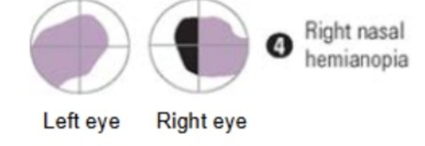
For a patient with left OR right nasal hemianopia, where would you see the damage occurring?
For damage to occur that would result in right or left nasal hemianopia, a growth or tumor would impact the lateral region of the optic chiasm (outer edge)
Nasal Hemianopia
Where do you lose vision?
Where do you still have vision?
Where does the damage occur to cause this issue?
- Loss of vision in inner visual fields (in right or left eye)
- you will either have a left nasal hemianopia or a right nasal hemianopia - you will NOT usually have them at the same time
*It is uncommon to have both at the same time as you would need two different tumors to be in those lateral regions of the optic chiasm at the same time
- results from lesion of the lateral (side) region of the optic chiasm
Scotomas
What is it?
What is it a result of?
small blind spots in the visual field
- usually caused by a small stroke
- visual system may be able to compensate for this blind spot
--> compensation occurs through the help of nystagmus which are small eye movements which aid V1 in filling in the blanks
What is nystagmus?
What is macular sparing?
- macular region of eye contains the fovea and tissue area around
Instead of complete homonymous hemianopia, sometimes people with experience some vision in fovea visual field but not perimeter
Believed that this area of occipital lobe may receive irrigation from middle cerebral arteries to maintain

Visual Agnosia
- typically a result of ventral stream damage
- DF suffered from this
- Inability to recognize objects despite intact vision
Apperceptive Agnosia
Inability to form a percept of whole objects (absence of perception
- involves damage to the LO (Lateral Occipital Cortical Area or V3)
- damage occurs early in the ventral stream (posterior inferior area)
Image: when asked to copy pictures down, they may get some features but never see the full image
What does it entail when a disorder or type of damage starts with an "A"?
This "A" identifies the absence of something
E.g., Apperceptive Agnosia
This means that there is an absence of perception
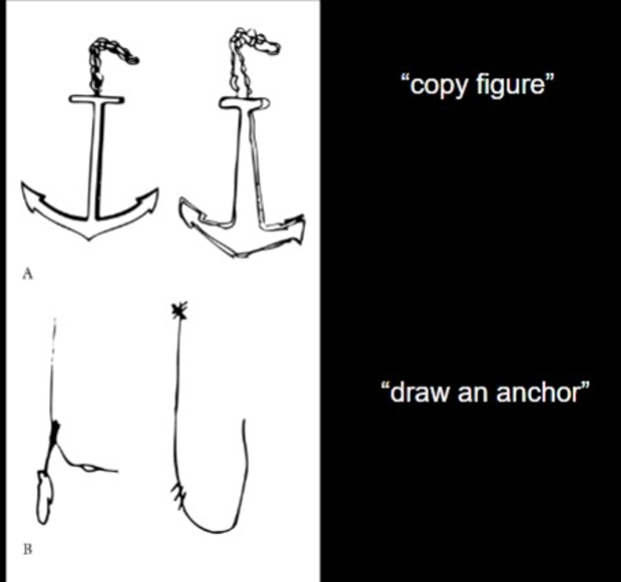
Associative Agnosia
Inability to link perception to knowledge
- they can put the object together but cannot link to what it actually is
- connection between ventral stream and memory is
- damage much later down ventral stream (more into anterior regions)
IMAGE: If they see an image and are asked to copy, they can form the image. They wont be able to tell you what it is.
When they are asked by memory to draw an anchor, they will not be able to do it
Prosopagnosia
Inability to recognize familiar faces but can still know a face is a face (because of LOC still functioning)
- damage to right hemisphere FFA
- bilateral damage to FFA is possible but primarily occurs in right hemi.
Category-Specific Agnosia
Know all objects except for those linked to a specific category e.g., Fruit
- this is a memory access disorder
Color Vision
Ability to perceive different colors.
V4
Cortex area primarily responsible for color vision.
V5 (MT)
Area involved in motion perception.
Visual Recognition
Process of identifying objects using visual input.
Visual Attention
Selective focus on specific visual stimuli.
Goodale & Milner Model
Distinguishes vision for perception from vision for action.
Perimetry Test
Assessment of visual field defects.
What is the Fusiform Face Area?
- involved in the configuration of faces (particularly in the right hemisphere)
- the LOC (Lateral Occipital Complex) can process faces but not as well as the FFA
- this region looks for eyes, then nose, then mouth
Thompson Illusion
a phenomenon where it becomes more difficult to detect local feature changes in an upside-down face, despite identical changes being obvious in an upright face.
- damage to the FFA in the right hemisphere:
Inverted performance - same as control partipants
Upright performance - control partipants do well but people with damage in FFA do not do very well in task (configuration info.)