Cellular Responses to CNS Injury
 Call Kai
 Learn
Learn Practice Test
 Spaced Repetition
 Match
Match Flashcards
 Knowt Play
1/16

There's no tags or description
Looks like no tags are added yet.
Name | Mastery | Learn | Test | Matching | Spaced | Call with Kai | Chat |
|---|
No analytics yet
Send a link to your students to track their progress
17 Terms
List the Cells of the CNS
Derived from? Sub types? Fxn?
CELLS OF THE CNS
Neurons:
Glia:
derived from neuroectoderm
Types:
Astrocytes:
supportive function for neurons,
repair cells,
Part of BBB
Oligodendrocytes
myelin production
Ependymal cells
line ventricles
Microglia
derived from the yolk sac
Fx: macrophage-like cells
Cells of the meninges and blood vessels


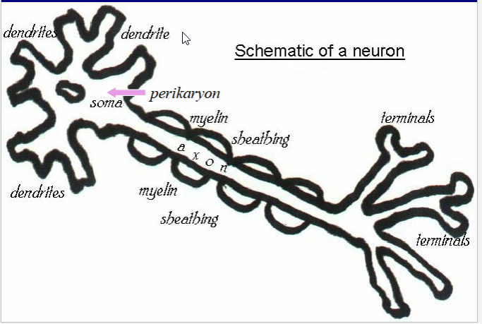
Draw out a schematic of the Neuron
Describe ASTROCYTES
Histo
Maintaining BBB mech?
Supportive function?
Response to Injury?
Different types
Histo:
H&E stain: only nuclei are seen
multiple branching cytoplasmic processes
Can be seen w/ metallic impregnation technique (e.g. Golgi method)
(+) for glial fibrillary acidic protein (GFAP)
Maintaining BBB Mech:
By wrapping cytoplasmic processes around capillary walls
Supportive Function:
structural & supply nutrients
Response to injury:
reactive gliosis
*** brain’s version of scar tissue**
Different types:
Gemistocytes: Reactive astrocytes
Astrocytomas: Tumors arising from astrocytes


Describe OLIGODENDROCYTES
Fxn
Histo
Alignment
Unlike Schwann Cells?
What are tumors called?
OLIGODENDROCYTES
Fxn:
Produce + maintain CNS myelin
(counterparts of Schwann cells in the PNS)
Histo:
few (oligo) dendrites
best visualized with a silver stain
Contain myelin basic protein (MBP)
Alignment:
In white matter, aligned along axons
Unlike Schwann cells
limited regenerative capacity
do not effectively remyelinate axons
oligodendrogliomas
Tumors arising from oligodendrocytes


Describe EPENDYMAL CELLS
What are they?
Histo?
Injury response?
What type of cells are Choroid plexus epithelium
What are tumors called?
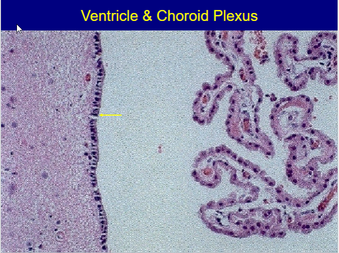
EPENDYMAL CELLS
What are they?
Single layer of columnar cells
Lines:
Ventricles
aqueduct of Sylvius
central canal of spinal cord
Histo
Has cilia
Rest on subependymal glia
Injury response:
Can’t Regen
Causes subependymal astrocytic proliferation -> can obstruct aqueduct -> hydrocephalus
“ependymal granulations”
Choroid plexus epithelium
form of ependyma specialized in production of CSF
Ependymomas
Tumors arising from ependymal cells

Describe MICROGLIA
Derived from?
Fxn?
Histo?
Response to injury?
Tumor?
MICROGLIA
Derived
yolk sac
Fxn:
Mobile phagocytic cells
Similar cell markers w/ monocytes/macrophages
(e.g. CR3 & CD68)
brain development
Histo:
only nuclei are seen on H&E stain
fine cytoplasmic processes w/ Silver Stains
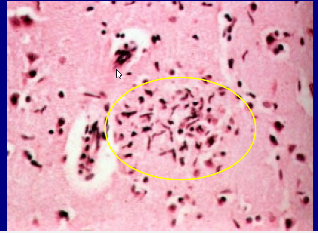
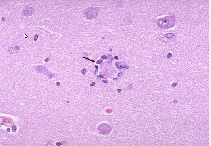
Response to Injury:
Reactive -> nuclei elongated (rod cells)
-> Microglial nodules
-> Neuronophagia
Lymphomas
Tumors arising from microglia


Describe these Neuronal Responses to Injury:
Axonal Reaction (Central Chromatolysis):
Causes/Effect
Acute Neuronal Injury
AKA?
Causes/Effect
Neurodegeneration
Causes/Effect
Neuronal Inclusions:
Causes/Effect
Axonal Reaction (Central Chromatolysis):
Causes
axonal injury
Effect:
reaction of perikaryon
Peripheral dispersion of Nissl substance
peripheral displacement of nucleus
Acute Neuronal Injury (Red Neurons):
Causes:
acute injury -> cell death
Effect:
shrinkage of cell body,
intense eosinophilia of cytoplasm,
pyknosis of nucleus
loss of Nissl substance
Neurodegeneration:
Causes:
subacute to chronic progressive neuronal injury
ALS & AD
Effect:
gradual death & loss of neurons,
reactive gliosis,
Abnormal protein aggregates
Neuronal Inclusions:
Causes/Effects (aggregates)
aging
(lipofuscin),
viral infections
Cowdry bodies – herpes,
Negri bodies - rabies
neurodegenerative diseases
neurofibrillary tangles – Alzheimer disease,
Lewy bodies – Parkinson disease


What can Accumulated misfolded proteins lead to?
Accumulated misfolded proteins can:
Interfere with cellular metabolism
initiation of the UPR
Describe the UPR process
