Chapter 6 Bones
 Call Kai
 Learn
Learn Practice Test
 Spaced Repetition
 Match
Match Flashcards
1/48

There's no tags or description
Looks like no tags are added yet.
Name | Mastery | Learn | Test | Matching | Spaced | Call with Kai | Chat |
|---|
No analytics yet
Send a link to your students to track their progress
49 Terms
Skeletal Stystem Functions
Support, movement, protection, mineral storage (calcium/phosphate), blood cell production (aka hematopoiesis)
Skeletal system Components
206 bones total
Axial Skeleton (126 Bones)
-Bones of Skull, thorax and vertebral column
Appendicular Skeleton (80 Bones)
-Bones of limbs and girdles that attach them to axilla skeleton

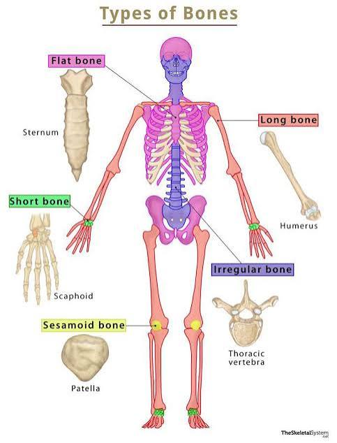
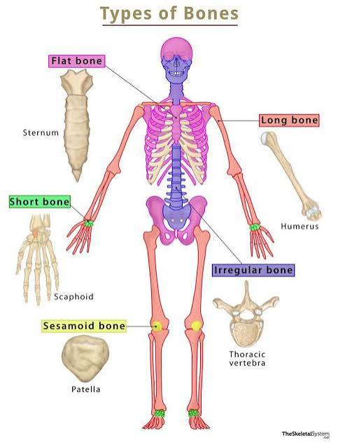
Flat Bones
Thin and curved bones that provide protection and a broad surface for muscle attachment, such as the skull and ribs.
Sutural Bones (wormian bones)
Small, irregular bones that develop between the joints (sutures) of the skull.

Long bones
Long and slender bones (loner than wide)
ex: various bones of the limbs (femur)

Irregular Bones
Bones with complex shapes that do not fit into the other categories, such as the vertebrae and some facial bones.

Sesamoid Bones
Small, flat and somewhat shaped like a sesame seed
Develop in tendons of keens, hands, and feet (don’t connect directly to other bones)
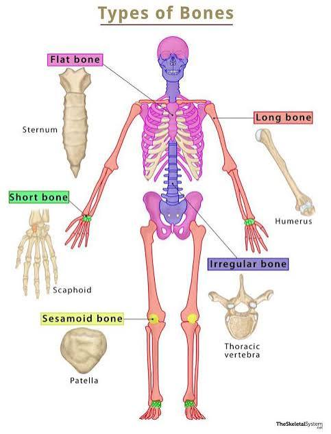
Short bones
Small and boxy bones
ex: carpals and tarsals
Bone surface features: Elevations/Projections
Characteristic markings on bones where tendons, ligaments, and other tissues attach, including tubercles, spines, and processes.
Bone surface features: Depressions/grooves/tunnels
Characteristics on bone surfaces that allow for the passage of blood vessels and nerves.
including fossae, sulci, and foramina.
Skull surface feature: Canal/meatus
A tube-like large passage within a bone
often serving as a conduit for nerves and blood vessels.
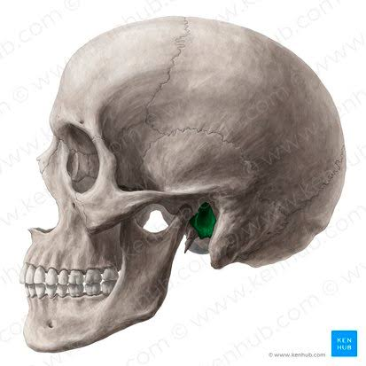
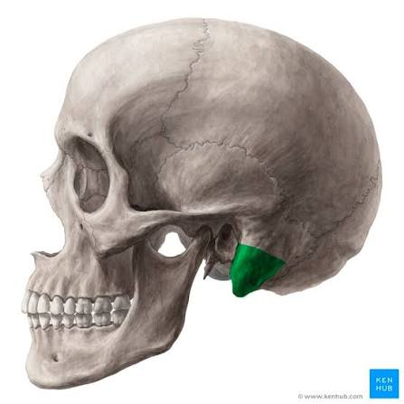
Skull surface feature: Process
A prominent projection or bump on a bone
serves as an attachment point for muscles, tendons, or ligaments.
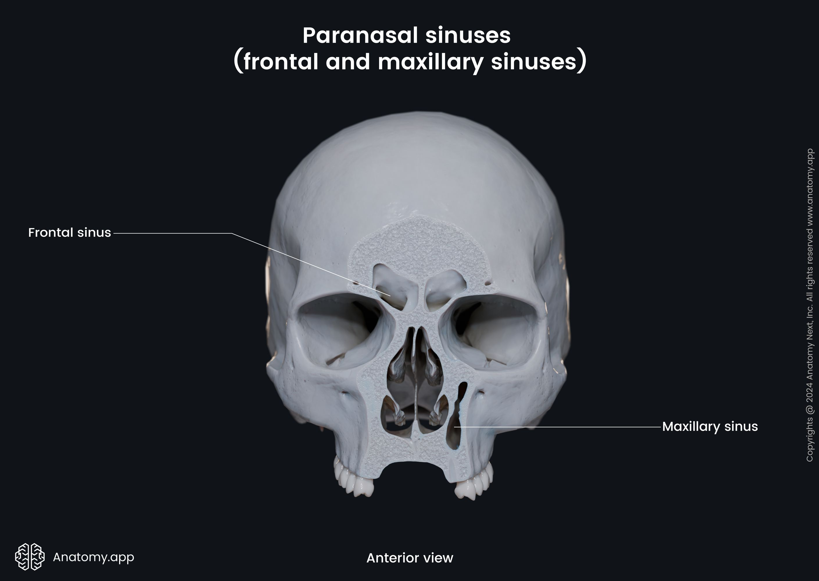
Skull surface feature: Sinus
A hollow cavity within a bone, typically filled with air, that helps reduce weight and improve resonance.
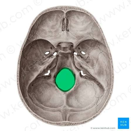
Skull surface feature: Foramen
A small rounded opening or hole in a bone
allows for the passage of nerves and blood vessels.
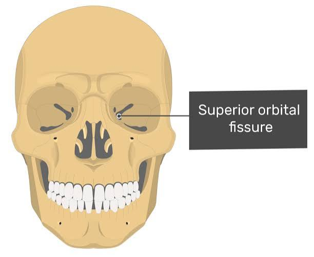
Skull surface feature: Fissure
A narrow slit-like opening in a bone
allows for the passage of nerves and blood vessels.
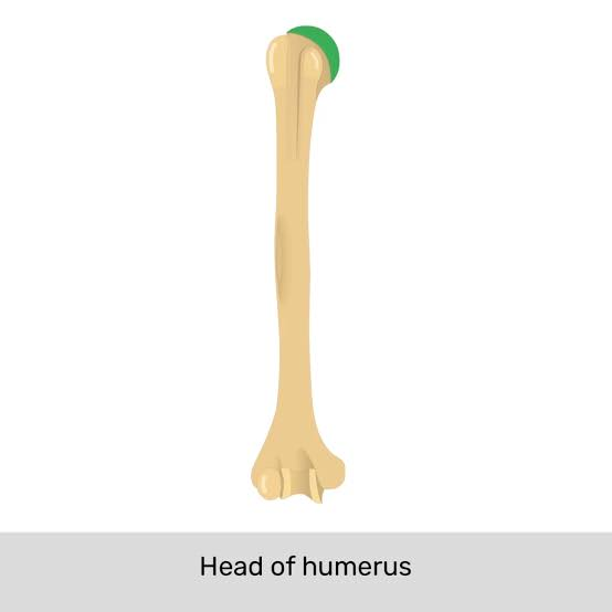
Humerus (long bone) Features: Head
The rounded expanded proximal end that forms part of joint
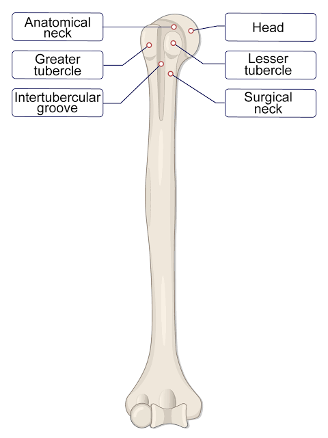
Humerus (long bone) Features: Tubercle
A small, rounded projection on a bone that serves as an attachment site for muscles and ligaments.
Humerus (long bone) Features: Suclus
A long deep narrow groove or furrow on the surface
accommodates tendons, nerves, or blood vessels.
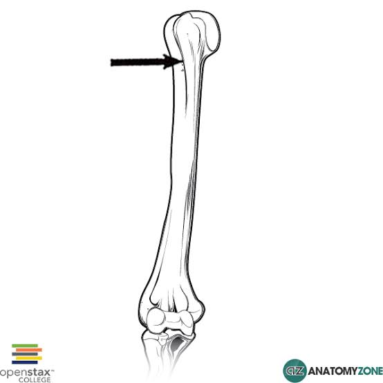
Humerus (long bone) Features: Tuberosity
Small rough projection that may occupy broad area

Humerus (long bone) Features: Diaphysis
Shaft of the bone

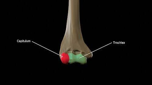
Humerus (long bone) Features: Trochlea
A smooth grooved articular process

Humerus (long bone) Features:Condyle
Smooth rounded articular process (cartilage and bone) that articulates with another bone.
Femur (long bone) Surface Features: Trochanter
Large, blunt projection on the femur.

Femur (long bone) Surface Features: Neck
The constricted part of the femur located just below the head (strong to resist force in many different directions), providing a point for muscle attachment and articulation.

Femur (long bone) Surface Features:Facet
Small, flat surface on a bone that forms a joint with another bone.

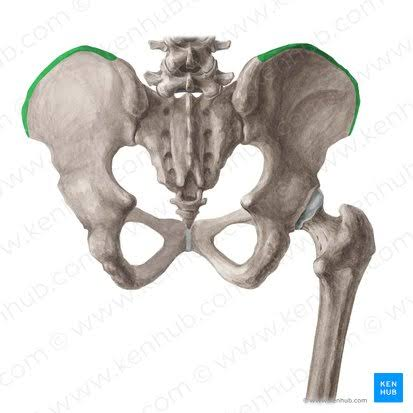
Pelvis Surface Features: Crest
The prominent ridge on the ilium of the pelvis, serving as an attachment point for muscles and ligaments.
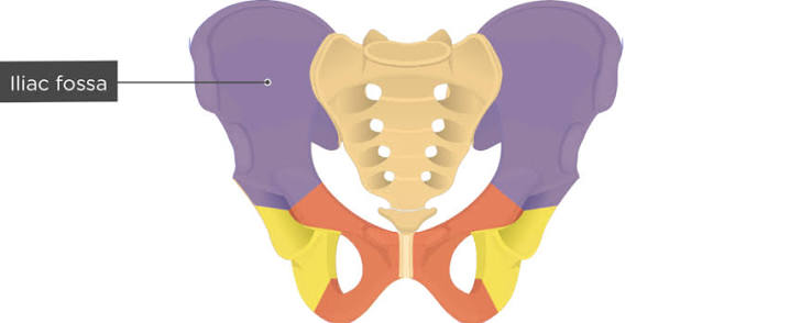
Pelvis Surface Features: Fossa
A shallow depression in a bone, typically serving as a hollow or socket for muscle attachment or articulation with another structure.
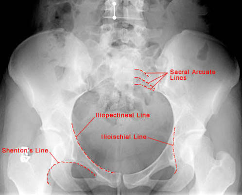
Pelvis Surface Features: Line
low ridge, more delicate than crest (where muscles attach)
Pelvis Surface Features: Spine
A sharp, slender projection of bone that serves as an attachment point for muscles and ligaments.

Pelvis Surface Features:Ramus
A branch or extension of a bone that typically connects to another part of the structure, often found in the pelvis. Makes an angle with the rest of the structure

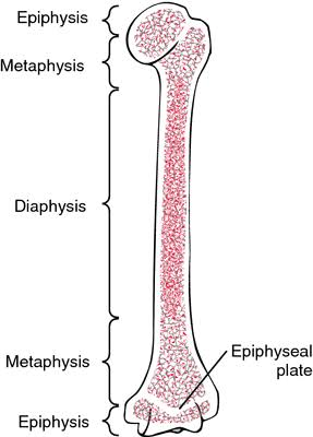
Epiphysis
The end part of a long bone, initially growing separately from the shaft. It is involved in the formation of joints and is where bones articulate with each other. (made of spongy (trabecular) bone)
Outer covering is compact bone

Articular cartilage
Covers portion of epiphysis (where articulation occurs)
Avascular (resources through synovial fluid)and provides a smooth, lubricated surface for joint movement.
Metaphysis
Connects epiphysis to shaft

Diaphysis
The main or central shaft of a long bone, providing structural support and containing the medullary cavity filled with bone marrow.
Red Marrow: makes RBC (usually in epiphysis)
Yellow Marrow: Adipose (fat) Storage (usually in diaphysis)
Nutrient Artery/Vein (bone)
Commonly, one on each bone. Nutrient foramen is a small opening in the bone that allows the nutrient artery and vein to enter and exit, supplying blood to the bone tissue.

Metaphyseal Artery/Vein
The blood vessels supplying the metaphysis region of a growing long bone, providing necessary nutrients and oxygen. Connects to epiphysis arteries/veins

Periosteum Features
A dense layer of connective tissue that covers the outer surface of bones, containing nerve fibers, blood vessels(supply superficial osteons), and osteoblasts for bone growth and repair. Lymphatic vessels collect lymph from bone and osteons

4 Bone cell types
osteoblasts, osteocytes, osteoclasts, and osteogenic/osteoprogenitor cells
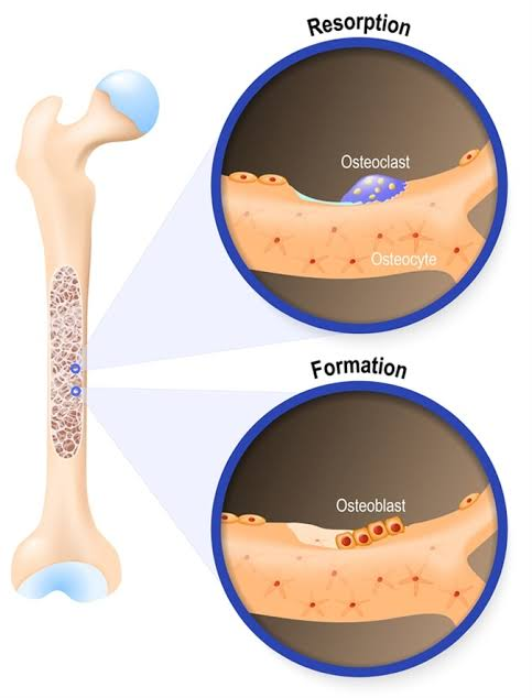
Osteocytes
Mature most numerous bone cells (cannot divide) that maintain bone tissue and communicate with other bone cells through tiny channels called canaliculi.

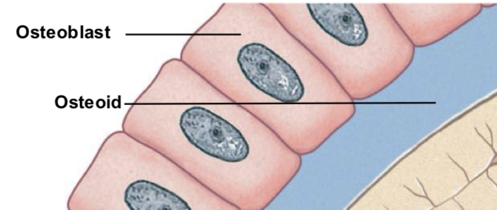
Osteoblasts
B for Build: Bone-forming cells (osteogenesis or ossification) that synthesize and secrete the bone matrix (osteoid=protein part) , responsible for bone growth and mineralization (deposit calcium salts to convert osteoid to bone)
Become Osteocyte once surrounded by bony matrix
Osteoclasts
C for Crush (or break)
Giant (50+ nuclei) Bone-resorbing cells that break down bone tissue by secreting acids and enzymes, playing a critical role in bone remodeling and calcium homeostasis.
Osteoprogenitor Cells
Bone stem cells that differentiate into osteoblasts, important in fracture repair
found in inner lining of periosteum, lining endosteum (walls of shaft: empty space) in medullary cavity, lining passageways containing blood cells

Osteoprogenitor Cells differ from Mesenchymal cells?
They are specific bone stem cells derived from mesenchymal cells, playing a key role in forming osteoblasts, whereas mesenchymal cells are multipotent and can differentiate into various cell types.