Microbiology Practical Exam 1 --- ACC Norris
1/130
There's no tags or description
Looks like no tags are added yet.
Name | Mastery | Learn | Test | Matching | Spaced | Call with Kai | Chat |
|---|
No analytics yet
Send a link to your students to track their progress
131 Terms
what can we throw away in the orange biohazard bags?
- petri dishes
- gloves/towels with bacteria
what do we disinfect with in the lab?
Lysol
- tryptic soy agar
- tryptic soy broth
what examples of undefined media should we know?
- associated with human disease
Bacteria biosafety level 2
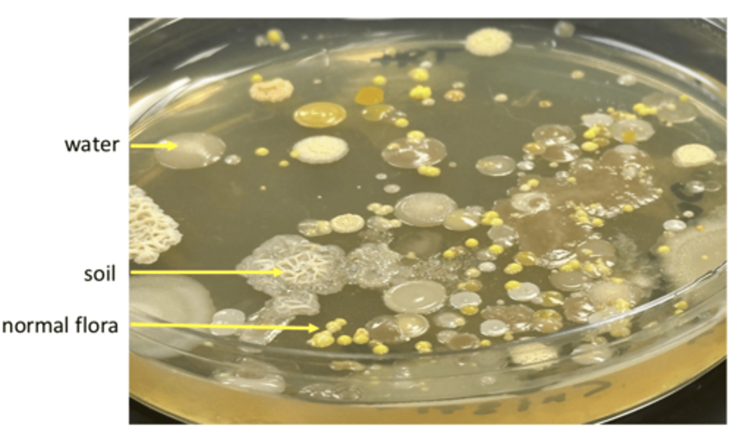
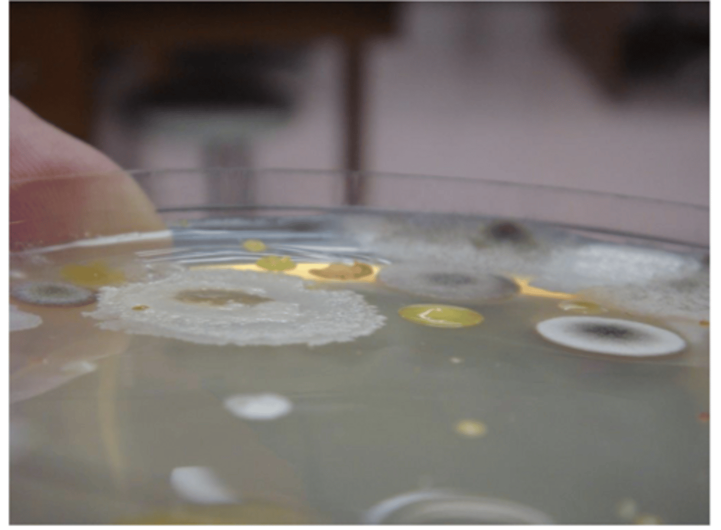
Environmental Assay Characteristics
glossy colonies = water
rough, whitish colonies = soil
yellow or whitish colonies = normal flora
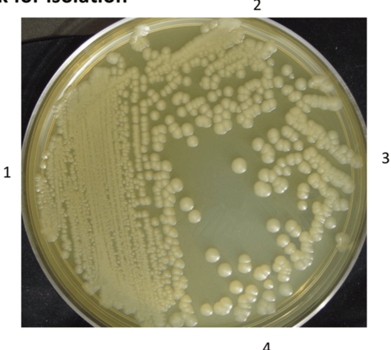
what is the purpose of a streak for isolation?
to separate a mixture of different bacteria into distinct, isolated colonies on a nutrient surface
what is step three of streak isolation?
- spread small amount from section 2 into section 3
- streak it parallel to quadrant 3 and do not go back into section 2
- flame loop once more
Where is the environment aerobic in a thioglycolate broth tube?
At the top of the broth (atmospheric oxygen)
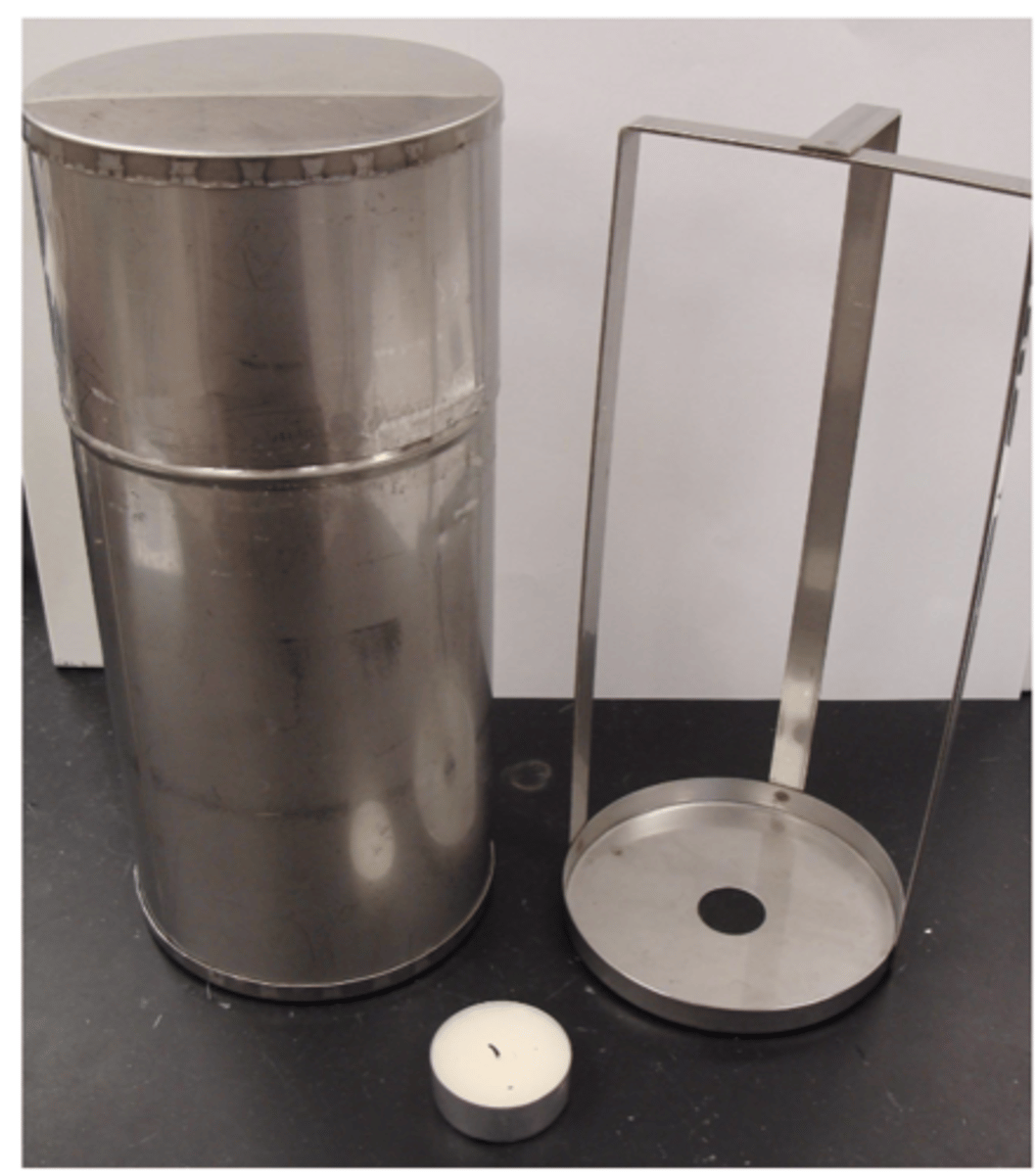
candle jar
- grows facultative and microaerophilic species
- clostridium does not grow - killed by exposure to o2
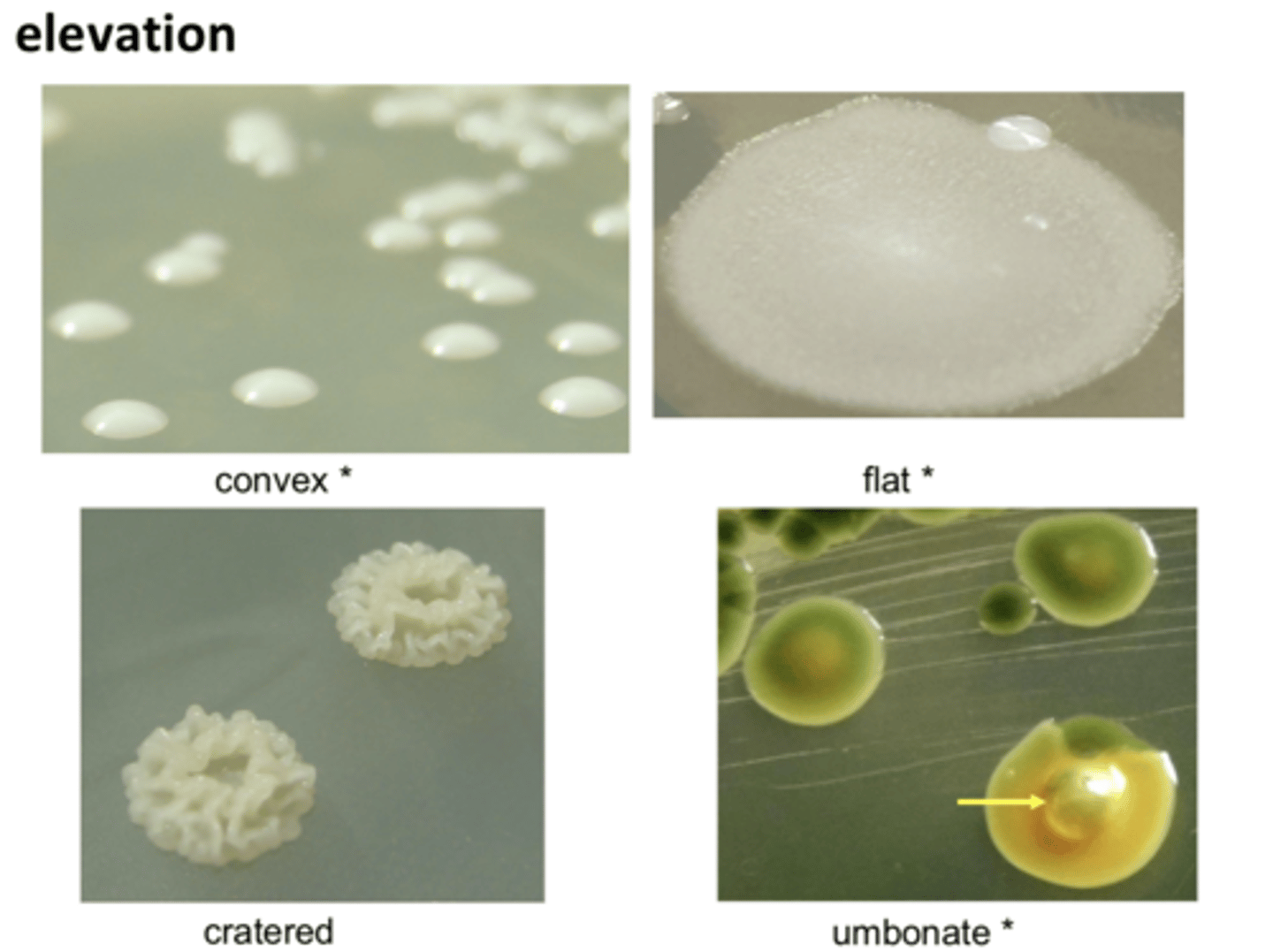
convex, flat, cratered, umbonate
colony elevation
- growth at the top
EX: bacillus
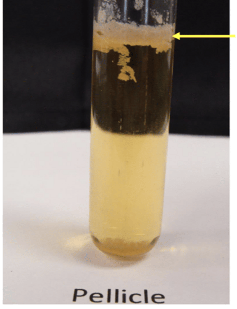
Pellicle
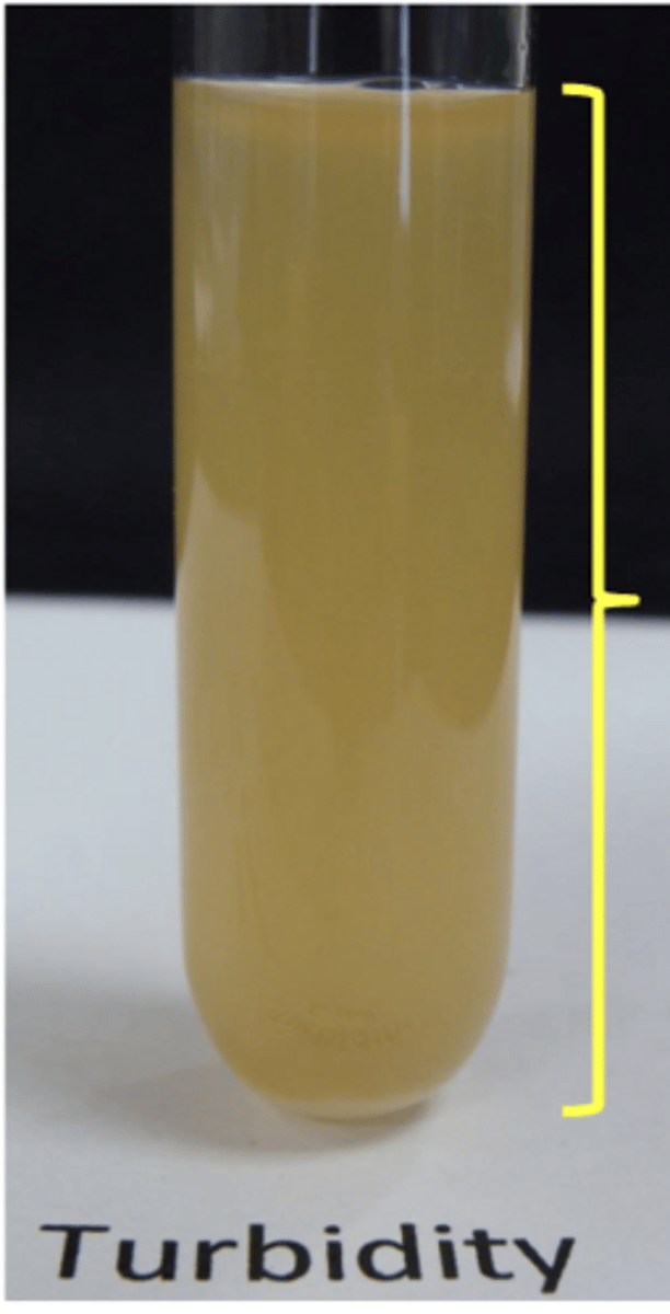
turbidity
- growth throughout
EX: E. Coli, staphylococcus
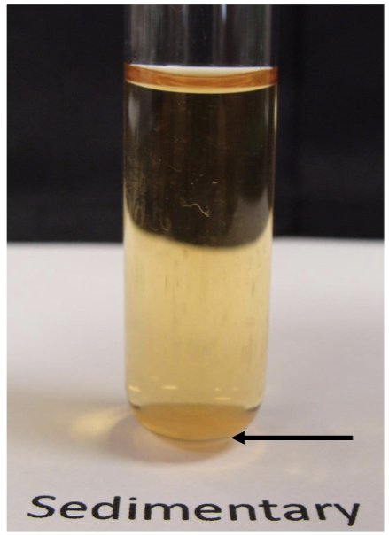
sedimentary
growth at bottom
EX: streptococcus
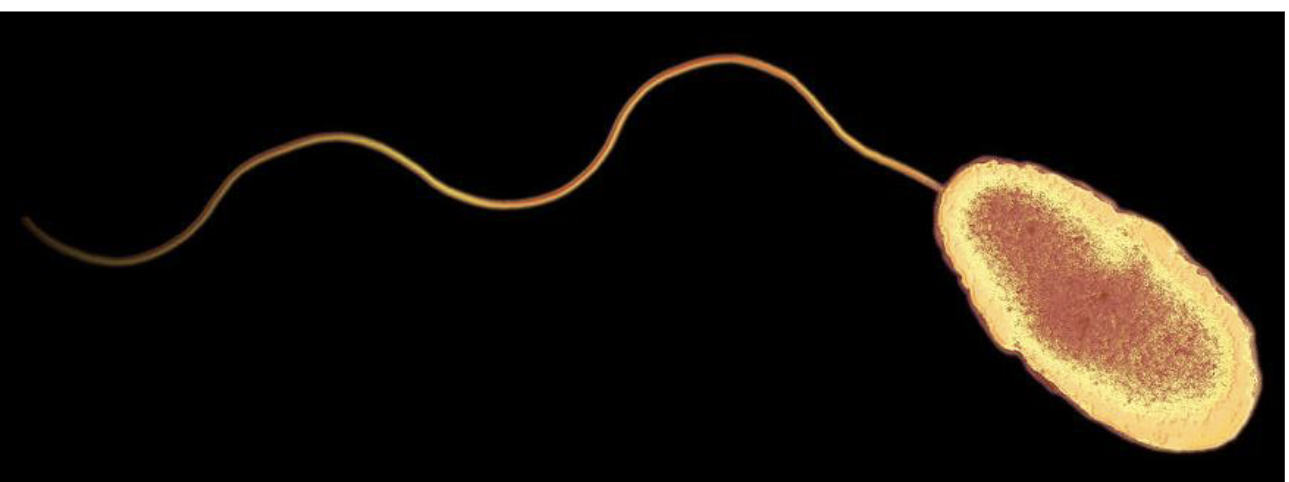
monotrichous flagella
- same as bacteria cell model
EX: pseudomonas flagella
steps of negative stain
- place a drop of nigrosin at one end of a clean slide
- mix a small loop of live bacteria into the drop (do not need to heat fix)
- use a second slide at an angle and spread mixture across slide, creating a smear
- let air dry
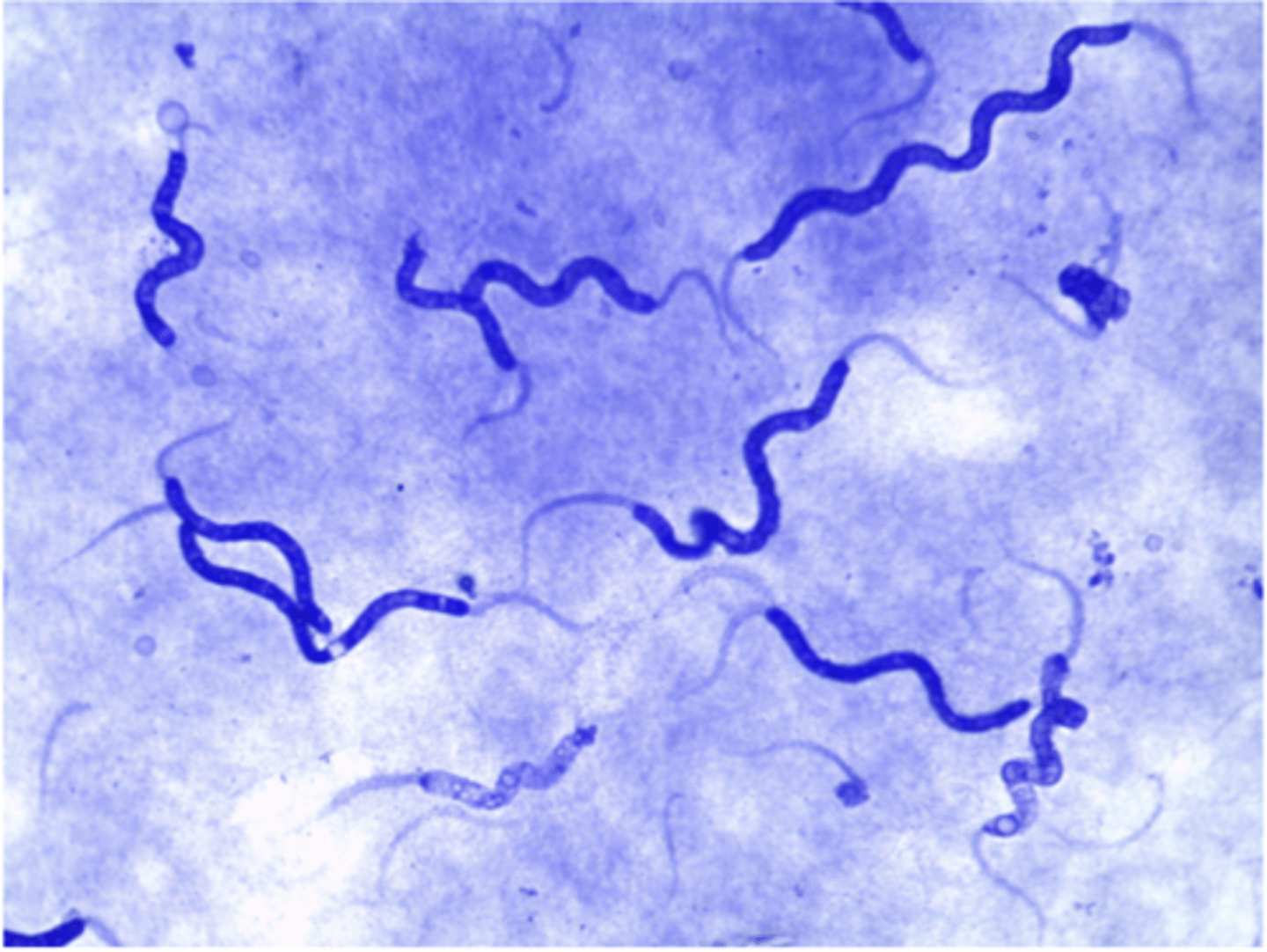
bacteria example of negative stain
- best for delicate organisms
- spirochetes
- separate bacteria into two major groups based on cell wall structure: Gram-positive or gram negative
- primary stain: crystal violet
- mordant: iodine
- destain: alcohol
- counterstain: safranin
Gram Stain characteristics
examples of acid fast
- mycobacterium tuberculosis
- mycobacterium leprae
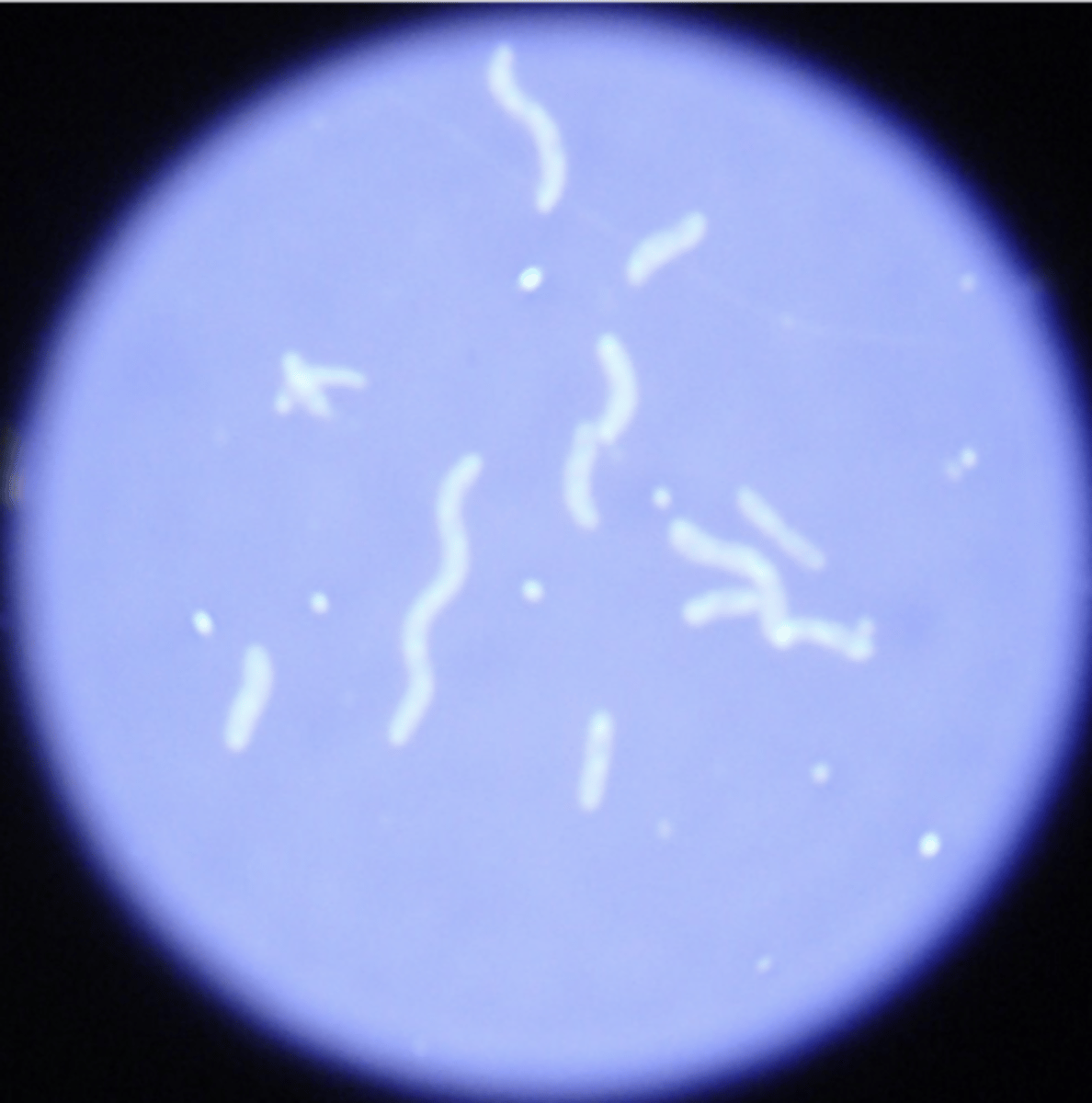
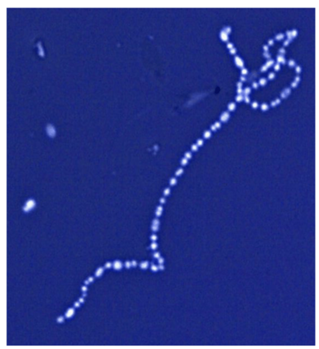
spirillum
spiral shaped bacteria
Staphylococcus in simple stain


streptococcus in a simple stain
endospore stain-
bacillus
endospores = blue/green
cell bodies = pink

capsule stain example 1
Staphylococcus aureus -- golden color
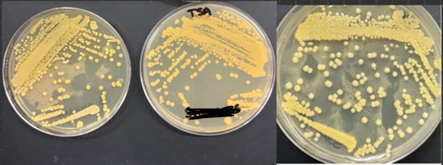
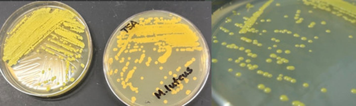
micrococcus luteus -- yellow
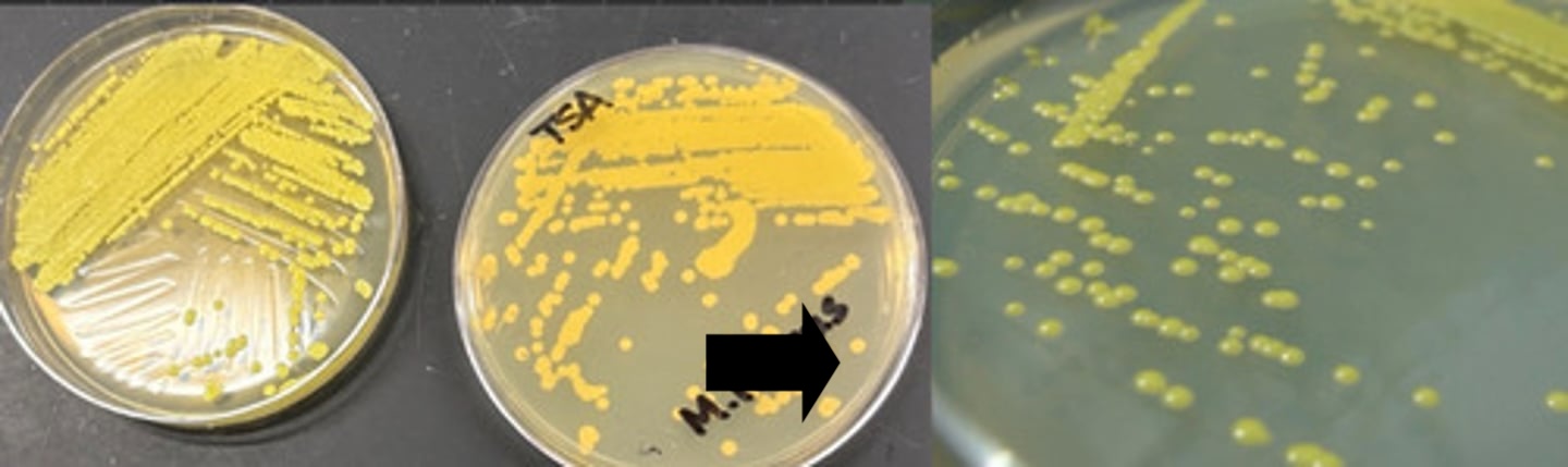

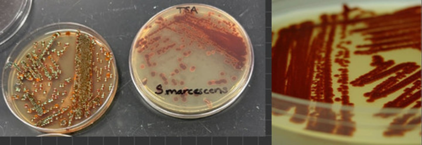
serratia marcescens
slide mount
flagellum
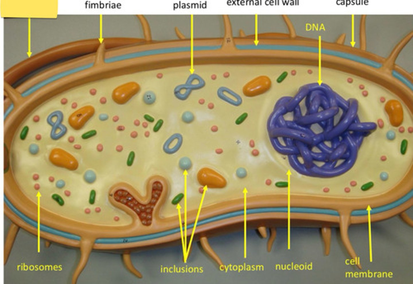
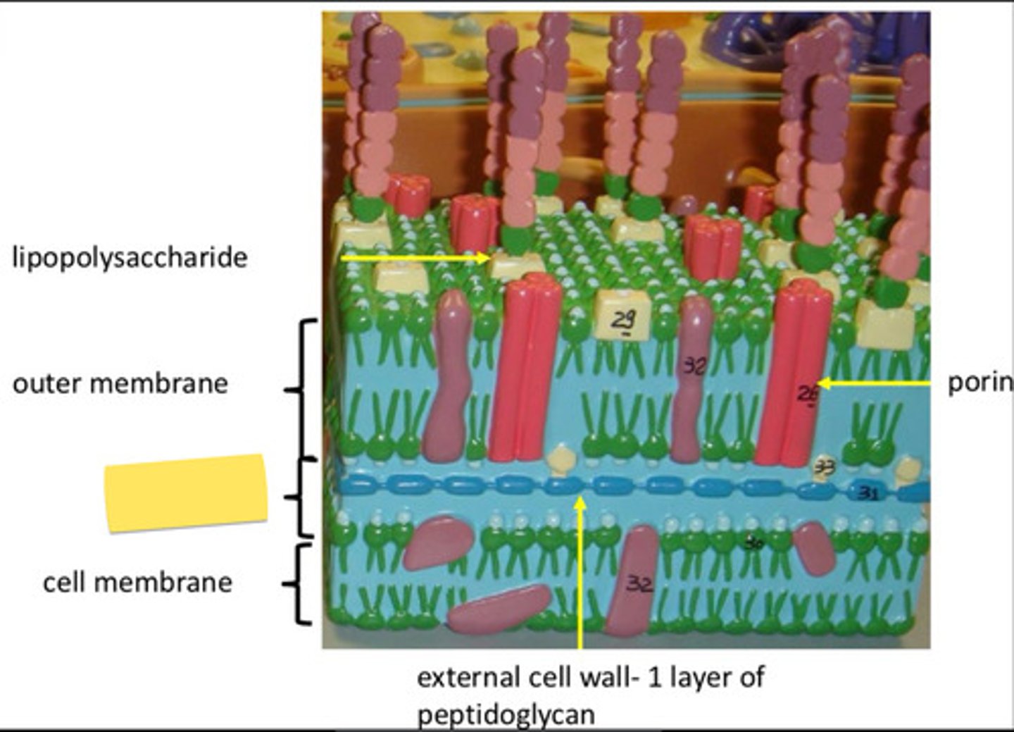
teichoic acid (+)
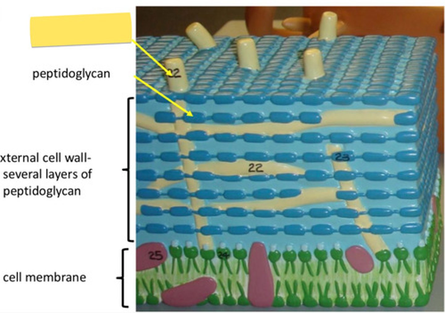
periplasm (-)
Bacillus cereus
what is the safe way of disposing items used in the lab?
through orange biohazard bag and white slant rack
what do we place in white slant rack?
contaminated test tubes
what are the different media types?
- undefined media
- differential media
-motility agar
- thioglycolate broth
what examples of differential media should we know?
how many bacteria biohazard safety levels are there?
two
biosafety level 1
- present minimal threat
- do not normally cause disease in healthy humans
bacillus, micrococcus, E. coli
what are some bacteria that can be found in biosafety level 1?
what are some examples in biosafety 2?
staphylococcus, streptococcus, Proteus, Salmonella
In the handwashing exercise we did four treatments and recorded their effectiveness:
- water alone: least effective
- sanitizer alone: better than water alone
- soap and water: better than sanitizer alone
- soap, water, and sanitizer: most effective
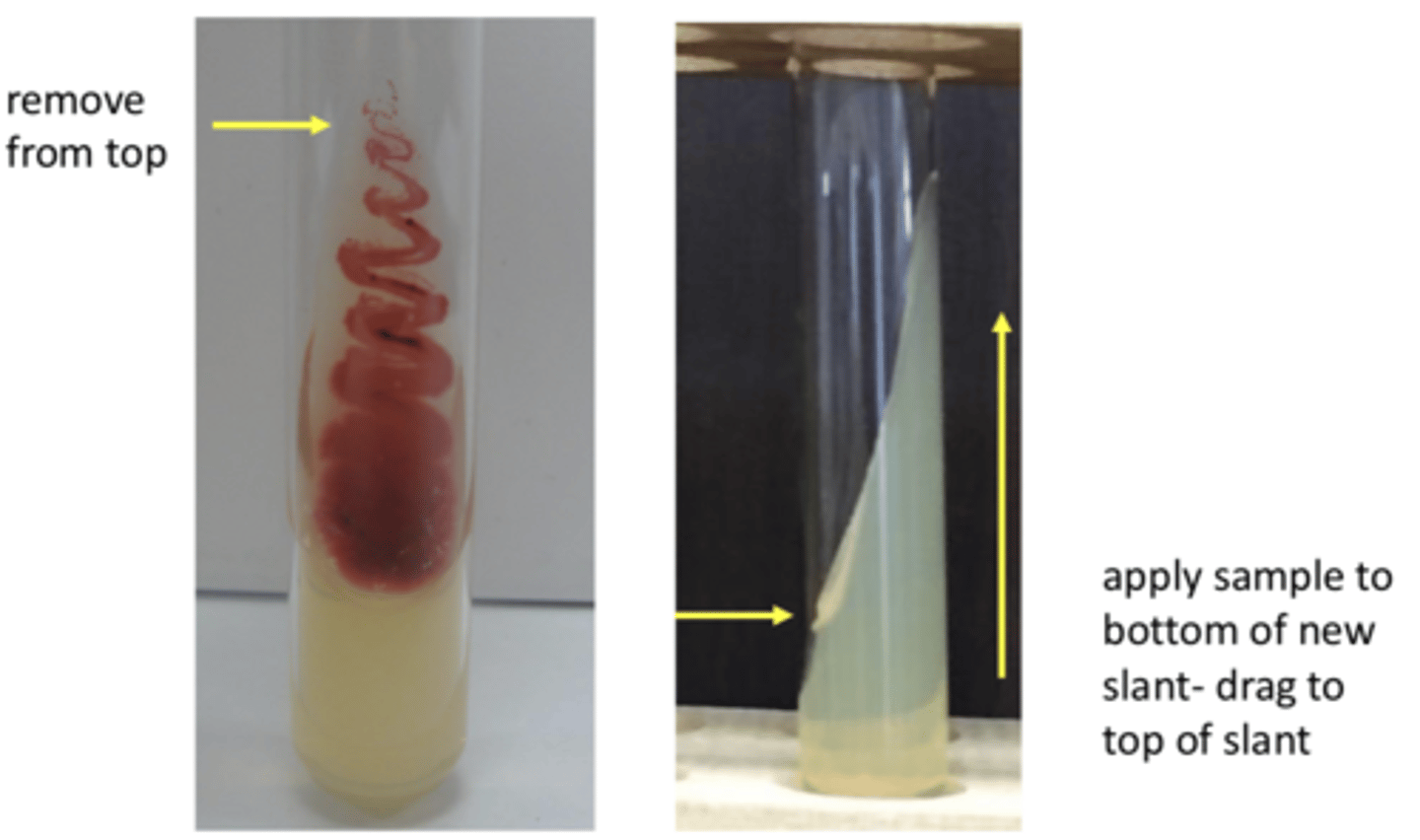
how do we aseptically transfer from slant to slant?
- remove from top of original slant
- apply to bottom of new slant and then drag from bottom of slant to top of it
- make sure you mention flaming loop and neck of tubes!!
what should we be ensuring we are doing to maintain aseptic techniques?
- when pulling a sample, we must open the tube and run it through flame before innoculating sample
- must run tube through flame again before closing the tube
- must run inoculating loop/needle through flame, starting from midpoint all the way to the end
slant to broth following aseptic technique
- remove from top of slant (same process as slant to slant)
- swirl loop in broth
- make sure you mention flaming loop and neck of tubes!!
What is step one of streak isolation plate?
- inoculate section 1 with sample from sample tube (following aseptic technique - flame loop and neck of tube)
- streak parallel to long axis (up and down) of section 1 and cover entire section
- flame loop
what is step two of streak isolation plate?
- spread small amount of sample from section 1 into section 2
- streak that parallel to section 2 quadrant and do not go back into section 1
- flame loop
what is final step of streak isolation plate?
- spread small amount from 3 into section 4
- streak parallel to quadrant four and make sure to not go back into section 3!
- flame loop for the last time
after completing plate how do we incubate them?
- incubate plates upside down in 37 degrees celsius (body temp)
- incubating plate upside down helps with moisture on lid
What type of medium is thioglycolate broth?
Differential medium
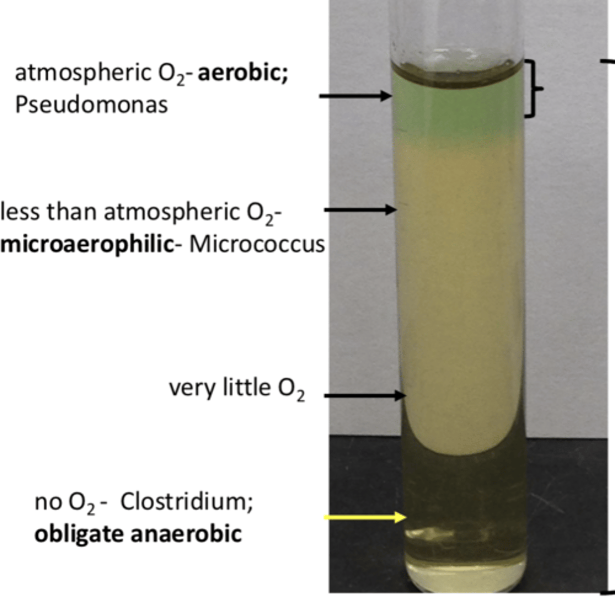
Where is the environment anaerobic in a thioglycolate broth tube?
At the bottom of the broth (no oxygen)
what does the green band at the top the broth indicate?
- indicates amount of atmospheric oxygen in tube/broth
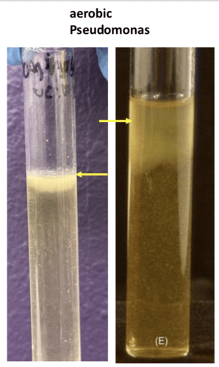
- top of the broth
- requires atmospheric oxygen
EX: pseudomonas
growth pattern of aerobic
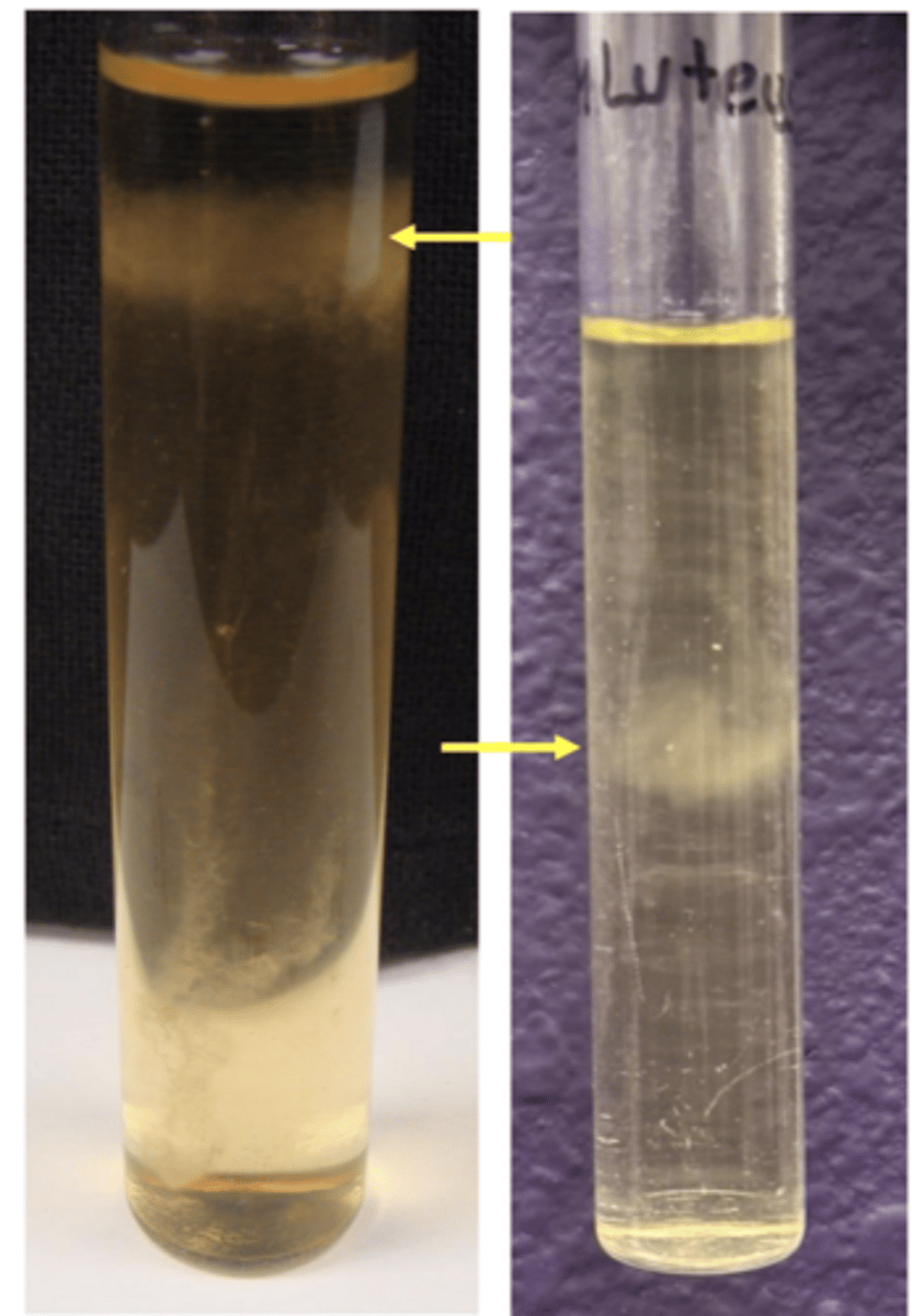
- below green brand
- requires less than atmospheric oxygen
EX: micrococcus
growth pattern of microaerophilic
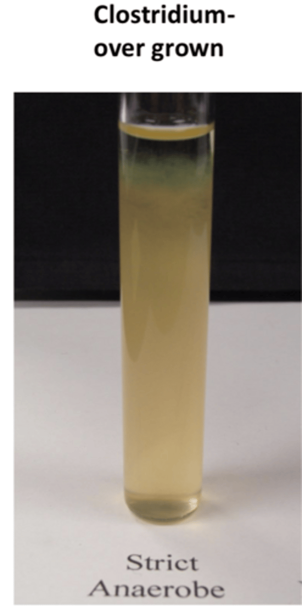
- bottom of broth
- requires no oxygen, killed by oxygen
EX: clostridium
growth pattern of obligate anaerobic
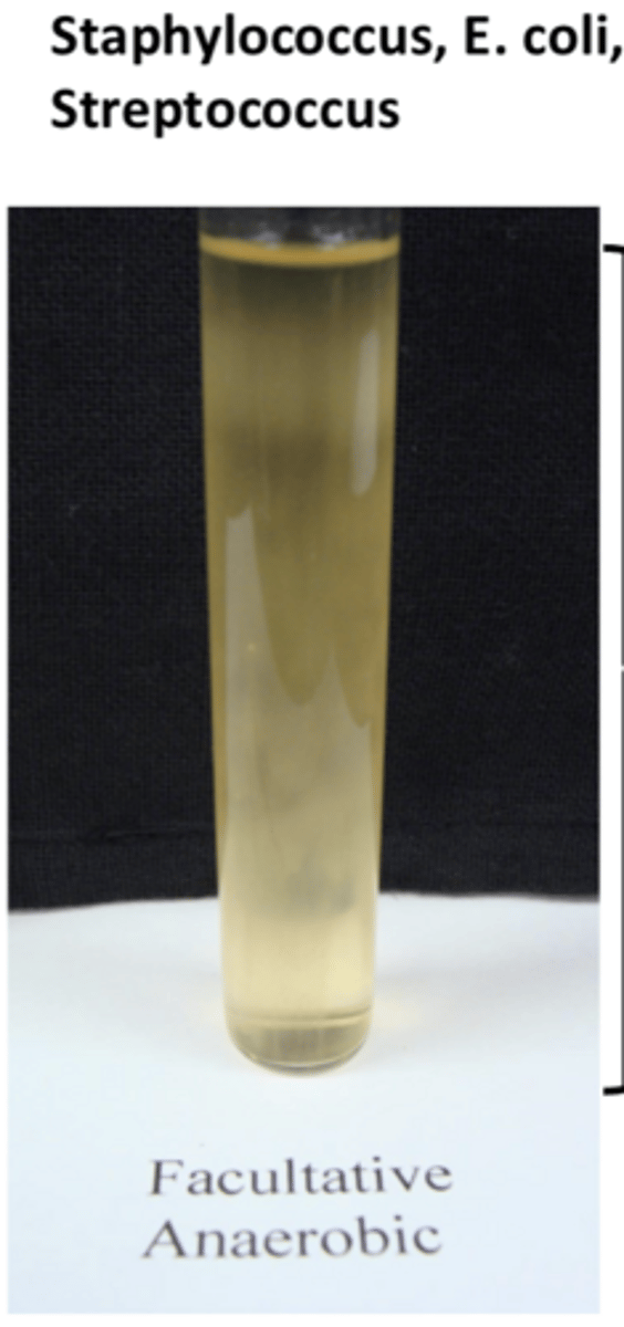
- throughout broth
- grows with or without oxygen
EX: E. coli, staphylococcus, streptococcus
growth pattern of facultative anaerobic
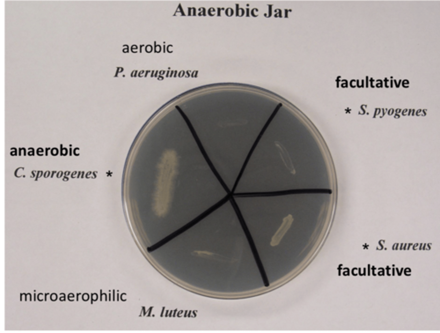
Brewer anaerobic container (no o2)
- clostridium and facultative species grow (strict anaerobe, facultative anaerobic)
- microaerophilic and aerobic species do not grow (require o2)
bacteria colony morphology on plates
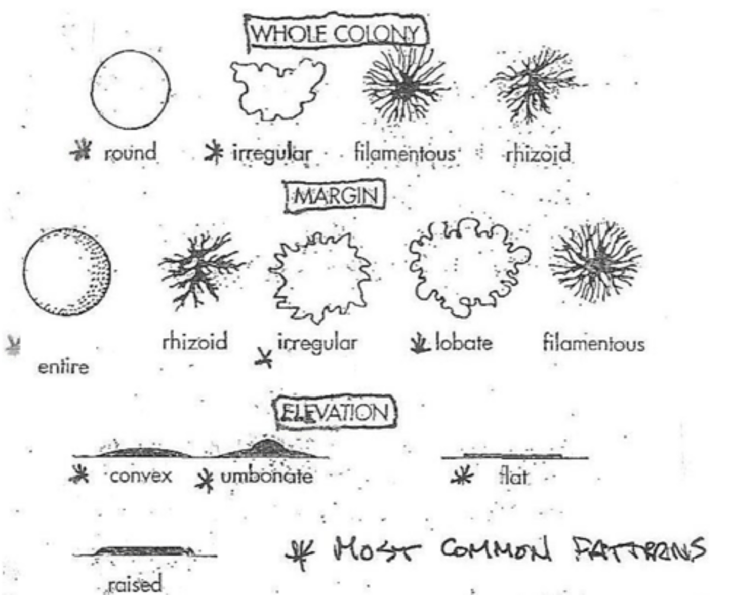
circular, irregular, lobate, pinpoint
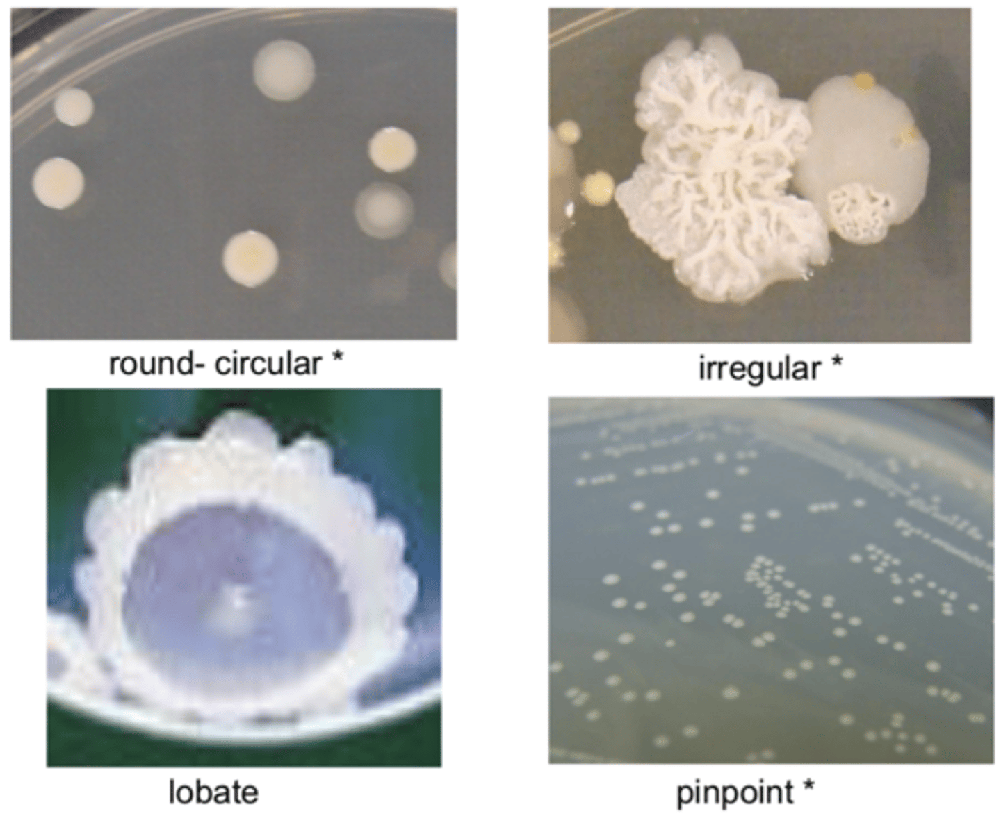
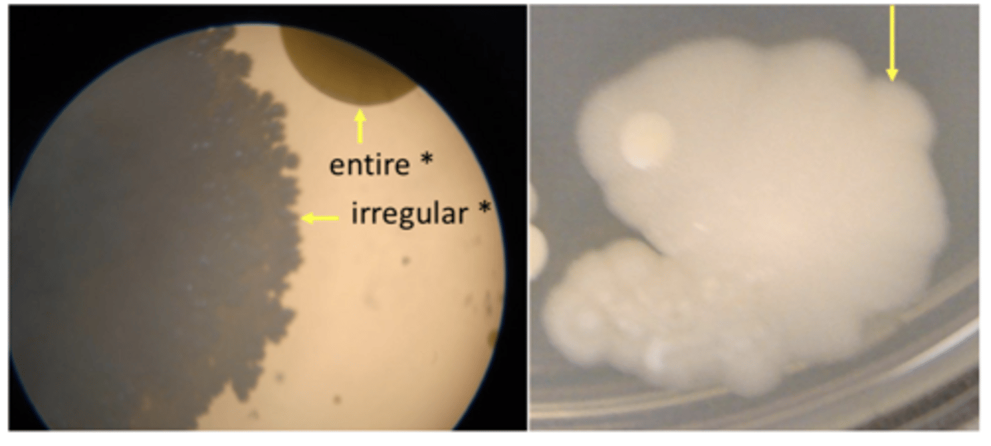
entire, irregular, lobate
colony margin
another colony elevation picture

- pellicle
- turbidity
- sediment
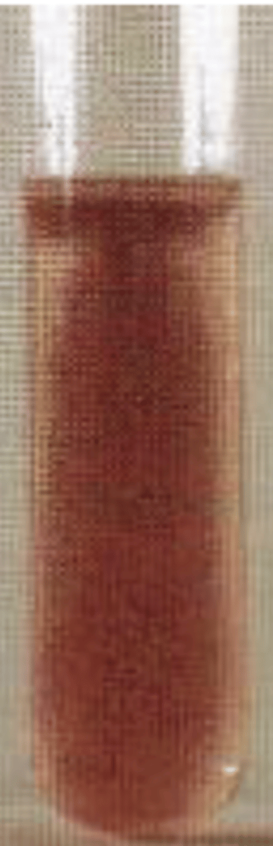
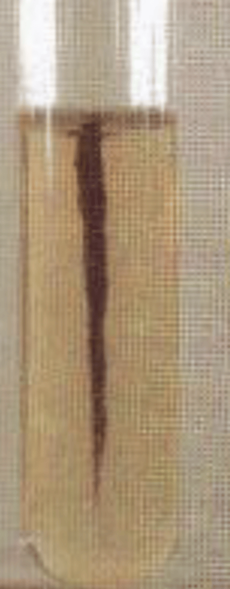
what are the different growth patterns in broth tubes?
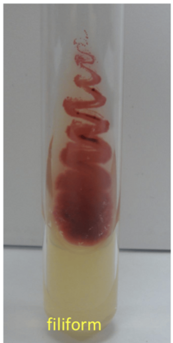
filiform
Growth pattern on a slant tube?
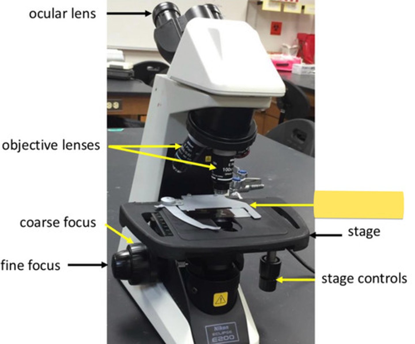
how do we calculate total magnification of microscope?
ocular lens x objective lens value
what is the relationship between field size and magnification?
field size decreases as magnification increases (inverse)
define resolution
ability to distinguish 2 things close together as separate
what increases resolution?
- oil immersion increases resolution bc they decrease refraction of light
- staining increases contrasts ultimately increasing resolution
E. coli
example of motile bacteria
staphylococcus
example of non motile
what are the different flagella patterns
- amphitrichous
- peritrichous
- monotrichous
- endoflagella
amphitrichous flagella
- spiral shape
EX: spirillum
peritrichous flagella- bacillus
EX: salmonella, proteus mirabilis

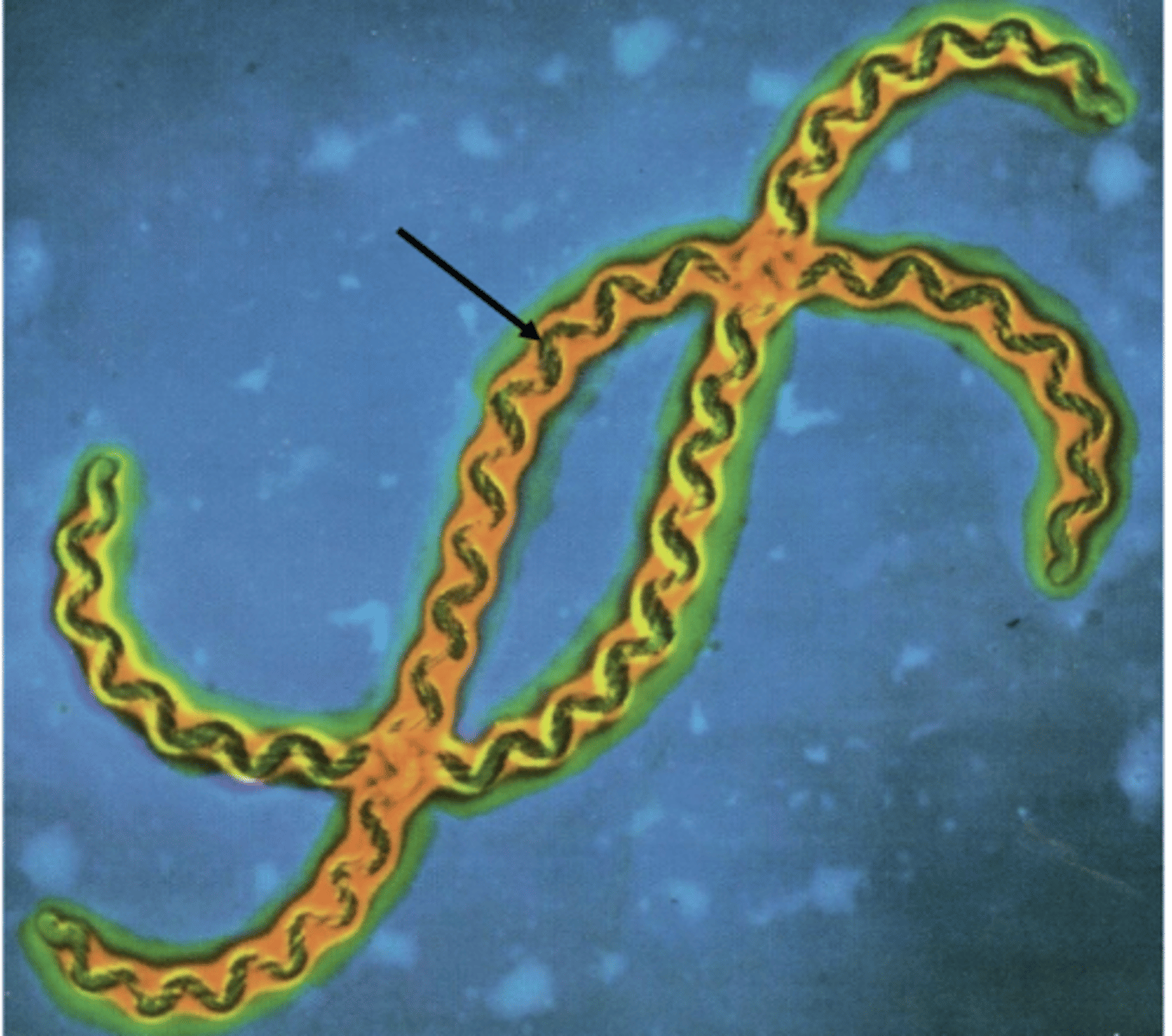
endoflagellum - axial filaments
Leptospira - spirochete bacterium
heat fixation
- functions to help adherence of primary stains onto bacterium
steps of heat fixation
- place small drop of water and mix with bacteria or use broth culture spreading the one you used evenly on slide
- full let air dry
- to heat fix, you pass underside of slide through upper flame of bunsen burner about three quick times
never use 100x immersion
- start on lowest lense (4x or 10x)
- lower condenser (cutting down light helps create shadows)
things to note while viewing wet mount under microscope
preparing wet mounts to view living eukaryotes in microscope
- apply sample on slide using pipette
- position coverslip by placing one edge flat against the slide at 45 degree angle right next to drop
- you can blot away excess water that is not under coverslip
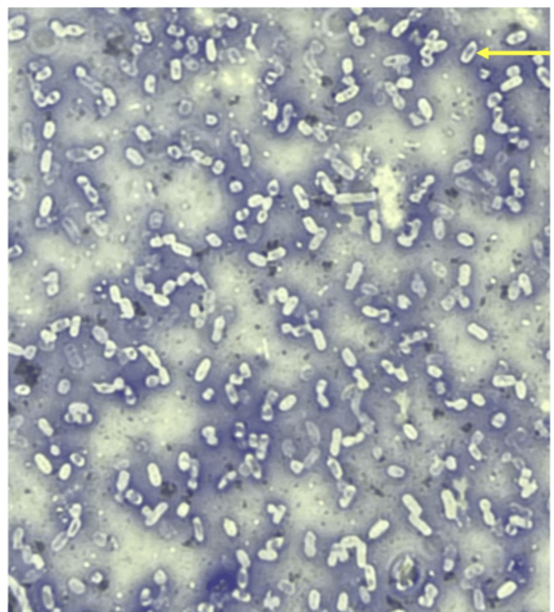
Simple stain purpose and characteristics
- purpose is to quickly determine cell shape, size, and arrangement
- primary stain: crystal violet
- has no mordant, destain, or counterstain
simple stain steps
- prepare heat fixed slide
- add crystal violet and let sit for 30-60 secs
- rinse with water
- blot dry and view
works on almost any standard bacteria
negative stain
- colors the background, which makes capsules more visible
- primary stain: nigrosin, no mordant, destain, or counterstain
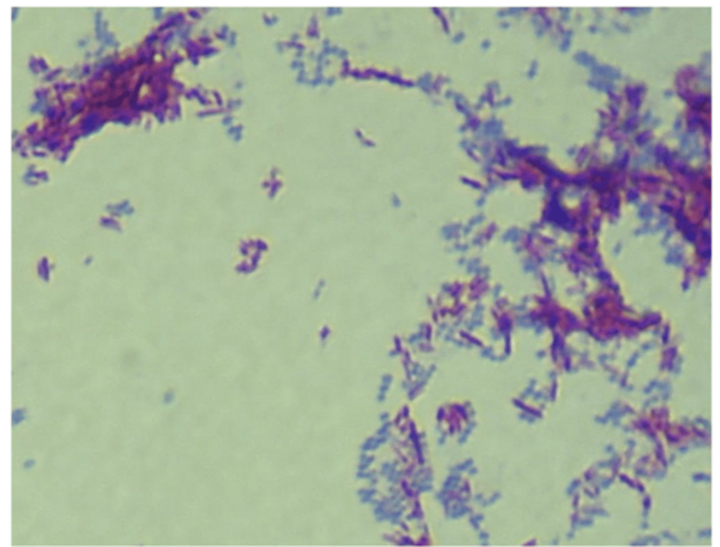
steps of gram stain
1. flood heat fixed smear with crystal violet for 1 min, rinse, blot dry
2. flood with iodine, sit for 1 min, rinse, blot dry
3. drip alcohol down tilted slide for 10-20 secs, immediately rinse with water, blot dry
4. flood with safranin for 1-2mins, rinse, blot dry, view
- gram negative (pink): e. coli, pseudomonas aeruginosa
- gram positive (purple): staphylococcus aureus, bacillus subtilis
examples of both kinds of gram stain results
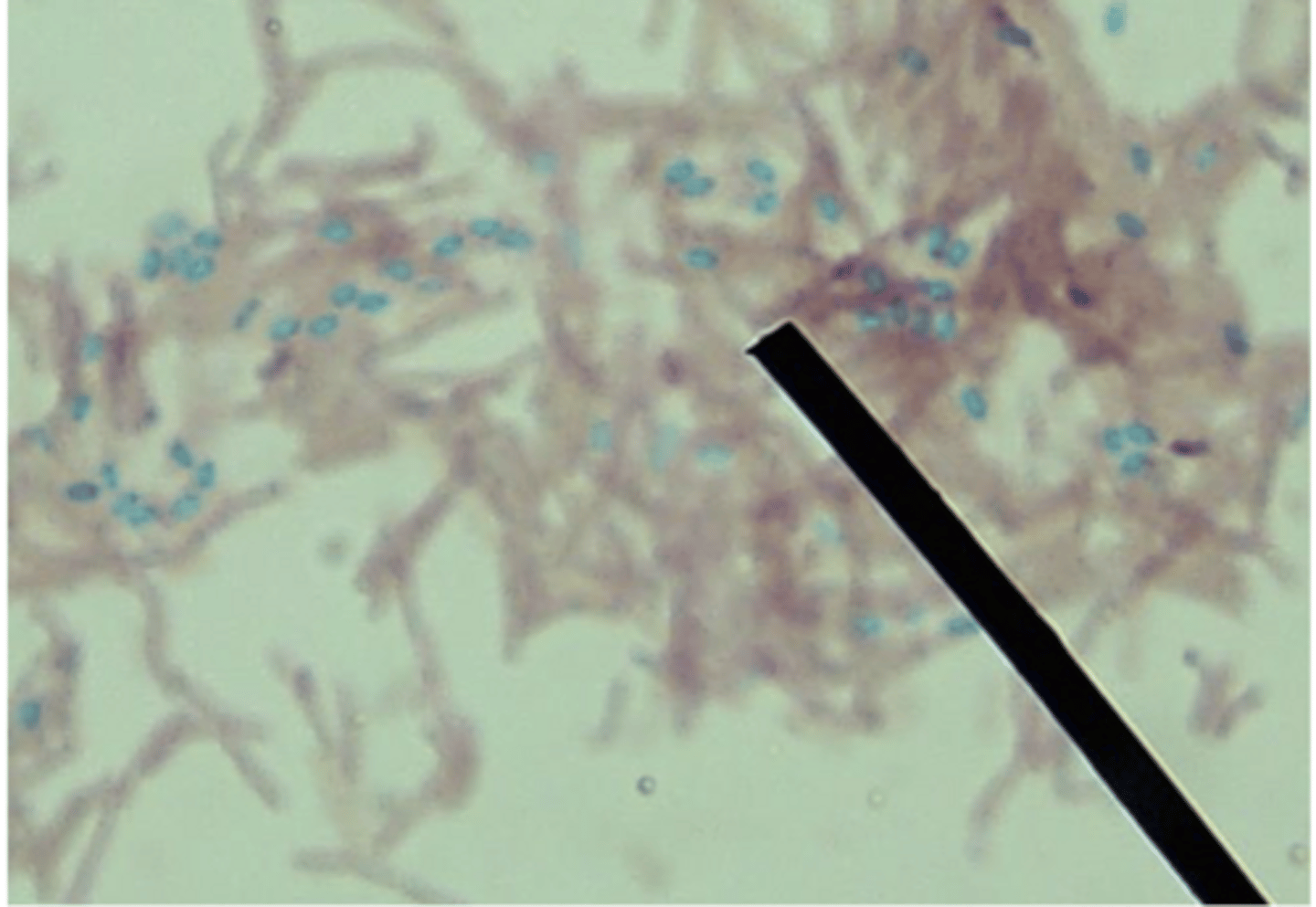
Acid Fast characteristics
- identify bacteria that possess waxy mycolic acid in their cell walls, which resists traditional staining methods.
- primary stain: carbolfuchsin
- mordant: heat --apply heat for 5 mins but not let it dry out
- destain: alcohol -- 15 to 30 secs/until run off clear, rinse
- counterstain: methylene blue, flood for 1 min, rinse, blot dry, and view
- differentiate highly resistant bacterial endospores (which stain green) from vegetative cells (which stain pink).
- primary: malachite
- counterstain: safranin
Endospore stain characteristics
endospore steps
- place piece paper towel over a heat fixed smear, flood with malachite green every time it starts to evaporate while applying heat for 5 mins (prevents drying out)
- remove paper towel, let cool, rinse with water fro 30 secs (decolorizer)
- flood with safranin for 1 min, rinse w water, blot dry, view
bacillus or clostridium
bacteria example of endospore
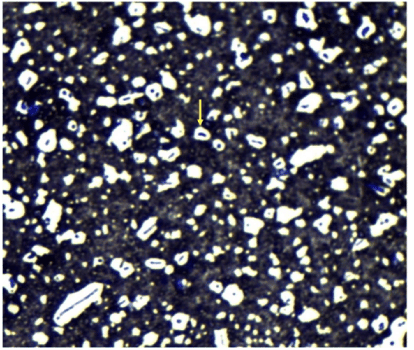
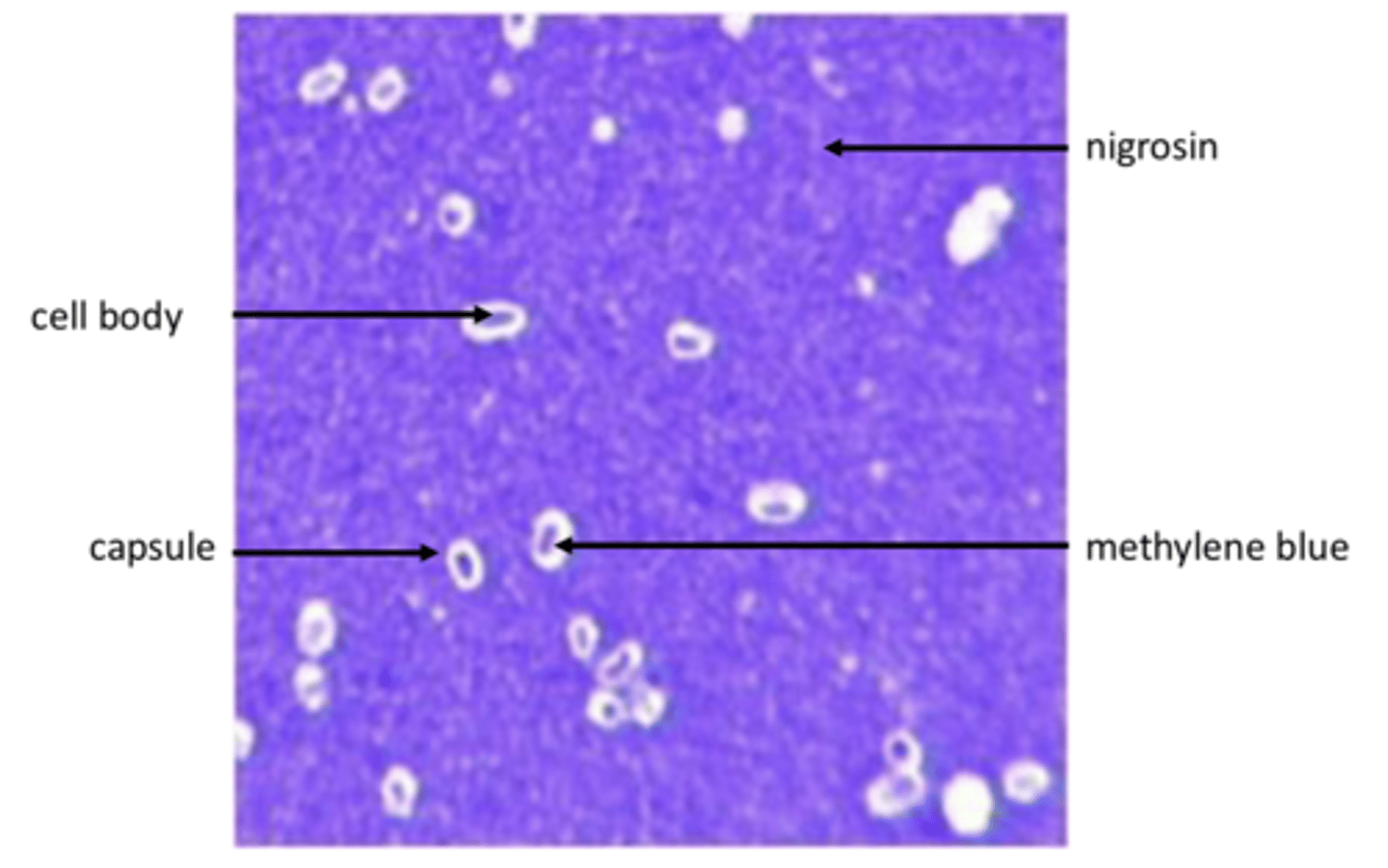
Capsule stain characteristics
- visualize extracellular capsules
- primary: nigrosin
- no mordant or destain
- counterstain: methylene blue
steps of capsule stain
- mix bacteria into drop of nigrosin on one end of slide (no heat fixing)
- spread mixture with second slide and allow to air dry
- flood dry slide with methylene blue for 1 min to stain interior cell body
- gently rinse with water or a light saline solution, air dry, view
capsule bacteria example
klebsiella pneumoniae, streptococcus pneumoniae
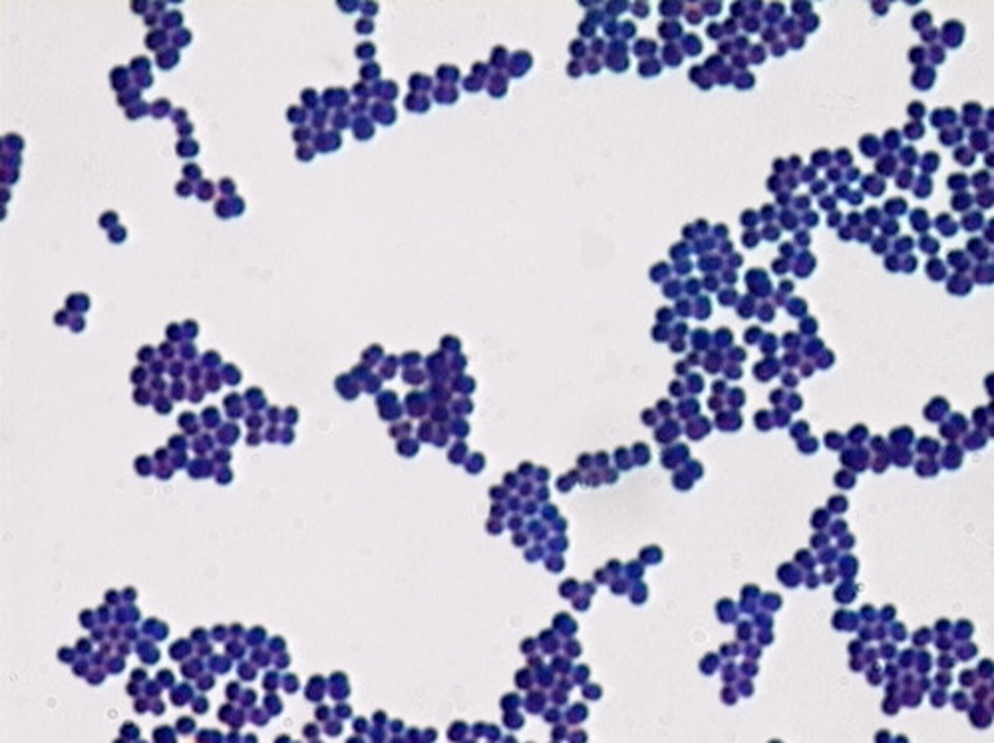
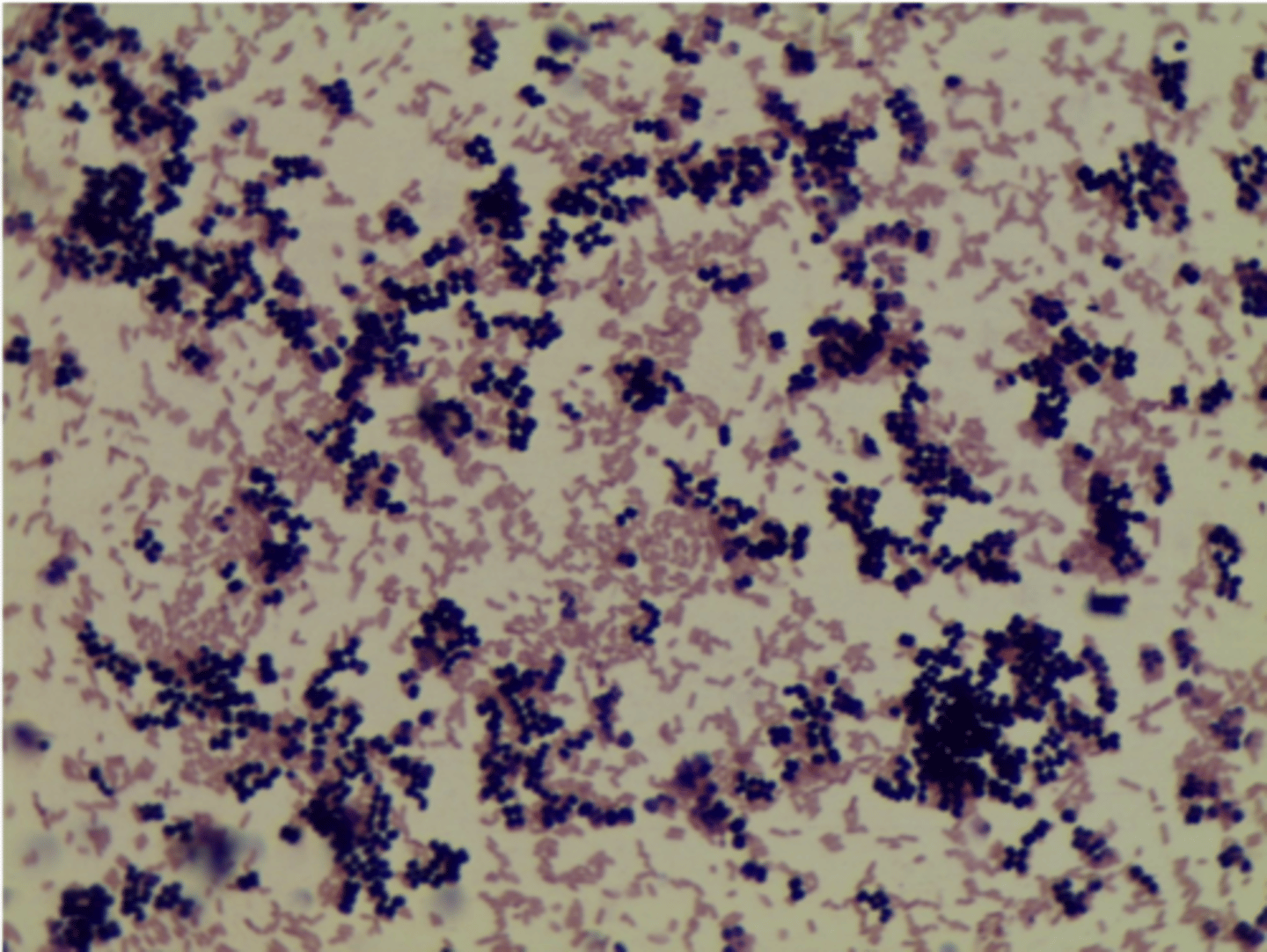
staphylococcus
cluster of coccus shaped cells
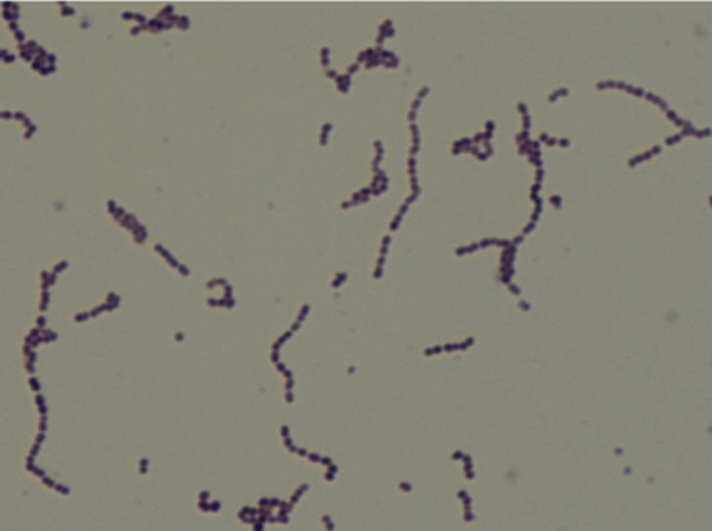
streptococcus
coccus cells in chains
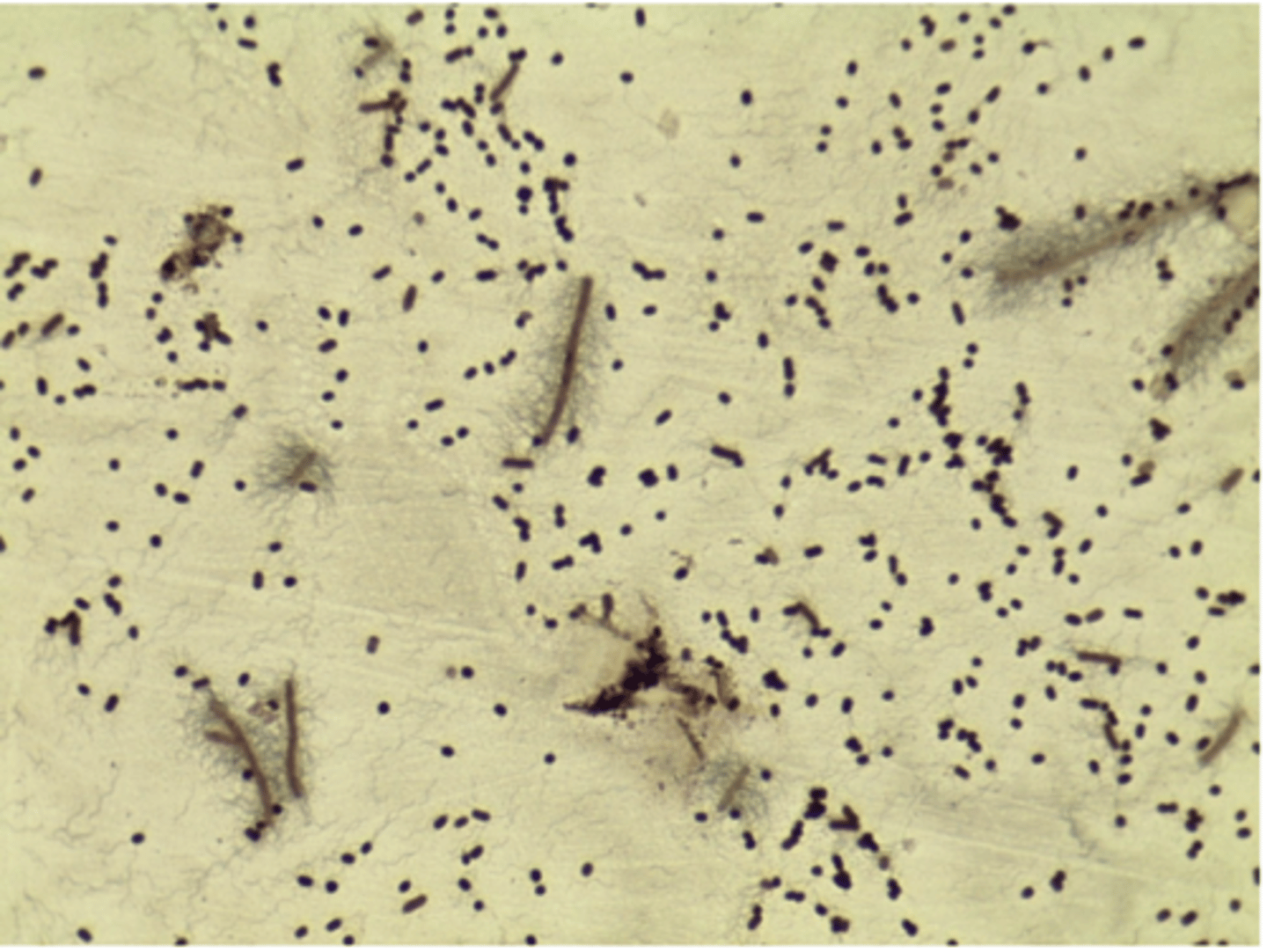
micrococcus
tetrad of coccus shaped cell
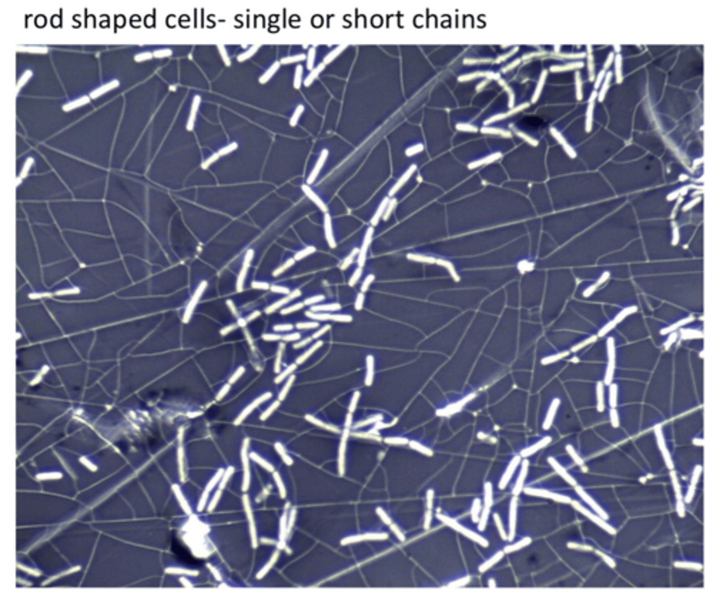
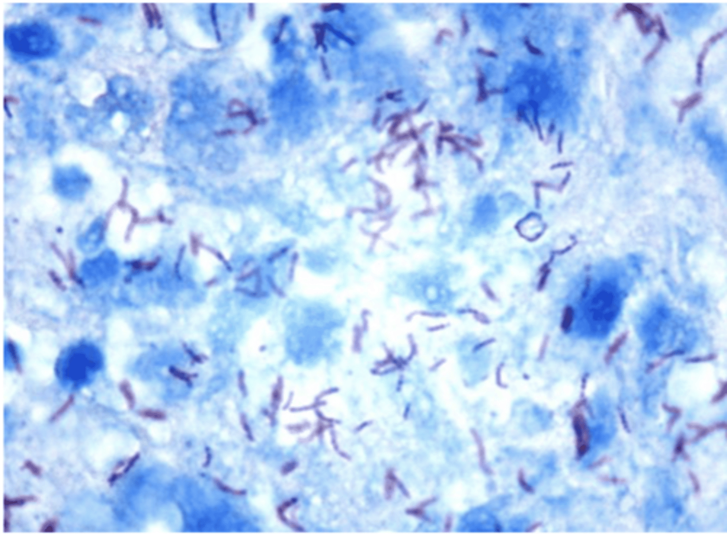
bacillus
large rod shaped cells
note: look for rods
bacillus - large rod shaped cell example
example of streptococcus
remember: in chains
example of micrococcus
tetrads
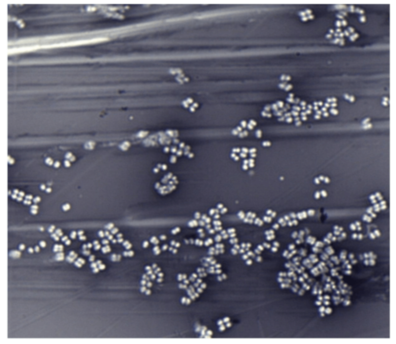
micrococcus in simple stain

Gram negative
-look for e. coli (pink) -- gram negative
-Bacillus- Gram positive; blue; bigger rods
acid fast stain - mycobacterium (dark red)
Staphylococcus- blue
second example of acid fast stain
capsule stain
- nigrosin- negative stain; shows cell shape
- basic stain- stains cell body; shows capsule thickness
example number 2
capsule stain example 3
