Parasitology

0.0(0)
Studied by 0 people
call kaiCall Kai
Locked
learnLearn
examPractice Test
spaced repetitionSpaced Repetition
heart puzzleMatch
flashcardsFlashcards
GameKnowt Play
Card Sorting

1/53

encourage image

There's no tags or description

Looks like no tags are added yet.

Last updated 2:44 PM on 7/21/26
Name
Mastery
Learn
Test
Matching
Spaced
Call with Kai
Chat

No analytics yet

Send a link to your students to track their progress

54 Terms

1
New cards

Plasmodium Species you need to know

  • P. vivax

  • P. malaria

  • P. falciparum

  • P. knowlesi

  • Babesia microti

2
New cards

Malaria

  • cause = sporazoa from plasmodium genus (P. malaria and P. falciparum

  • found in tropical and subtropical areas.

  • causes paroxysms that cause RBCs to rupture via release of merozoites (toxins)

  • symptoms of paroxysm phase = headache, deep bone pain, nausea or flu like symptoms, and a violent shaking chill that last 10 to 15 minutes initiates the paroxysm, which is followed by a fever with the temperature

  • long-term effects of malaria = damage to the liver and spleen caused by deposits of malarial pigments, otherwise known as hemozoin

3
New cards

Clinical signs of Malaria

  • Ring form refers to a ring like appearance of a malarial parasites following the invasion of a previously healthy red blood cell. The typical ring consists of a blue cytoplasmic circle connected with or to a red chromatin dot. The space inside is a vacuole.

  • The appearance of the developing trophozoite varies among the plasmodium species.

  • Mature schizonts are characterized by the emergence of merozoites. The number and arrangement of the merozoites vary within each malarial species.

  • The gametocyte can be divided into microgametocytes and macrogametocytes.

  • The microgametocytes is roundish in shape, with the exception of Plasmodium falciparum, which is banana or crescent shape.

  • Macrogametocytes range in shape from round to oval, with the exception of Plasmodium falciparum again, which is crescent shaped.

  • the gametocytes stain pink/purple.

4
New cards

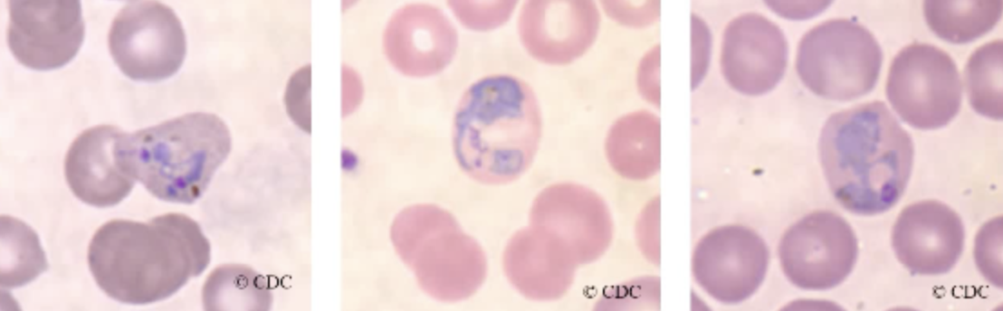
Plasmodium vivax: ring form

  • Red blood cells infected with plasmodium vivix tend to become enlarged and distorted in response to the presence of the growing parasites.
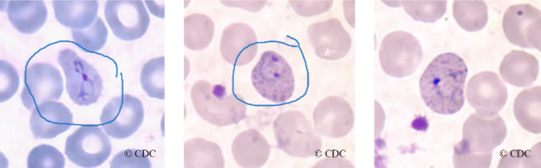
<ul><li><p><span style="background-color: transparent;">Red blood cells infected with plasmodium vivix tend to become enlarged and distorted in response to the presence of the growing parasites.</span></p></li></ul><p></p>
5
New cards

Plasmodium vivax: Trophozoite form

  • a single large chromatin dot is present among the cytoplasmic material.

  • The vacuole remains visible and basically intact.

  • The presence of brown pigments, which is a remnant of the parasite feeding on the red blood cells hemoglobin, becomes apparent in the site of cytoplasm.

  • Schuffner’s dots may also be present.

  • take on amoeboid shape

<ul><li><p><span style="background-color: transparent;">a single large chromatin dot is present among the cytoplasmic material.</span></p></li><li><p><span style="background-color: transparent;">The vacuole remains visible and basically intact.</span></p></li><li><p><span style="background-color: transparent;">The presence of brown pigments, which is a remnant of the parasite feeding on the red blood cells hemoglobin, becomes apparent in the site of cytoplasm.</span></p></li><li><p><span style="background-color: transparent;">Schuffner’s dots may also be present.</span></p></li><li><p><span style="background-color: transparent;">take on amoeboid shape</span></p></li></ul><p></p>
6
New cards

Plasmodium vivax: schizont

  • Mature schizonts generally contain about 12 to 24 merozoites.

  • Merozoites are created by the division of the chromatin

  • Brown pigment and Schuffner’s dots may also be present.
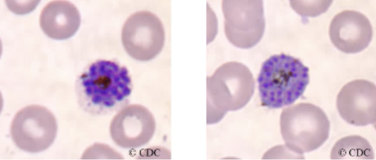
<ul><li><p><span style="background-color: transparent;">Mature schizonts generally contain about 12 to 24 merozoites.</span></p></li><li><p><span style="background-color: transparent;">Merozoites are created by the division of the chromatin</span></p></li><li><p><span style="background-color: transparent;">Brown pigment and Schuffner’s dots may also be present.</span></p></li></ul><p></p>
7
New cards

Plasmodium Vivax: Gametocyte

  • Plasmodium vivax gametocyte consists of a large pink to purple chromatin mass, which is surrounded by a colorless pale halo.

  • Evenly distributed cytoplasmic brown pigments is usually visible.
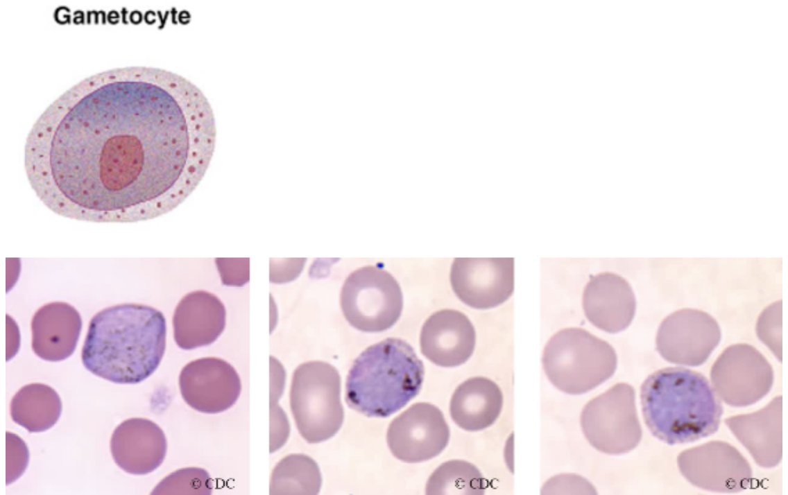
<ul><li><p><span style="background-color: transparent;">Plasmodium vivax gametocyte consists of a large pink to purple chromatin mass, which is surrounded by a colorless pale halo.</span></p></li><li><p><span style="background-color: transparent;">Evenly distributed cytoplasmic brown pigments is usually visible.</span></p></li></ul><p></p>
8
New cards

Plasmodium vivax Clinical Significance

  • found in tropical and subtropical areas

  • transmitted by the anopheles mosquito

  • infectious form = sporozoite

  • causes benign tertian malaria

  • gametocyte fills and enlarges RBCs

9
New cards

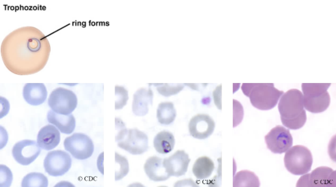
Plasmodium malaria: Ring form

  • only infect mature RBCs (smaller cells) and have lower parasitic load.

  • Delicate cytoplasmic ring ⅙ RBC diameter connected by a heavy chromatin dot

  • Vacuole appears “filled in”

  • Pigment forms early

  • RBCs are not distorted

<ul><li><p>only infect mature RBCs (smaller cells) and have lower parasitic load. </p></li><li><p><span style="background-color: transparent;">Delicate cytoplasmic ring ⅙ RBC diameter connected by a heavy chromatin dot</span></p></li><li><p><span style="background-color: transparent;">Vacuole appears “filled in”</span></p></li><li><p><span style="background-color: transparent;">Pigment forms early</span></p></li><li><p><span style="background-color: transparent;">RBCs are not distorted</span></p></li></ul><p></p>
10
New cards

Plasmodium malaria: trophozoite

  • The formation of a non-amoeboid solid cytoplasm that may assume an oval, band or bar shape is characteristic for the plasmodium malaria.

  • The cytoplasm consists of a coarse dark-brown pigment that often mask the chromatin material.

  • Vacuoles are absent in the mature forms of this stage.

  • The band and oval shapes are diagnostic
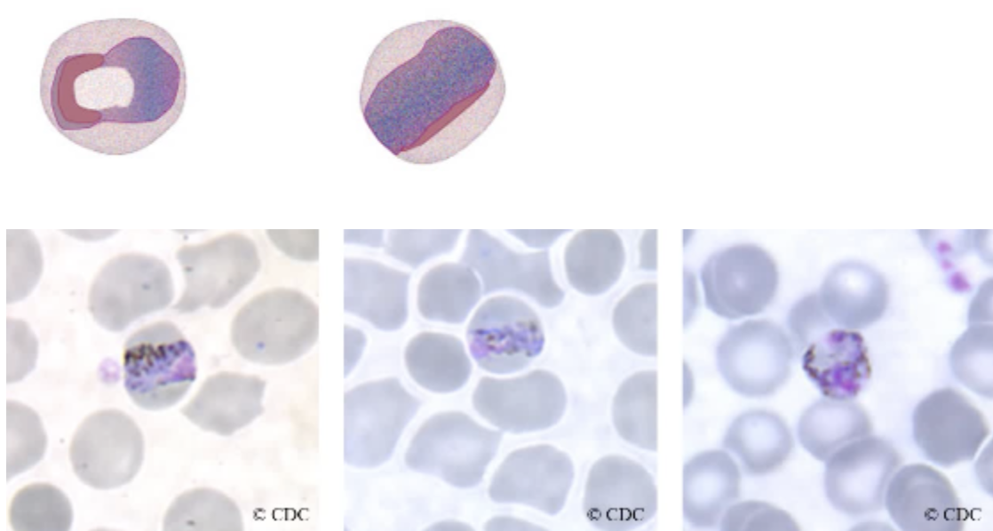
<ul><li><p><span style="background-color: transparent;">The formation of a non-amoeboid solid cytoplasm that may assume an oval, band or bar shape is characteristic for the plasmodium malaria.</span></p></li><li><p><span style="background-color: transparent;">The cytoplasm consists of a coarse dark-brown pigment that often mask the chromatin material. </span></p></li><li><p><span style="background-color: transparent;">Vacuoles are absent in the mature forms of this stage.</span></p></li><li><p><span style="background-color: transparent;"><strong><u>The band and oval shapes are diagnostic</u></strong></span></p></li></ul><p></p>
11
New cards
<p>Plasmodium malaria: Schizont</p>

Plasmodium malaria: Schizont

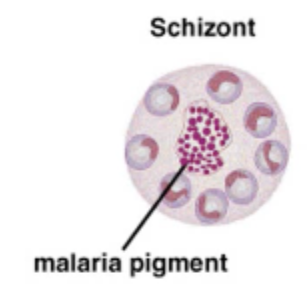
  • Plasmodium malaria schizont typically contains about 6 to 12 merozoites that are generally arranged in rosettes or irregular clusters.

  • A central arrangement of brown-green pigment may also be visible at this stage
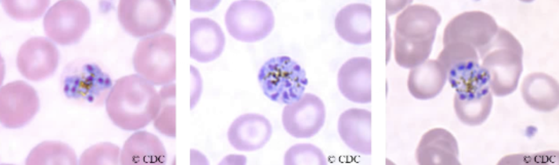
<ul><li><p><span style="background-color: transparent;">Plasmodium malaria schizont typically contains about 6 to 12 merozoites that are generally arranged in <u>rosettes</u> or irregular clusters.</span></p></li><li><p><span style="background-color: transparent;">A central arrangement of brown-green pigment may also be visible at this stage</span></p></li></ul><p></p>
12
New cards

Plasmodium malaria: gametocyte

  • The plasmodium malaria gametocyte resembles that of plasmodium vivax, but it’s smaller. 

  • The brown pigment is darker and coarser than that of Plasmodium vivax.

  • Other forms of this stage also tend to assume an oval shape.

  • There are no Schuffner’s dots present in this form in plasmodium malaria.
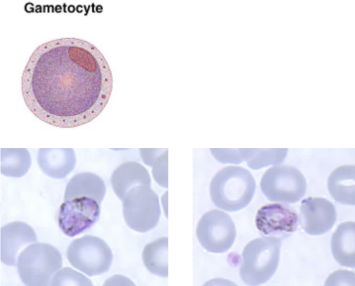
<ul><li><p><span style="background-color: transparent;">The plasmodium malaria gametocyte resembles that of plasmodium vivax, but it’s smaller.&nbsp;</span></p></li><li><p><span style="background-color: transparent;">The brown pigment is darker and coarser than that of Plasmodium vivax.</span></p></li><li><p><span style="background-color: transparent;">Other forms of this stage also tend to assume an oval shape.</span></p></li><li><p><span style="background-color: transparent;">There are no Schuffner’s dots present in this form in plasmodium malaria.</span></p></li></ul><p></p>
13
New cards

Clinical Significance of Plasmodium malaria

  • causes quartan malaria or malarial malaria

  • found in tropic, subtropic, and temperate regions of the world

  • attacks only mature RBCs

  • paroxysms every 72 hours, repeated attacks over 20 years

  • spontaneous recovery may occur.

  • may lead to nephrotic syndrome, which arises from the deposition of circular circulating immune complexes of the malarial antigen and antibody on the basement membrane of the glomerulus, which is causing an auto immune reaction in the patient.

14
New cards

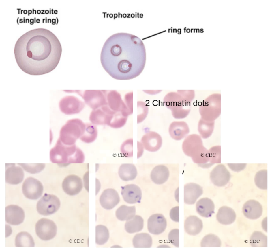
Plasmodium falciparum: Ring form

  • Plasmodium falciparum, or falciparum, can infect red blood cells of all ages, so the parasitic load of this parasite tends to be the highest.

  • The typical small, delicate ring form of Plasmodium falciparum consists of scanty cytoplasm connected to one or two small chromatin dots.

  • A small vacuole is also always visible.

  • Multiple rings in the red blood cells are frequently seen, and you can see applique forms where the ring form lines up against the edge of the red blood cells.

  • multiple rings = key characteristic

<ul><li><p><span style="background-color: transparent;">Plasmodium falciparum, or falciparum, can infect red blood cells of all ages, so the parasitic load of this parasite tends to be the highest.</span></p></li><li><p><span style="background-color: transparent;">The typical small, delicate ring form of Plasmodium falciparum consists of scanty cytoplasm connected to one or two small chromatin dots.</span></p></li><li><p><span style="background-color: transparent;">A small vacuole is also always visible.</span></p></li><li><p><span style="background-color: transparent;">Multiple rings in the red blood cells are frequently seen, and you can see applique forms where the ring form lines up against the edge of the red blood cells.</span></p></li><li><p><span style="background-color: transparent;">multiple rings = key characteristic</span></p></li></ul><p></p>
15
New cards

Plasmodium falciparum: Trophozoite

  • The Plasmodium falciparum developing trophozoite is characterized by the presence of heavy ring forms.

  • Fine pigment and granules may also be visible.

  • Mature stages of this form are not routinely seen in the peripheral blood.
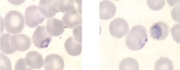
<ul><li><p><span style="background-color: transparent;">The Plasmodium falciparum developing trophozoite is characterized by the presence of heavy ring forms.</span></p></li><li><p><span style="background-color: transparent;">Fine pigment and granules may also be visible.</span></p></li><li><p><span style="background-color: transparent;">Mature stages of this form are not routinely seen in the peripheral blood.</span></p></li></ul><p></p>
16
New cards

Plasmodium falciparum: Schizont

  • The schizont of Plasmodium falciparum is the only visible form in patients with severe infection.

  • The typical schizont contains 24 merozoites, but it can contain anywhere from 8-36, and the cluster arrangement may also be present, but it is rarely seen.
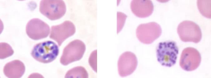
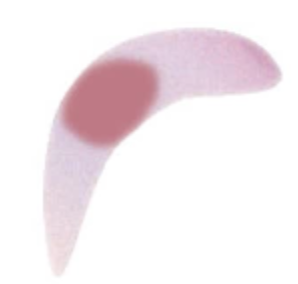
<ul><li><p><span style="background-color: transparent;">The schizont of Plasmodium falciparum is the only visible form in patients with severe infection.</span></p></li><li><p><span style="background-color: transparent;">The typical schizont contains <strong>24 merozoites</strong>, but it can contain anywhere from 8-36, and the cluster arrangement may also be present, but it is rarely seen.</span></p></li></ul><p></p>
17
New cards
<p>Plasmodium falciparum: gametocyte </p>

Plasmodium falciparum: gametocyte

  • The gametocyte of Plasmodium falciparum is typically sausage- or crescent-shaped. 

  • The chromatin is usually compact, and black pigment surrounds the chromatin, which may be visible.

  • This form is diagnostic for Plasmodium falciparum, meaning that is not seen with other Plasmodium species.
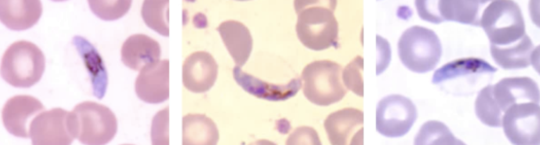
<ul><li><p><span style="background-color: transparent;">The gametocyte of Plasmodium falciparum is typically sausage- or crescent-shaped.&nbsp;</span></p></li><li><p><span style="background-color: transparent;">The chromatin is usually compact, and black pigment surrounds the chromatin, which may be visible.</span></p></li><li><p><span style="background-color: transparent;"><strong><u>This form is diagnostic</u></strong> for Plasmodium falciparum, meaning that is not seen with other Plasmodium species.</span></p></li></ul><p></p>
18
New cards

Plasmodium falciparum: Clinical Significance

  • ruptures RBCs at irregular intervals

  • gametocyte form is diagnostic

  • invades RBCs at any stage

  • causes malignant tertian malaria, most deadly of all plasmodium species.

  • infective form = sporozoite from anopheles mosquito

  • The primary complication is central nervous system involvements, and the organism reduces oxygen delivery to the tissues resulting in tissue anoxia, the patient ends up having severe headaches may be confused and ultimately lapses into a coma if untreated.

  • A second but less common complication of infection is called blackwater fever. Black urine due to mass intravascular hemolysis and hemoglobinuria. Renal complications/failure can occur.

19
New cards

Plasmodium ovale

morphologically similar to P. vivax, difficult to differentiate.

20
New cards

Plasmodium knowlesi

  • resembles P. malaria, and may resemble P. falciparum

  • causes life-threatening malaria

  • primarily zoonotic, affecting primates

  • if detected early, infections in humans can be treated

21
New cards

Babesia microti Clinical Significance

  • Found worldwide, but most commonly in Northeastern US (New England), Wisconsin, Connecticut

  • Symptoms = typically develop a hemolytic anemia and experience jaundice, weakness, fever, chills, sweating, headache, and hepatosplenomegaly. Renal failure is possible. Most cases are self-limiting.

  • Vector = Ixodes scapularis tick (deer tick)

  • official term is babesiosis, but look for hemolytic anemia.

  • prevention = avoid tick-infested areas, insect repellent, long sleeves and pants.

  • treat with Quinine and Clindamycin

22
New cards

Lab diagnosis

  • ring form is similar to P. falciparum

  • travel history and clinical signs

23
New cards
<p>Babesia microti: Ring form</p>

Babesia microti: Ring form

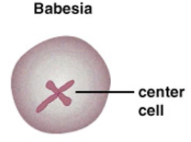
  • The rings of Babesia consists of a small chromatin dots and a scant amount of cytoplasm.

  • More mature versions may exhibit two or more chromatin dots.

  • Infected red blood cells usually contain 2 to 4 rings.

  • The stippling and malarial pigments are absent in Babesia

  • multiple rings with center cross-form is diagnostic
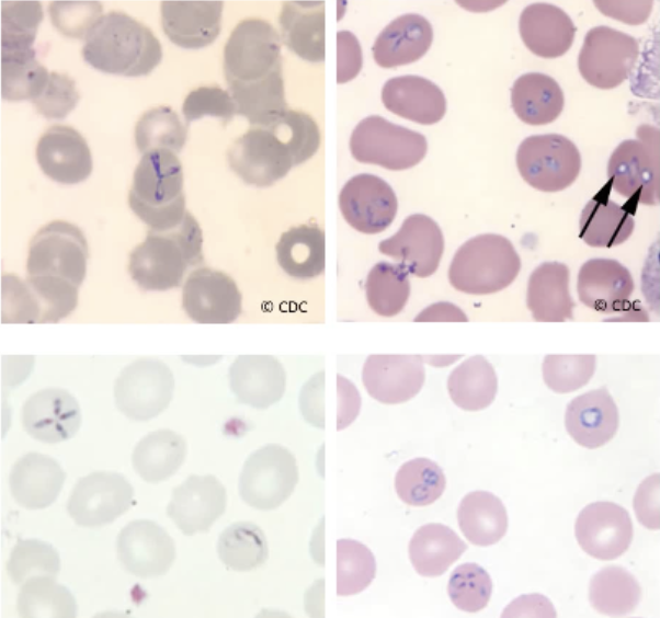
<ul><li><p><span style="background-color: transparent;">The rings of Babesia consists of a small chromatin dots and a scant amount of cytoplasm.</span></p></li><li><p><span style="background-color: transparent;">More mature versions may exhibit two or more chromatin dots.</span></p></li><li><p><span style="background-color: transparent;">Infected red blood cells usually contain 2 to 4 rings.</span></p></li><li><p><span style="background-color: transparent;">The stippling and malarial pigments are absent in Babesia</span></p></li><li><p><span style="background-color: transparent;"><strong><u>multiple rings with center cross-form is diagnostic</u></strong></span></p></li></ul><p></p>
24
New cards

Isospora belli/Cytoisospora belli: Oocyst

  • oocyst = oval and transparent, 25-35 um

  • The polyvinyl alcohol-preserved sediment makes it difficult to see the oocyst because the cyst wall is very thin and retractile.

  • They're variably stained in the modified acid-fast stain; some will stain light pink to purple, while others may remain unstained.

  • Their internal structure will not be seen, and some oocysts may appear collapsed or distorted on one side.
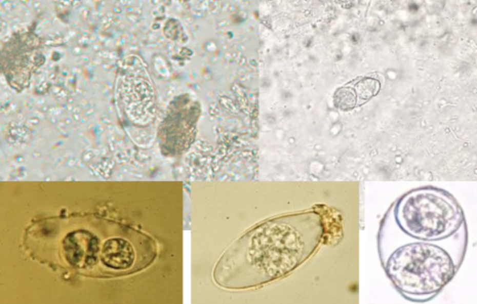
<ul><li><p>oocyst = oval and transparent, 25-35 um</p></li><li><p><span style="background-color: transparent;">The polyvinyl alcohol-preserved sediment makes it difficult to see the oocyst because the cyst wall is very thin and retractile.</span></p></li><li><p><span style="background-color: transparent;">They're variably stained in the modified acid-fast stain; some will stain light pink to purple, while others may remain unstained.</span></p></li><li><p><span style="background-color: transparent;">Their internal structure will not be seen, and some oocysts may appear collapsed or distorted on one side.</span></p></li></ul><p></p>
25
New cards

Isospora belli/Cytoisospora belli: Clinical Significance

  • infective form = oocyst

  • it is spread via fecal-oral route human-to-human, and contaminated food/water

  • humans are the only host

  • infection is confined to intestinal tract, and many people are asymptomatic

  • Symptoms: anorexia, nausea, abdominal pain, diarrhea, and possible malabsorption syndrome.

  • Treatment = trimethoprim-sulfamethoxazole

26
New cards

Cryptosporidium parvum

  • 4-5 um, round, thick cell wall

  • 4 sporozoites

  • easily mistaken as yeast

  • modified acid-fast stain is used for diagnosis, otherwise an enzyme immunoassay for Ag detection or molecular diagnostic tests.

27
New cards

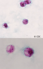
Cryptosporidium parvum: Oocyst

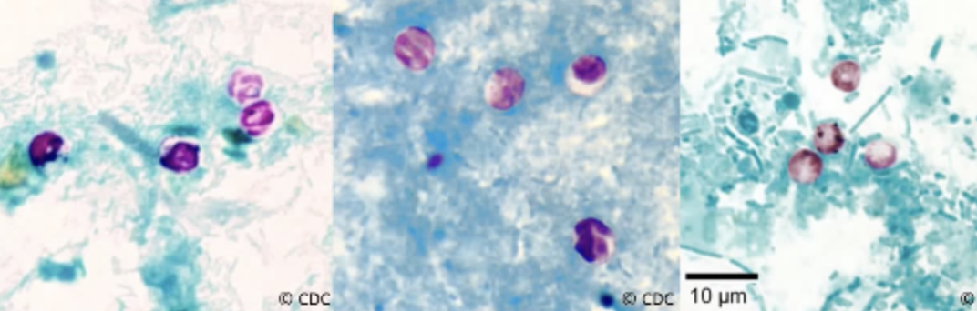
  • often have distinct walls and stain from a light pink to bright red color

  • Mature oocyst have discernible sporozoites (up to four), with a blue background and the pink or magenta color staining oocyst, on your modified acid fast stain.

  • the infective form = oocyst

<ul><li><p><span style="background-color: transparent;">often have distinct walls and stain from a light pink to bright red color</span></p></li><li><p><span style="background-color: transparent;">Mature oocyst have discernible sporozoites (up to four), with a blue background and the pink or magenta color staining oocyst, on your modified acid fast stain.</span></p></li><li><p><span style="background-color: transparent;"><strong>the infective form = oocyst</strong></span></p></li></ul><p></p>
28
New cards

Cryptosporidium parvum: Clinical Significance

  • spread through fecal-oral route, contaminated food/water

  • common in midwest

  • domestic animals harbor oocysts

  • An infection can be fatal for patients that are severely immunocompromised

  • treatment often not recommended, but use paromomycin.
29
New cards
<p>Cyclospora cayetanensis: Oocyst</p>

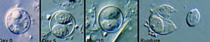
Cyclospora cayetanensis: Oocyst

  • resembles cryptosporidium

  • 7-10 um diameter, round-oval, transparent, when mature contains 2 sporocysts with 2 sporozoites each. May contain granules and have a wrinkled appearance.

  • The oocysts are variably stained on a modified acid fast stain, some will stain the light pink to deep purple, while others may remain unstained.

  • modified acid-fast stain and molecular diagnostics used for diagnosis
<ul><li><p>resembles cryptosporidium</p></li><li><p>7-10 um diameter, round-oval, transparent, when mature contains 2 sporocysts with 2 sporozoites each. May contain granules and have a wrinkled appearance.</p></li><li><p><span style="background-color: transparent;">The oocysts are variably stained on a modified acid fast stain, some will stain the light pink to deep purple, while others may remain unstained.</span></p></li><li><p><span style="background-color: transparent;">modified acid-fast stain and molecular diagnostics used for diagnosis</span></p></li></ul><p></p>
30
New cards

Cyclospora cayetanensis: Clinical Significance

  • The infectious form for Cyclospora cayetanensis is the oocyst through fecal-oral contamination.

  • Transmitted by contaminated food or water.

  • Infection causes self-limiting diarrhea that can take 3 to 4 days to resolve itself, but relapses can occur over a period of 2 to 3 weeks.

  • Antidiarrheal medications can provide relief of the symptoms, and Trimethoprim-sulfamethoxazole can reduce the possibility of relapse.

31
New cards

Size Comparison of Cryptosporidium parvum, Cyclospora, and Isospora belli
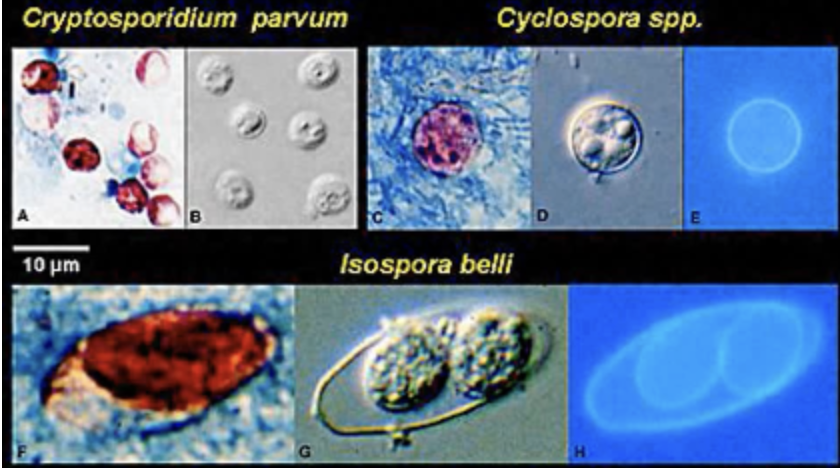
knowt flashcard image
32
New cards

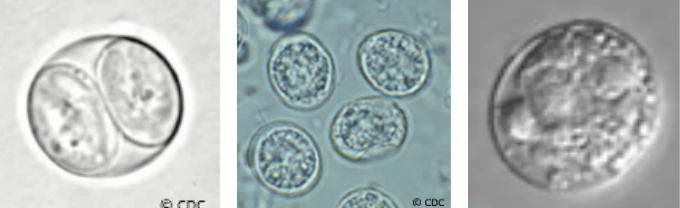
Toxoplasma gondii

  • sporozoan parasite that infects nucleated cells of almost all animals and birds

  • non-specific invasion, difficult to diagnose

  • Diagnosis involves detection of IgG and IgM ab by enzyme immunoassay.
<ul><li><p>sporozoan parasite that infects nucleated cells of almost all animals and birds</p></li><li><p>non-specific invasion, difficult to diagnose</p></li><li><p>Diagnosis involves detection of IgG and IgM ab by enzyme immunoassay.</p></li></ul><p></p>
33
New cards

Toxoplasma gondii: Clinical Significance

  • Humans become infected by ingesting the infective oocyst in contaminated food or drink or by accidental hand-to-mouth transmission of contaminated soil or cat litter.

  • If an immunocompromised person is infected, their symptoms can present like mono with swollen lymph nodes, fever, fatigue, hepatosplenomegaly, or an enlarged spleen, and absolute lymphocytosis. If severe enough, it causes encephalitis, myocarditis, hepatitis, pneumonia, and/or eye inflammation depending on location of organism.

  • If a fetus were to be infected, it's called congenital toxoplasmosis, so a fetus can be infected by toxoplasma by crossing the placenta from the mother.

  • Symptoms in the infant can include hydrocephaly, microcephaly, intracerebral calcification, chorioretinitis, convulsions, and psychomotor disturbances.

  • The diagnosis in infants is made by detection of IgM antibodies in an enzyme immunoassay method from the infant themselves.

  • generally, causes congenital toxoplasmosis.

34
New cards

Microsporidia

  • They are tissue parasites and are recognized to cause infections in immunocompromised patients that include enteritis, encephalitis, nephritis, and keratoconjunctivitis.

  • Diagnosis of the different species of Microsporidium varies.

  • Serologic tests look for antibody production, and tests are available for the detection of some species.
35
New cards
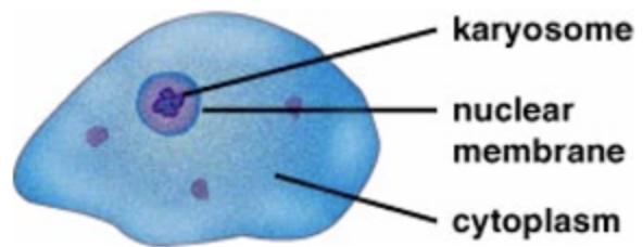
<p>Entamoeba histolytica: Trophozoite</p>

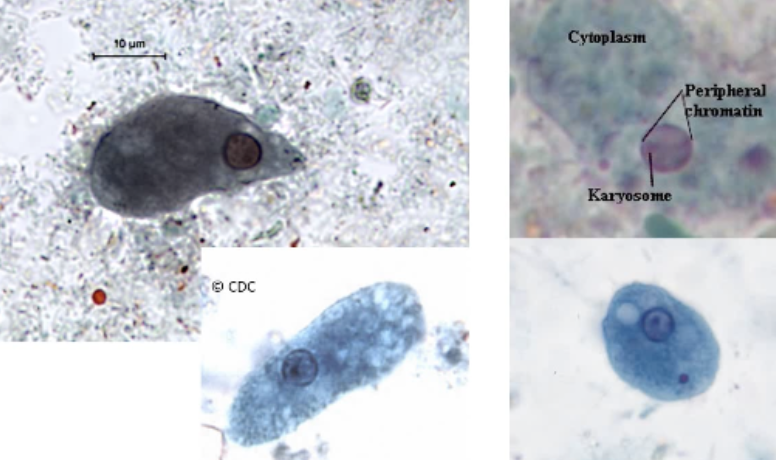
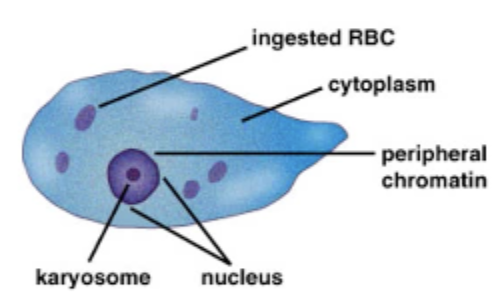
Entamoeba histolytica: Trophozoite

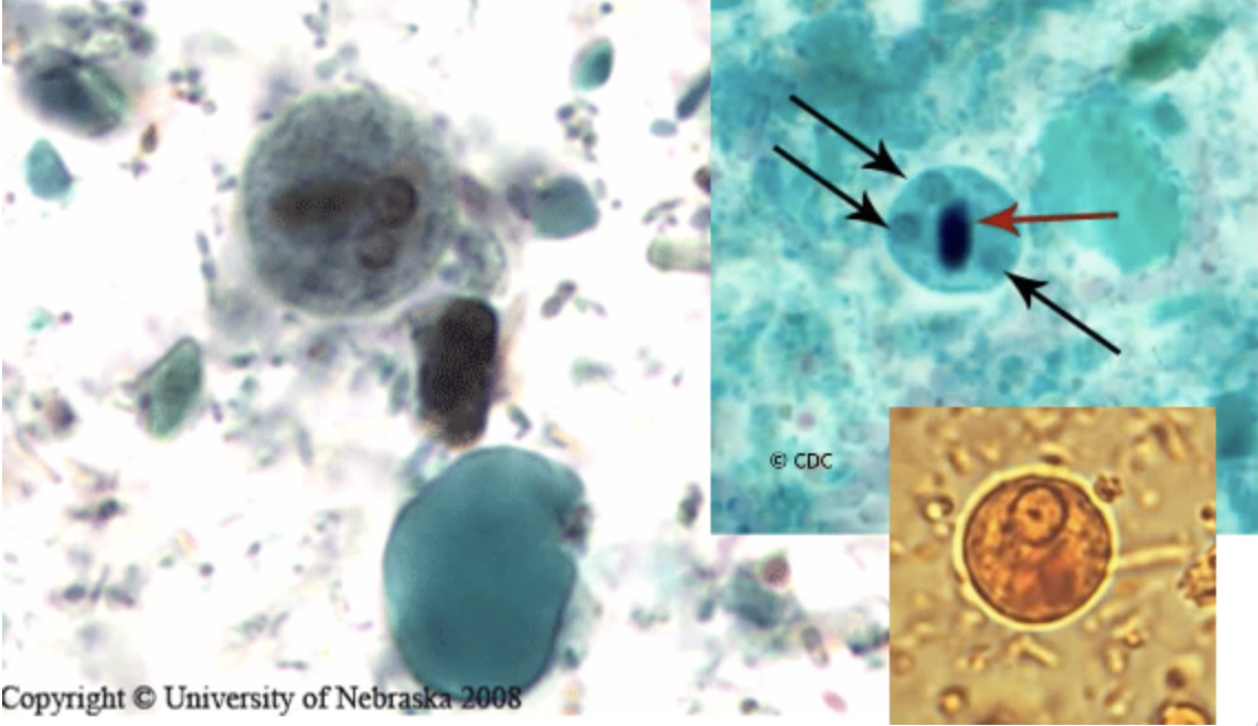
  • Motility is progressive and rapid. 

  • Finger-like pseudopods

  • 12-60 um

  • 1 nucleus

  • PC = fine

  • K = central

  • Cytoplasm appears green. Vacuoles can be present.

  • Can have inclusions (RBCs)→ good differentiator btw histolytica and dispar.
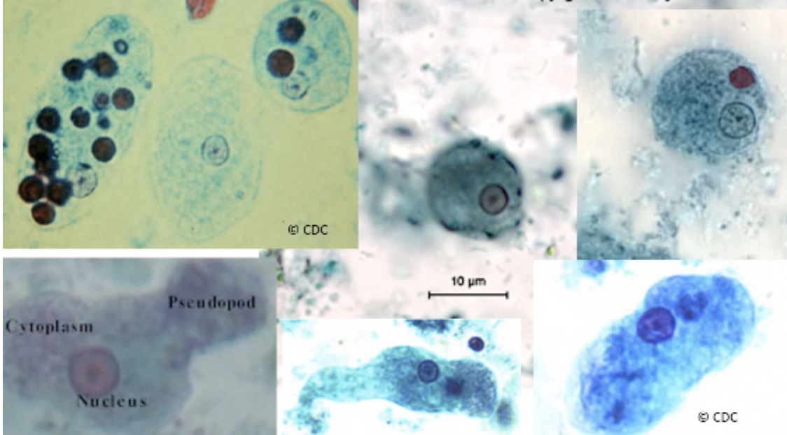
<ul><li><p><span style="background-color: transparent;">Motility is progressive and rapid.&nbsp;</span></p></li><li><p><span style="background-color: transparent;">Finger-like pseudopods</span></p></li><li><p><span style="background-color: transparent;">12-60 um</span></p></li><li><p><span style="background-color: transparent;">1 nucleus</span></p></li><li><p><span style="background-color: transparent;">PC = fine</span></p></li><li><p><span style="background-color: transparent;">K = central</span></p></li><li><p><span style="background-color: transparent;">Cytoplasm appears green. Vacuoles can be present.</span></p></li><li><p><span style="background-color: transparent;">Can have inclusions (<strong>RBCs</strong>)→ good differentiator btw histolytica and dispar.</span></p></li></ul><p></p>
36
New cards
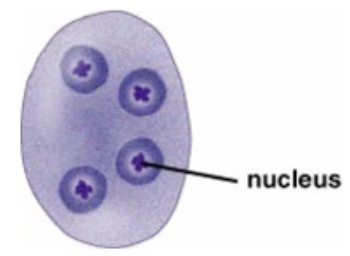
<p>Entamoeba histolytica: Cyst</p>

Entamoeba histolytica: Cyst

  • 10-20 um

  • Spherical in shape

  • Mature = nucleus

  • Immature = 4 nuclei

  • PC = fine, een

  • K = small, compact, central

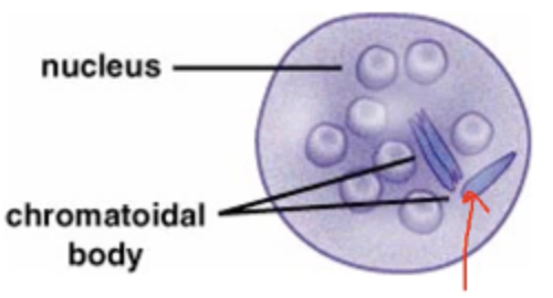
  • Cytoplasm = sometimes chromatid bodies are present

  • Elongated with blunt or rounded smooth edges.

  • Glycogen granules may be present

  • infective form!

<ul><li><p><span style="background-color: transparent;">10-20 um</span></p></li><li><p><span style="background-color: transparent;">Spherical in shape</span></p></li><li><p><span style="background-color: transparent;">Mature = nucleus</span></p></li><li><p><span style="background-color: transparent;">Immature = 4 nuclei</span></p></li><li><p><span style="background-color: transparent;">PC = fine, een</span></p></li><li><p><span style="background-color: transparent;">K = small, compact, central</span></p></li><li><p><span style="background-color: transparent;">Cytoplasm = sometimes chromatid bodies are present</span></p></li></ul><ul><li><p><span style="background-color: transparent;">Elongated with blunt or rounded smooth edges.</span></p></li></ul><ul><li><p><span style="background-color: transparent;">Glycogen granules may be present</span></p></li><li><p><span style="background-color: transparent;"><strong>infective form!</strong></span></p></li></ul><p></p>
37
New cards

Entamoeba histolytica: Clinical Significance

  • Found in subtropical and tropical areas of the world + Alaska, Russia, and Canada

  • Poor sanitation practices in areas that use human waste as fertilizer contribute to the infection

  • Infection occurs through ingestion of contaminated food or drink.

  • difficult to differentiate btw histolytica and dispar, so often notated as histolytica/dispar

Amoebic dysentery

  • Infective form: cyst

  • Intestinal ulceration

  • Diarrhea, bloody stool, dehydration, dehydration, abdominal cramping

  • Diagnosis: observe cysts and trophs

  • Differentiate from Shigella dysentery

  • resistant to chlorine

  • The organism can easily or can spread to the liver; it can spread the other body sites, primarily the liver, brain and lungs.

38
New cards

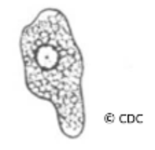
Entamoeba histolytica vs Shigella

  • white blood cells, or PMN. Entamoeba histolytica generally does not have very many whites present in the stool, where Shigella does, and they're often degraded.

  • Entamoeba histolytica generally doesn't have a lot of mucus in the stool, where Shigella does.
39
New cards
<p>Entamoeba hartmanni: trophozoite</p>

Entamoeba hartmanni: trophozoite

  • 5-12 um

  • Non-progressive motility

  • One nucleus

  • PC = fine granules, even distribution

  • K = small, central

  • Clean cytoplasm

  • No inclusions!!

<ul><li><p><span style="background-color: transparent;">5-12 um</span></p></li><li><p><span style="background-color: transparent;">Non-progressive motility</span></p></li><li><p><span style="background-color: transparent;">One nucleus</span></p></li></ul><ul><li><p><span style="background-color: transparent;">PC = fine granules, even distribution</span></p></li><li><p><span style="background-color: transparent;">K = small, central</span></p></li></ul><ul><li><p><span style="background-color: transparent;">Clean cytoplasm</span></p></li><li><p><span style="background-color: transparent;"><strong>No inclusions!!</strong></span></p></li></ul><p></p>
40
New cards

Entamoeba hartmanni: Cyst

  • 5-10 um

  • Spherical/round 

  • 2-4 nuclei

  • PC = fine, present

  • K = small, central

  • Cytoplasm = chromatid bodies

  • Glycogen vacuoles sometimes present

<ul><li><p><span style="background-color: transparent;">5-10 um</span></p></li><li><p><span style="background-color: transparent;">Spherical/round&nbsp;</span></p></li><li><p><span style="background-color: transparent;">2-4 nuclei</span></p></li></ul><ul><li><p><span style="background-color: transparent;">PC = fine, present</span></p></li><li><p><span style="background-color: transparent;">K = small, central</span></p></li></ul><ul><li><p><span style="background-color: transparent;">Cytoplasm = chromatid bodies</span></p></li><li><p><span style="background-color: transparent;">Glycogen vacuoles sometimes present</span></p></li></ul><p></p>
41
New cards

Entamoeba hartmanni: Clinical Significance

  • commensal, non-pathogenic

  • patients are asymptomatic and not treated
42
New cards
<p>Entamoeba coli: Trophozoite</p>

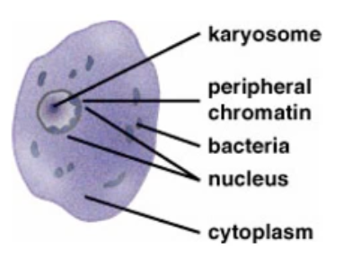
Entamoeba coli: Trophozoite

  • 15-50 um

  • Largest of the amoeba

  • Motile, sluggish, non-directional due to blunt pseudopods

  • 1 nucleus

  • PC = clumped, uneven

  • K = large, non-central

  • Cytoplasm = dirty appearance. Granular, vacuolated.

  • Inclusions = bacteria, yeast, debris

<ul><li><p><span style="background-color: transparent;">15-50 um</span></p></li><li><p><span style="background-color: transparent;">Largest of the amoeba</span></p></li><li><p><span style="background-color: transparent;">Motile, sluggish, non-directional due to blunt pseudopods</span></p></li><li><p><span style="background-color: transparent;">1 nucleus</span></p></li></ul><ul><li><p><span style="background-color: transparent;">PC = clumped, uneven</span></p></li><li><p><span style="background-color: transparent;">K = large, non-central</span></p></li></ul><ul><li><p><span style="background-color: transparent;">Cytoplasm = dirty appearance. Granular, vacuolated.</span></p></li><li><p><span style="background-color: transparent;">Inclusions = bacteria, yeast, debris</span></p></li></ul><p></p>
43
New cards
<p>Entamoeba coli: Cyst</p>

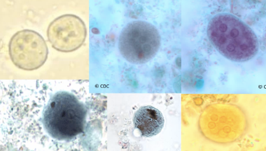
Entamoeba coli: Cyst

  • 35 um

  • Spherical or oval

  • Up to 8 nuclei

  • PC = coarse, granular

  • K = large, non-central

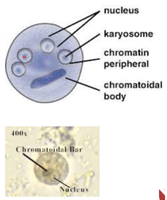
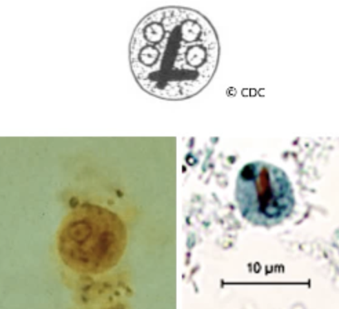
  • Cytoplasm = chromatoidal bodies present, generally splinter-shaped with rough pointed ends.

  • This is the only amoeba you can definitively identify with the cyst only
<ul><li><p><span style="background-color: transparent;">35 um</span></p></li><li><p><span style="background-color: transparent;">Spherical or oval</span></p></li><li><p><span style="background-color: transparent;"><strong><u>Up to 8 nuclei</u></strong></span></p></li></ul><ul><li><p><span style="background-color: transparent;">PC = coarse, granular</span></p></li><li><p><span style="background-color: transparent;">K = large, non-central</span></p></li></ul><ul><li><p><span style="background-color: transparent;">Cytoplasm = chromatoidal bodies present, generally splinter-shaped with rough pointed ends.</span></p></li><li><p><span style="background-color: transparent;"><strong><u>This is the only amoeba you can definitively identify with the cyst only</u></strong></span></p></li></ul><p></p>
44
New cards

Entamoeba coli: Clinical Significance

  • commensal, non-pathogenic, not treated

  • reported on stool report as Ent. coli

45
New cards
<p>Endolimax nana: Trophozoites</p>

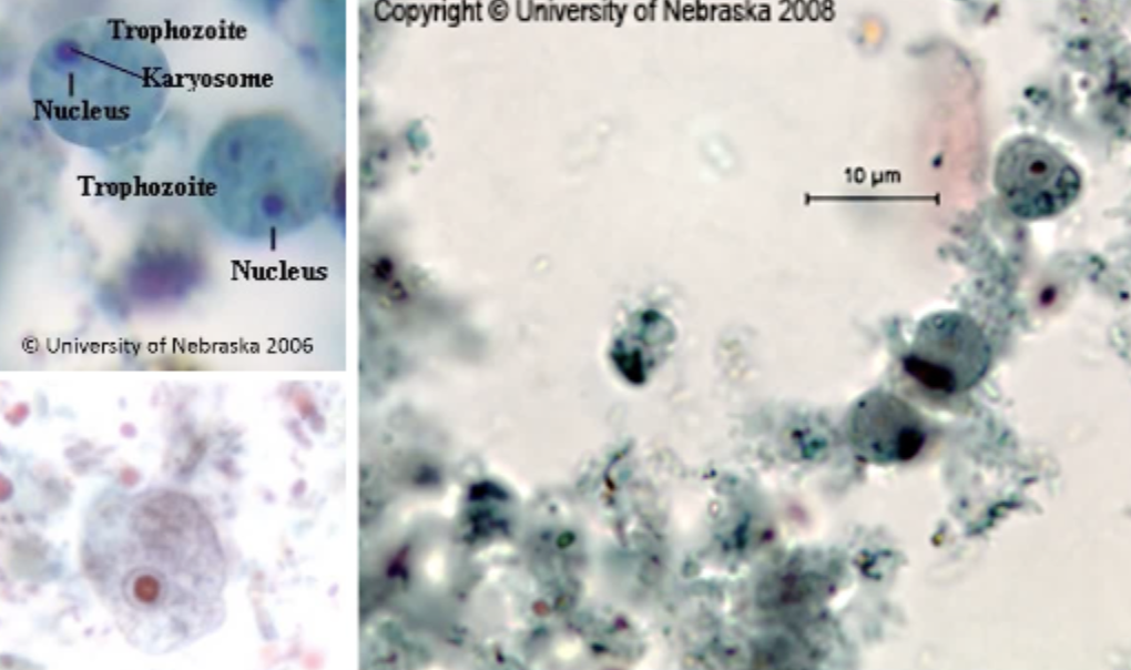
Endolimax nana: Trophozoites

  • 6-12 um

  • Sluggish motility, non-progressive

  • 1 nucleus

  • PC = none

  • K = very large, like a blot

  • Cytoplasm = granular, vacuolated, possible ingested bacteria
<ul><li><p><span style="background-color: transparent;">6-12 um</span></p></li><li><p><span style="background-color: transparent;">Sluggish motility, non-progressive</span></p></li><li><p><span style="background-color: transparent;">1 nucleus</span></p></li></ul><ul><li><p><span style="background-color: transparent;">PC = none</span></p></li><li><p><span style="background-color: transparent;">K = very large, like a blot</span></p></li><li><p><span style="background-color: transparent;">Cytoplasm = granular, vacuolated, possible ingested bacteria</span></p></li></ul><p></p>
46
New cards
<p>Endolimax nana: Cyst</p>

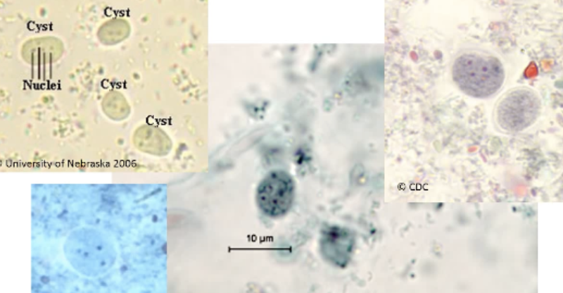
Endolimax nana: Cyst

  • 5-10 um

  • Oval, round

  • Nucleus: 2-4

  • PC = absent

  • K = smaller than in troph, but larger than Entamoeba

  • Cytoplasm = chromatid bodies

  • Glycogen is usually diffuse if present
<ul><li><p><span style="background-color: transparent;">5-10 um</span></p></li><li><p><span style="background-color: transparent;">Oval, round</span></p></li><li><p><span style="background-color: transparent;">Nucleus: 2-4</span></p></li></ul><ul><li><p><span style="background-color: transparent;">PC = absent</span></p></li><li><p><span style="background-color: transparent;">K = smaller than in troph, but larger than Entamoeba</span></p></li></ul><ul><li><p><span style="background-color: transparent;">Cytoplasm = chromatid bodies</span></p></li><li><p><span style="background-color: transparent;">Glycogen is usually diffuse if present</span></p></li></ul><p></p>
47
New cards

Endolimax nana: Clinical Significance

  • commensal, non-pathogenic, not treated

  • it’s reported when found in stool

  • Typically lives in warm, moist regions with poor hygiene
48
New cards
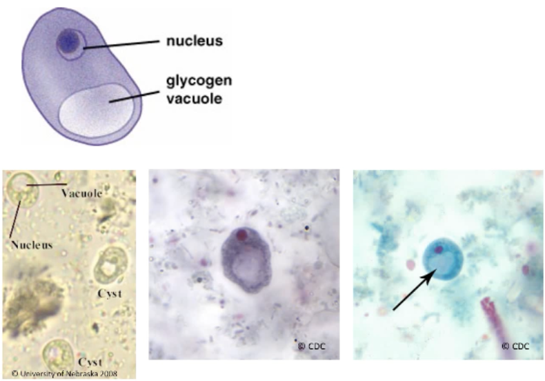
<p>Iodamoeba butschlii: Trophozoite</p>

Iodamoeba butschlii: Trophozoite

  • Motility = sluggish, non-progressive

  • 8-20 um

  • 1 nucleus

  • PC = none

  • K = large, can be surrounded by refractile granules

  • Cytoplasm = granular, heavily vacuolated. 
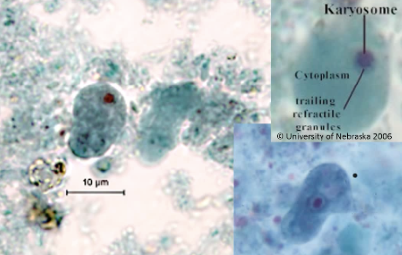
<ul><li><p><span style="background-color: transparent;">Motility = sluggish, non-progressive</span></p></li><li><p><span style="background-color: transparent;">8-20 um</span></p></li><li><p><span style="background-color: transparent;">1 nucleus</span></p></li></ul><ul><li><p><span style="background-color: transparent;">PC = none</span></p></li><li><p><span style="background-color: transparent;">K = large, can be surrounded by refractile granules</span></p></li></ul><ul><li><p><span style="background-color: transparent;">Cytoplasm = granular, heavily vacuolated.&nbsp;</span></p></li></ul><p></p>
49
New cards

Iodamoeba butschlii: Cyst

  • 13 um

  • Nucleus = 1

  • PC = absent

  • K = large, eccentric

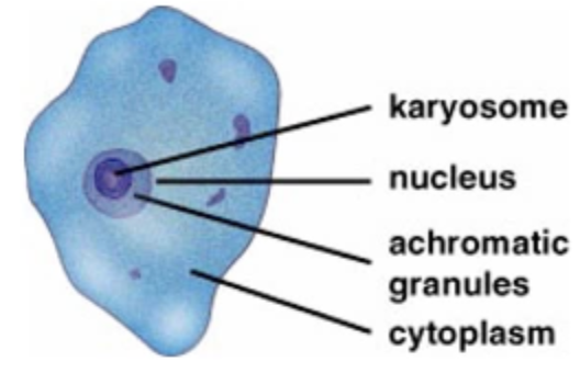
  • Achromatic granules

  • Glycogen vacuole

<ul><li><p><span style="background-color: transparent;">13 um</span></p></li><li><p><span style="background-color: transparent;">Nucleus = 1</span></p></li></ul><ul><li><p><span style="background-color: transparent;">PC = absent</span></p></li><li><p><span style="background-color: transparent;">K = large, eccentric</span></p></li></ul><ul><li><p><span style="background-color: transparent;">Achromatic granules</span></p></li><li><p><span style="background-color: transparent;">Glycogen vacuole</span></p></li></ul><p></p>
50
New cards

Iodamoeba butschlii: Clinical significance

  • commensal, non-pathogenic

  • asymptomatic, not treated

  • report if seen on O&P

51
New cards

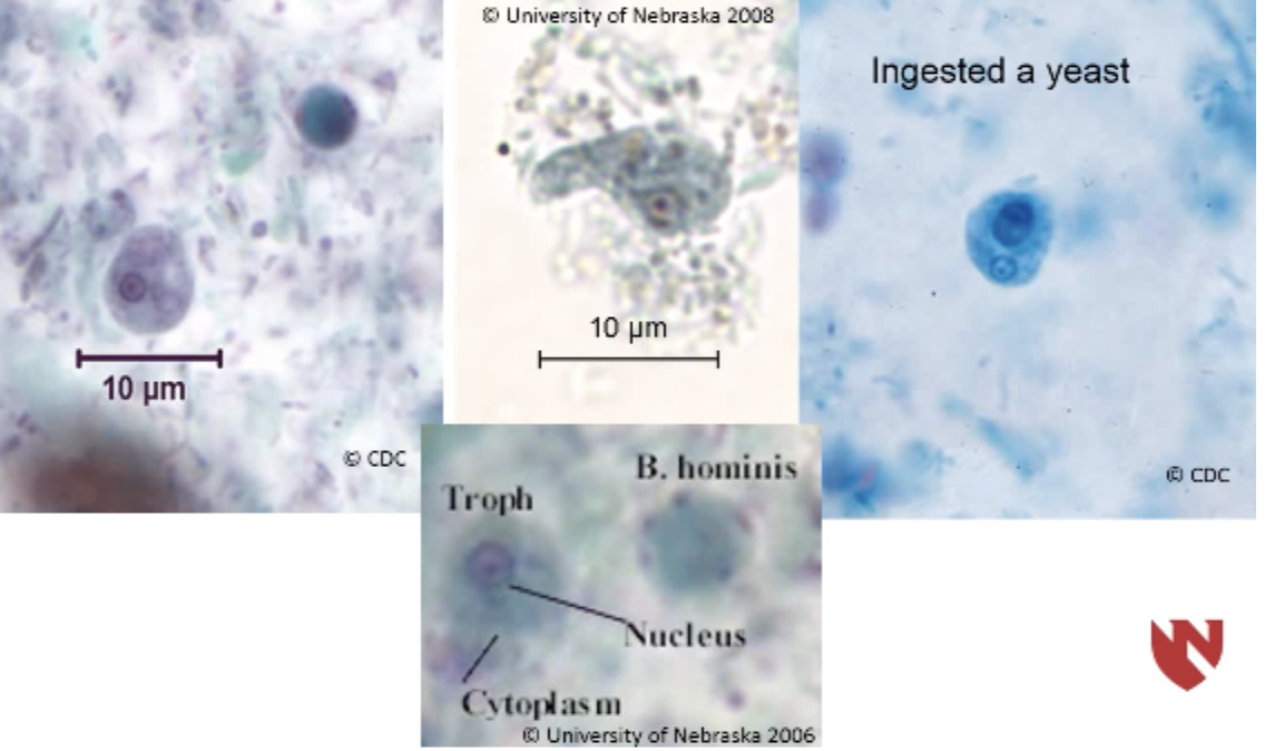
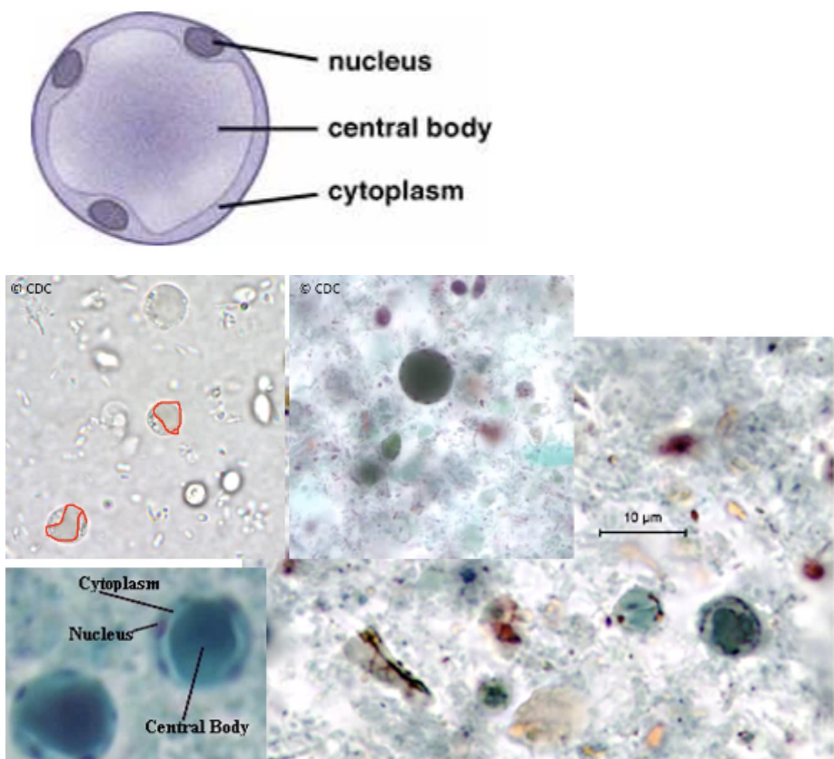
Blastocystis hominis:

  • Trophozoite

  • Clinical significance

Trophozoite

  • Nucleus = 2-6

  • Cyst-like structure

  • 5-30 um

  • Large central body

Clinical Significance

  • Can cause gastrointestinal disease in travelers and AIDS patients

  • typically commensal and non-pathogenic

  • spread fecal-oral

<p>Trophozoite</p><ul><li><p><span style="background-color: transparent;">Nucleus = 2-6</span></p></li><li><p><span style="background-color: transparent;">Cyst-like structure</span></p></li><li><p><span style="background-color: transparent;">5-30 um</span></p></li><li><p><span style="background-color: transparent;">Large central body</span></p></li></ul><p>Clinical Significance</p><ul><li><p><span style="background-color: transparent;">Can cause gastrointestinal disease in travelers and AIDS patients</span></p></li><li><p><span style="background-color: transparent;">typically commensal and non-pathogenic</span></p></li><li><p><span style="background-color: transparent;">spread fecal-oral</span></p></li></ul><p></p>
52
New cards
53
New cards
54
New cards