CREDIT 1 content.
1/174
Earn XP
Description and Tags
All content from credit 1, heart, vessels, brain and spinal cord.
Name | Mastery | Learn | Test | Matching | Spaced | Call with Kai |
|---|
No analytics yet
Send a link to your students to track their progress
175 Terms
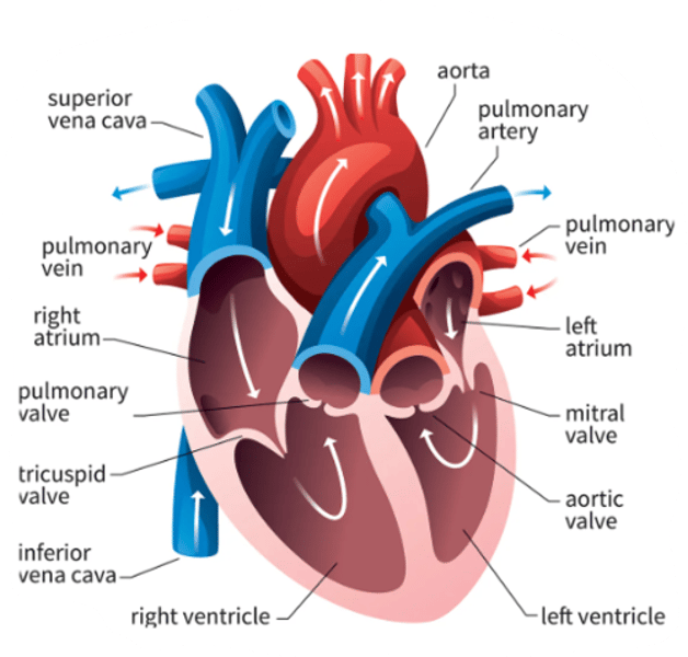
describe the heart. What is its function, location, chambers and position?
central organ, a 4 chambered muscular pump. It pumps blood through vessels by rhytmic contractions. It is enclosed by pericardium.
Location: in thoracic cavity, in the middle mediastinum.
function: pumps blood. oxygenated blood is sent from the lungs, to the left atrium --> left ventricle which pumps it through the aorta into the body. Right atrium gets deoxygenated blood from the vena cava, --> right ventricle --> pumped through pulmonary trunk --> to the lungs for reoxygenation.
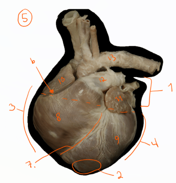
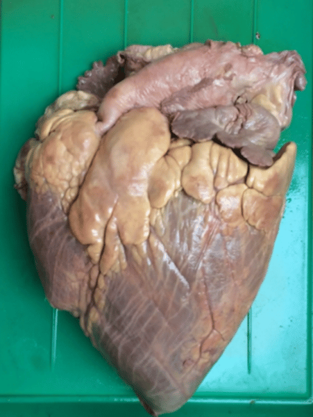
identify the structures of the heart seen here. (dog heart)
1. basis cordis- by atria, craniodorsally
2. apex cordis- by left ventricle
3. margo ventricularis dexter
4. margo centricularis sinister
5. facies auricularis (left)
6. sulcus coronarius - separates atria+ventricles.
7. sulcus interventricularis paraconalis - on f. auricularis, directed to margo ventricularis dexter.
8. ventriculus dexter
9. ventriculus sinister
10+11: auricula dextra et sinistra
12. Truncus pulmonalis
13. aorta.
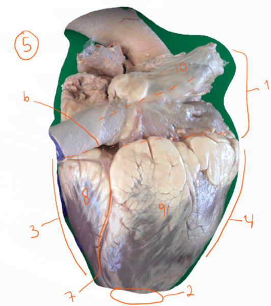
which side of the heart is this?
facies atrialis (right side).
Vena cava caudalis et cranialis seen here.
7. sulcus interventricularis subsinousus.
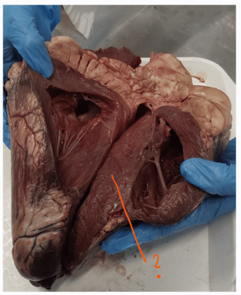
what is the structure seen called?
1. septum interventriculare (interventricular septum) - separates the ventricles. This structure is cutted right in the middle, therefore there is two structures as the wall is cutted through.
identify the auricula atrii.
auricula atrii - auricles of atrium.
blind diverticulus on left and right atrium
1. auricula dextra
2. auricula sinistra
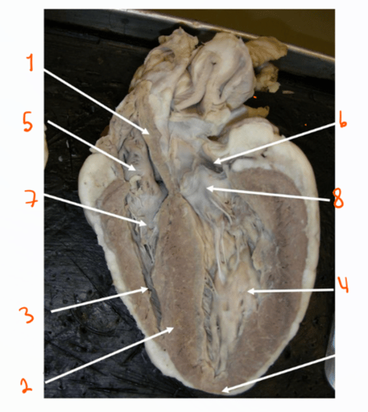
identify the structures, nr. 1-2 and 7-8. What are the names of the communication places between the atrium and ventricles?
1. septum interatriale (interatrial septum) - separates the atria
2. septum interventriculare (interventricular septum) - separates the ventricles.
3. ventriculus dexter.
4. ventriculus sinister - thicker walls.
5. atrium dextrum
6. atrium sinistrum
7. tricuspid valve
8. bicuspid valve (aka. mitral)
openings between the atrium and ventricles:
- ostium atrioventriculare sinistrum et dextrum.
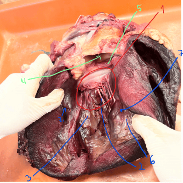
identify the structures of atrium dextrum. What is its function?
atrium dextrum - right atrium
function: gets venous blood from the body by means of the vena cava caudalis et cranialis.
1. shows the atrium - cutted into.
2. Sinus coronarius - opens below vena cava caudalis. This leads venous blood from the heart.
3. musculi pectinati (pectinate muscles) - forms the interior of the atrium.
4. Vena cava cranialis (a) et caudalis (b).
5. auricula dextra (right auricle) - is formed, directed to the left. (the piece here shown)
6. ventriculus dexter shown.
identify the structures of atrium sinistrium. What is its function?
atrium sinistrium - left atrium.
- characteristic: thicker walls.
function: gets blood from the lungs through the venae pulmonales (5-8 stk of these).
1. atrium sinistrium
2. musculi pectinati (pectinate muscles) - forms internal surface.
3. auricula sinistra (left auricle) - left, from truncus pulmonalis.
- the bigger hole (right side of picture) is showing the atrium dextrum on the side of the vena cava (in middle here).
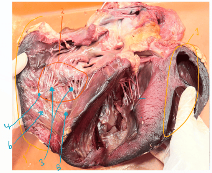
Identify the structures of ventriculus dexter. How does it communicate with right atrium?
shows heart - cutted through the septum interventriculare.
1. Ventriculus dextrum
2. valva tricuspidalis- this closes the ostrium atriocentriculare dextrum. Consist of 3 cusps, connected by chordae tendinae,
3. chordae tendinae
4. musculi papillares - the chords are connected to this muscle:
m. papillaris mm. papillares parvi
mm. papillaris magnus
m. papillaris subarteriosus
5. trabecula septomarginalis - crosses cavity, from septal to external wall
6. trabeculae carneae (irregular ridges)
identify these structures.
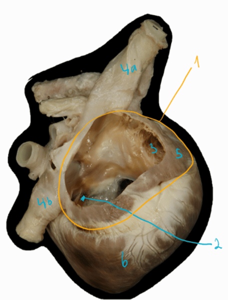
picture of opened right ventricle and truncus pulmonalis.
1. truncus pulmonalis (pulmonary trunk) - arises from ventriculus dexter.
2. valvulae semilunares (3 cusps) which forms the valva trunci pulmonalis (pulmonary valve) closing the opening of pulmonary trunk.
3. ostium trunci pulmonalis (opening of truncus).
4. septum interventriculare is seen here.
5. trabecula septomarginalis (muscular fibre) seen here.
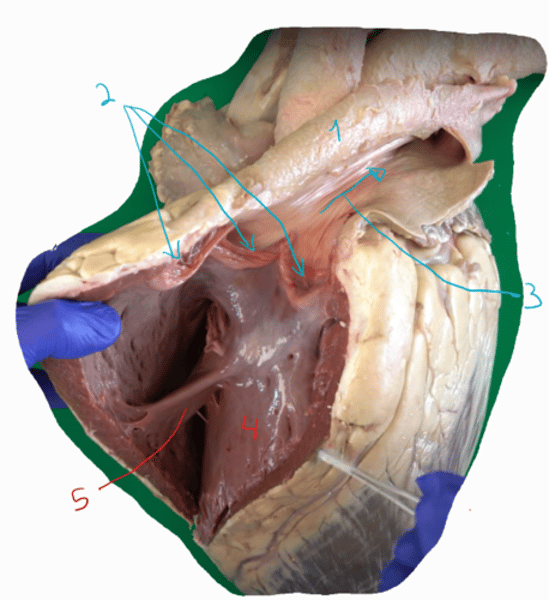
identify the structures of ventriculus sinister. How does it communicate with left atrium?
picture shows ventriculus sinistrum as the middle part. ventriculus sinistrum-2-3x thicker wall than the right ventricle wall.
1. valva bicuspidalis/mitral - closes the opening of ostium atrioventriculare sinistrum. It consist of 2 cusps.
2. chordae tendinae (the strings)
3. musculi papillares (2stk)
m. papillaris subatrialis
m. papillaris subauricularis
4. ostium aortae
5. 3 valvulae semilunares forming the valva aortae closing the opening.
6. trabeculae carneae - less
7. trabeculae septomarginalis (2)
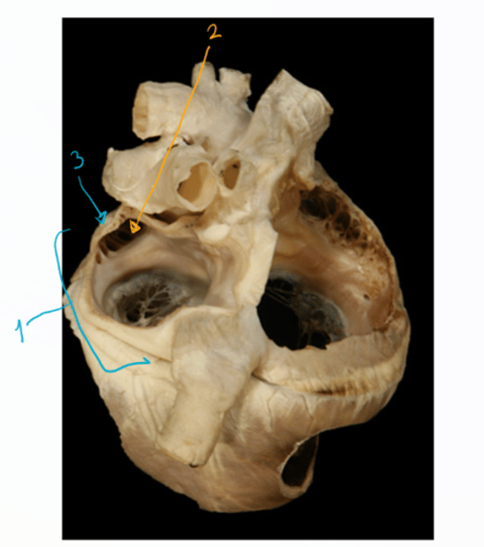
what is the composition of the heart wall?
1. epicardium (most external layer)
2. myocardium (2nd thickest layer) - made of muscle cells
3. endocardium - covers the heart chambers and cardiac valves.
what is the tissue component that surrounds the ostium aortae, ostium trunci pulmonales and ostium atrioventriculare dextrum et sinistrium?
Anuli fibrosi (fibrous rings) - partly formed of cartilage/bone.
what is the tissue component located between the ostium aortae and ostium atrioventriculare dextrum et sinistrium?
trigona fibrosa (fibrous triangles) - partly formed of cartilage/bone.
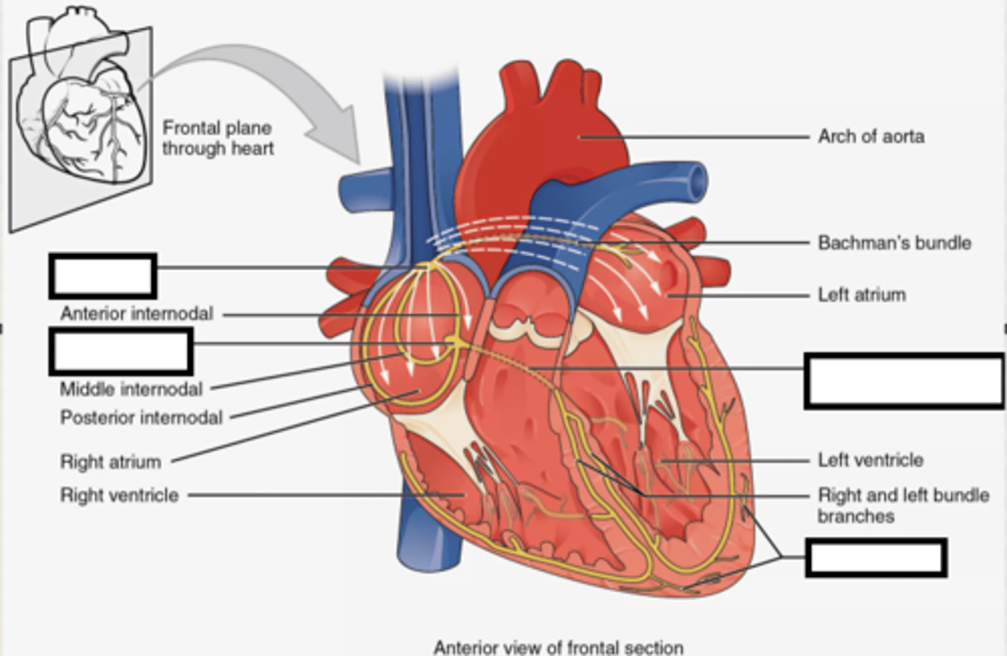
what does the conduction system of heart consist of? (4)
conduction system - keeps the heart beating.
1. nodus sinutrialis - located in the wall of atrium dextrum ventrally to opening of vena cava cranialis
2. nodus atrioventricularis - located within the septum interatrialis
3. fasciculus atrioventricularis (bundle)- originates from the nodus atrioventricularis - crus dextrum et sinistrum.
4. purkinje fibers - terminal end for septum interventriuclare which the crus dex. + sinistrum goes into.
what does the coronary arteries do? and what types are there?
arteriae coronariae (coronary arteries), gives oxygenated blood to the heart muscles. origin is bulbus aortae.
1. arteria coronaria sinister - larger, reaches the coronary groove, divides into ramus circumflexa + ramus interventricularis paraconalis
2. arteria coronaria dexter (right coronary a). - divides into ramus circumflexa + ramus interventricularis subsinuosus
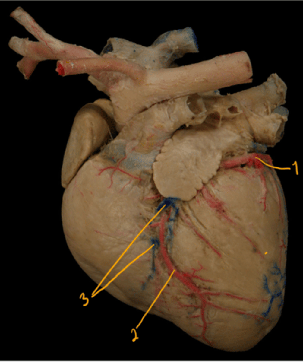
what are the vessels seen here?
1. ramus circumflexa - of a. coronaria sinister.
2. ramus interventricularis paraconalis - of a. coronaria sinister.
3. vena cordis magna (great cardiac vein)
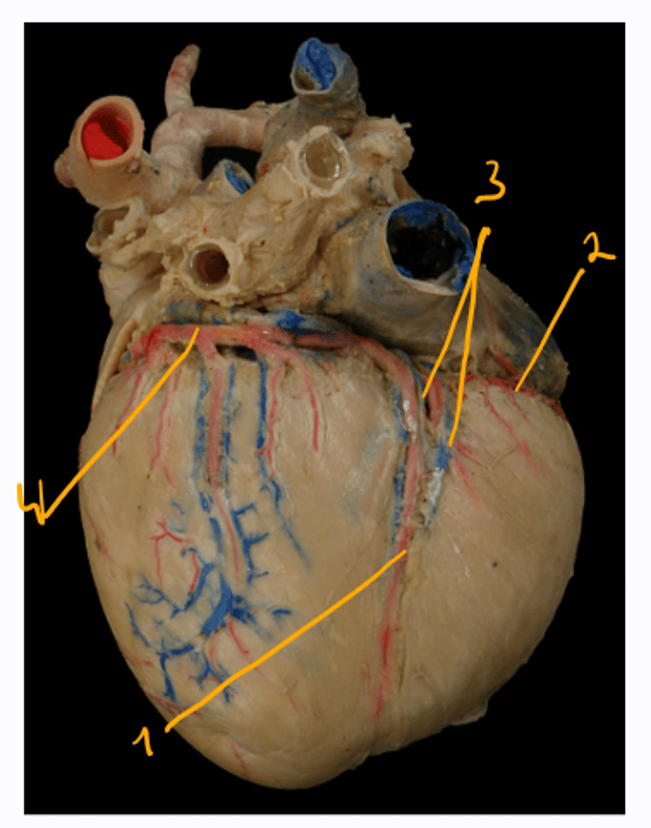
what are the vessels seen here?
1. ramus interventricularis subsinosus - of a. coronaria dexter.
2. ramus circumflexa - of a. coronaria dexter.
3. venis cordis media (middle cardiac vein)
4. ramus circumflexa - of a. coronaria sinister.
What are the veins of the heart?
1. vena cordis magna (great cardiac vein) - leads main part of the deoxygenated blood from left surface of heart.
2. vena cordis media (middle cardiac vein) leads deoxygenated blood from the right surface into the sinus coronarius. It empties into the right atrium.
3. vena cordis minimae (smallest cardiac veins) - opens directly into the heart chambers.
What layers does the pericardium consist of? And what does the pericardium contain?
contains: heart + initial parts of aorta, truncus pulmonalis, venae cavae and venae pulmonales.
3 layers:
1. pleura pericardiaca - part of pleura parietalis, most external layer.
2. pericardium fibrosum - 2nd thickest layer.
3. pericardium serosum - formed by lamina parietalis (fused with pericardium fibrosum) and lamina visceralis (covers the myocardium, known as the epicardium).
- cavum pericardii - located between the lamina parietalis et visceralis.
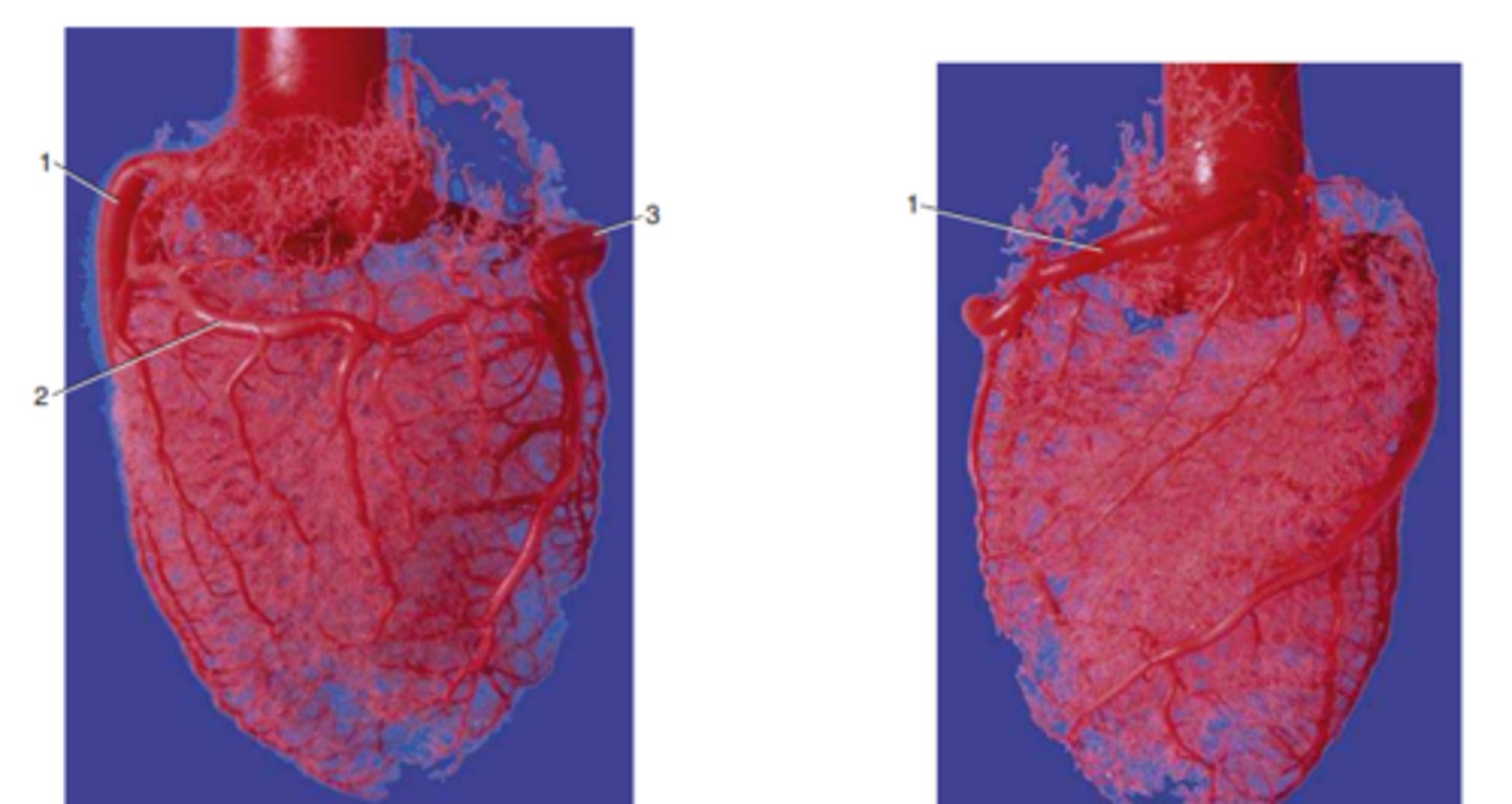
identify the structures.
structures seen on corrosion cast of aorta - pig.
1. a. coronaria sinister
2. ramus circumflexa
3. a. coronaria dexter
right picture: 1. a. coronaria dexter.
What are blood vessels (vasa sanguinea) divided into according to structure and function? What directs blood flow?
divided according to structure and function into:
1. arteries - transport the blood from the heart. It has thick, solid and flexible walls. It divides and then passes onto arterioles, then capillaries.
2. capillaries - in between the arteries and veins.
3. veins - comes from the capillary network, and gradually fuses together. It has thin walls and are larger than arteries of same type.
- valvular venosae (venous valves) - located at certain distances and directs the blood flow.
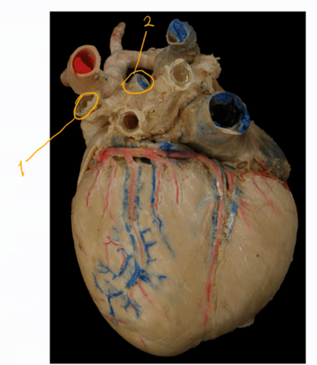
vessels of the great circulation, What does the truncus pulmonalis do? what arteries does it have?
truncus pulmonalis carries the venous blood from the ventriculus dexter into the lungs.
includes: 1. arteria pulmonalis dextra, 2. arteria pulmonalis sinistra - these carry deoxygenated blood from right ventricle and pulmonary trunk for gas exchange (continuation)
- each artery runs to the hilus of lung, then divides acc. to species.
what ligament connects the arteria pulmonalis dextra et sinistra to the truncus pulmonalis and aorta?
- Ligamentum arteriosum (arterial ligament) - connects them.
function of pulmonary veins?
venae pulmonales (pulmonary veins)
- 5-8 in numbers (varies)
- function: sends oxygenated blood from the lungs to the left atrium.
identify the parts. what is the main structure?
Aorta - main structure. It starts from ventriculus sinister.
1. bulbus aortae - enlargement of aorta, 2. aorta ascendens - runs craniodorsally, 3. arcus aortae - the truncus brachiocephalicus arises from the arcus.
- In su + car: left subclavian artery also arises.
4. aorta descendens - caudally, 2 parts, aorta thoracica + abdominalis (after passing hiatus aorticus)

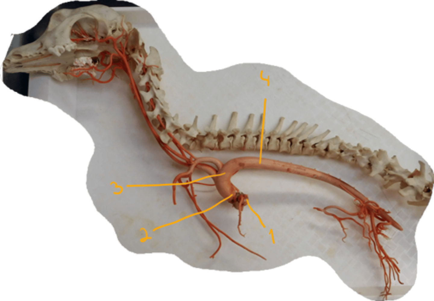
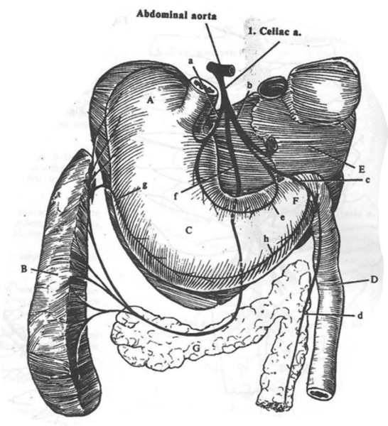
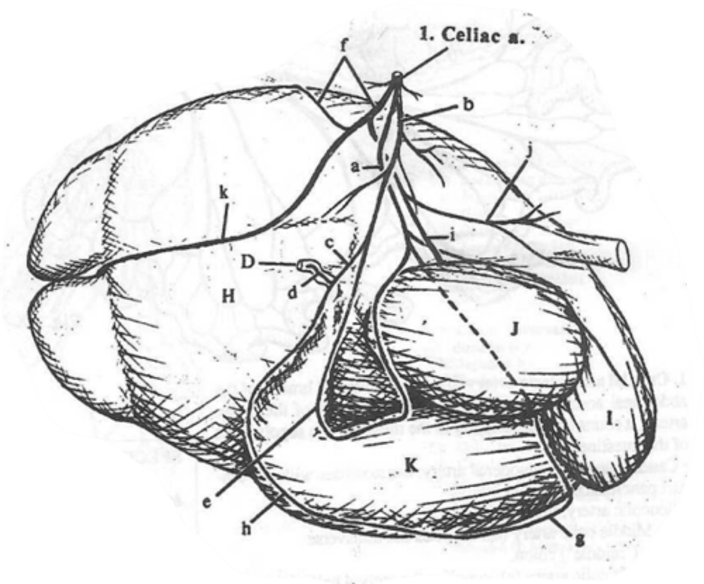
what are the 3 main branches of a. celiaca?
f: a. lienalis (splenic) - branches to spleen, pancreas + stomach. Continues along curvatura major of stomach as a. gastroepiploica sinistra.
in ru: a. ruminalis dextra et sinistra + a. reticularis. - has these.
a: a. gastrica sinistra: supplies left surface of stomach.
in ru: a. reticularis accessoria and a. gastroepiploica sinistra.
b: a. hepatica: nutritive branch for liver, supplies stomach, duodenum + pancreas. divides into some branches.
what are the further branches of a. hepatica?
e: a. gastrica dextra - supplies right side of stomach. Communicates with the a. gastrica sinistra.
continuation after origin of these branches is:
c: a. gastroduodenalis which branches into the
(h) a. gastroepiploica dextra (runs along curvatura major, communicates with (g) a. gastroepiploica sinistra) and (d) a. pacreaticoduodenalis cranialis (supplies duodenum + pnacreas)
- a. hepatica runs through porta hepatis and terminally divides inisde the liver parenchyma.
what are the branches of a. mesenterica caudalis?
unpaired branch dividing into:
1. a. colica sinistra - supplies middle part of descending colon.
2. a. rectalis cranialis - supplies caudal part of descending colon + rectum.
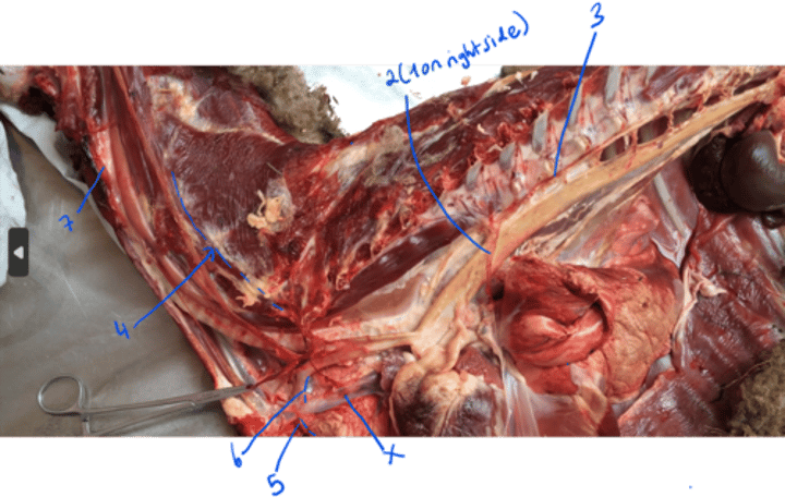
identify/mention the vena cava cranialis and its branches.
X: v. cava cranialis collects the venous blood from the head, neck, thoracic limbs and thorax. branches are:
1. v. azygos dextra - in eq, car + ru. Collects blood from intercostal spaces, from the level of first lumbar vertebrae.
2. v. azygos sinistra - in Ru + Su. Empties in sinus coronarius.
3. v. costocervicalis - with truncus.
4. v. vertebralis - with a.
5. v. thoracica interna - with a.
6. v. subclavia-with a.
7. truncus bijugularis - by fusion of v. jugularis externa sinister et dexter.
identify the vena cava caudalis and its branches.
v. cava caudalis collects venous blood from the postdiaphragmatic part of body. Located in the abd. cavity, enters thoracic cavity by foramen venae cavae. Branches in abd. cavity:
1. vv. phrenicae craniales. - cranially to hepatic v.
2. vv. hepaticae - enters v. cava on caudal border of liver.
3. vv. phrenicae caudales - caud. to hepatic v.
4. vv. abdominales craniales.
5. vv. lumbales
6. vv. renales
7. vv. adrenales - in eq + bo
8. vv. testiculares/vv. ovaricae
9. common iliac vv. - biggest
What is the location of vv. adrenales? In what species are they found?
together with the aa. adrenales - going to adrenal glands situated behind the kidneys. in bovine + equine.
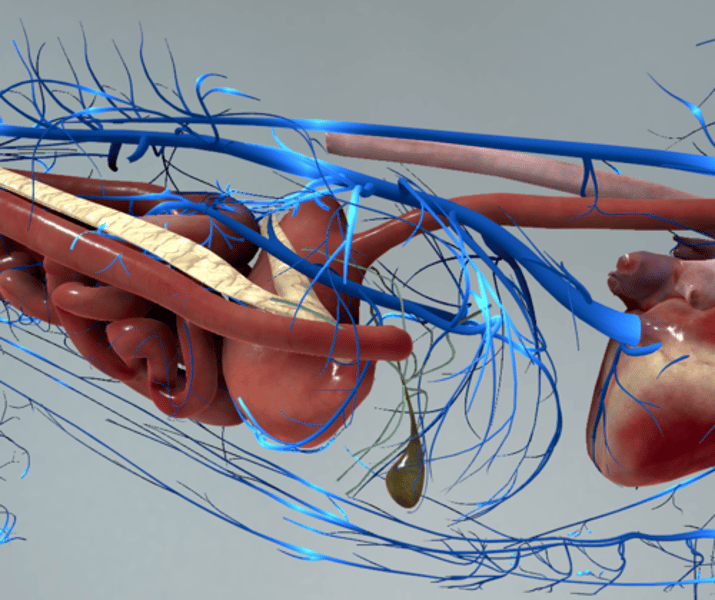
describe the vena portae.
v. portae: collects venous blood from the unpaired organs of abd. cavity (stomach, intestines, spleen and pancreas --> sends to liver.
It enters liver by porta hepatis and forms a dense network inside the liver parenchyma.
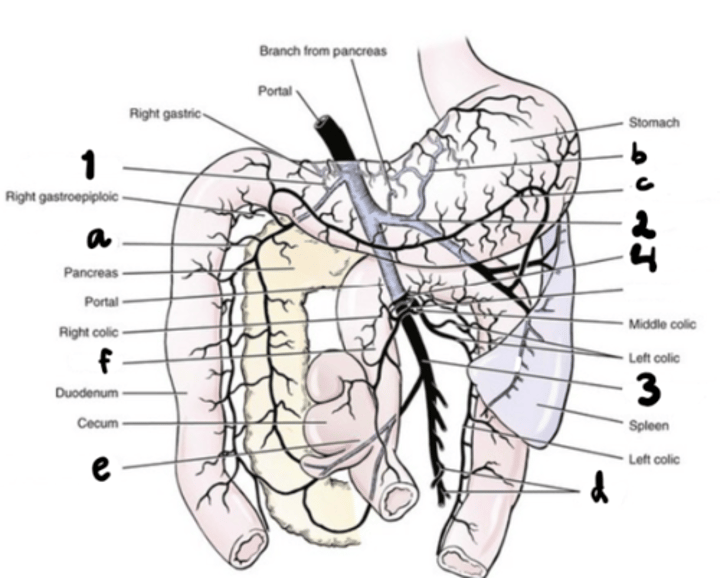
identify the vessels that forms the portal vein by fusion.
1. v. gastroduodenalis - blood from stomach, duodenum. From v. pancreaticoduodenalis cran.
2. v. lienalis - collects from spleen, forestomachs + of stomach, pancreas. Arises by multiple branches from spleen, gets blood from v. gastrica sinistra + v. gastroepiploica sinistra.
3. v. mesen. cranialis-Forms from junction of severeal vv. jejunales, gets blood from v. pancreaticoduodenalis cuad. + vv. ileocoliaca. joins nr. 4
4. v. mesen. caudalis - blood from colon. continues from cranial rectal v.
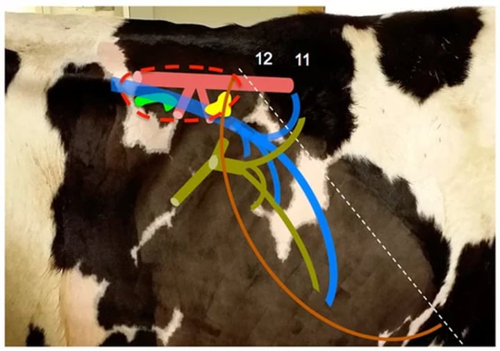
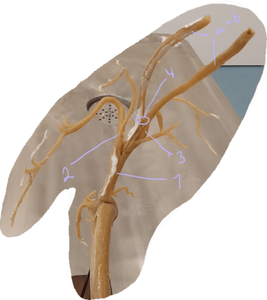
branches of a. carotis communis, identify j, k, m.
j: a. thyroidea caudalis - reaches the thyroid gland on caudal pole. - some differences.
2. muscular branches
k: a. thyroidea cranialis - reaches thyroid gland on cranial pole.
m: a. laryngea cranialis - supplies thorax in ru+su.
3. a. pharyngea ascendens - supplies palate, tonsils, pharynx RU. - around same location as laryngea.
4. a. palatina ascendens - palate, in small RU.
5. a. carotis interna et externa - terminal division at level of pharynx.
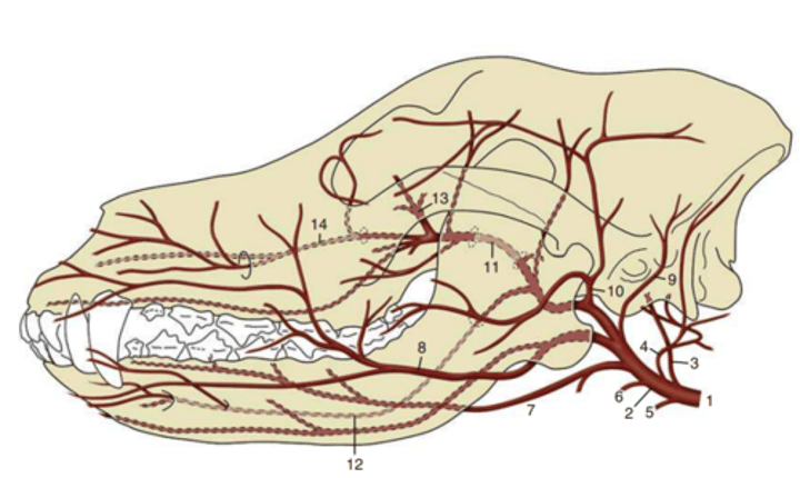
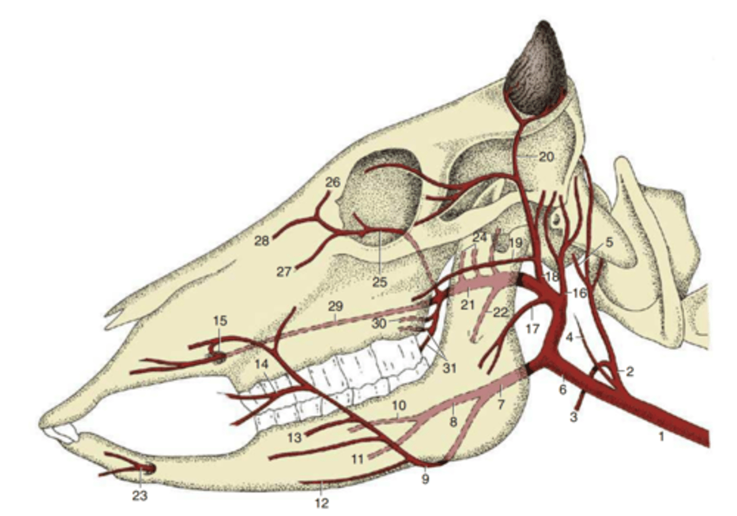
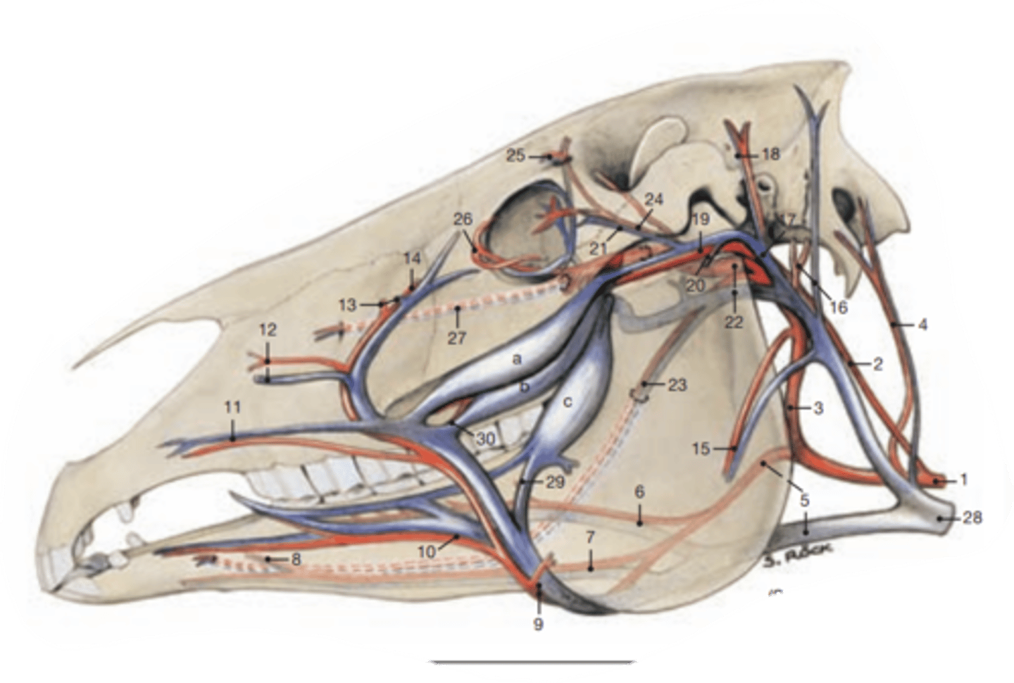
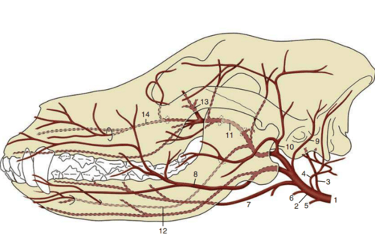
identify the branches of a. carotis externa.
4. a. occipitalis: directed into fossa condylaris ventralis, supplies muscles, ear, meninges.
7. a. lingualis: tongue,
bo+eq - arises in a truncus linguofacialis with a. facialis
8. a. facialis: runs medially of mandible, rostrally to the angle of mandible, bends. absent in ru. supplies surrounding place.
9. a. auricularis caudalis: ear + muscles.
10. a. temporalis superficialis: directed rostr. to m. acusticus externus into temporal region.
locate the a. maxillaris. and the branch arising from its mandibular part.
this artery is the continuation of the a. carotis externa towards the base of skull, after the origin of a. temporalis superficialis, its branches includes:
o: a. alveolaris inferior - arises from mandibular part. Goes through foramen mandibulare into canalis mandibularis, giving off branches supplying teeth. Leaves through foramen mentale, supplying the chin + lower lip.
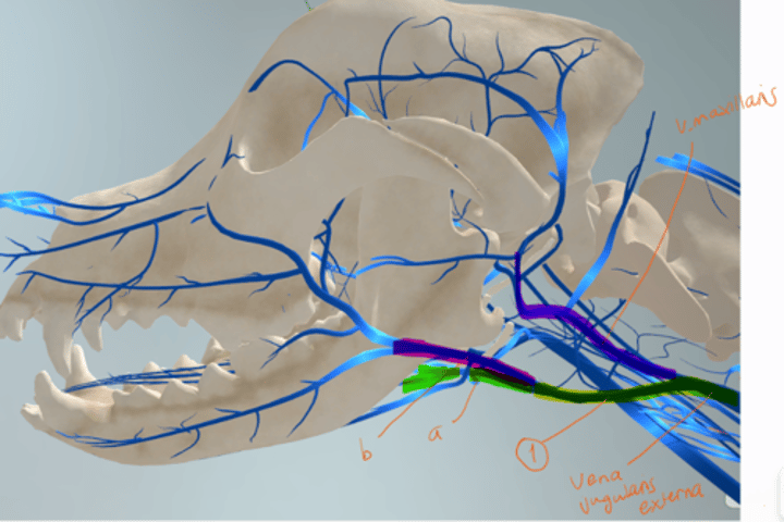
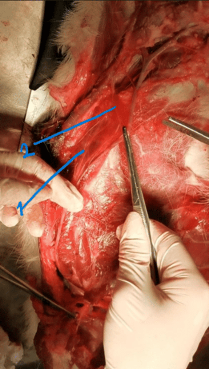
what is the main trunk (vein) that conveys the venous blood from the head and neck?
- V. jugularis externa - formed by fusion of v. linguofacialis and v. maxillaris. Located under the skin on lateral surface of neck in the jugular groove.
the branches of v. jugularis externa (5)
1. v. cervicalis superficialis - collects venous blood from the prescapular region and caudal region of neck.
2. v. cephalica - represents the superficial venous drainage system of thoracic limb
3. v. omobrachialis - present only in ca (dog), located before cephalic + cervi. superficialis veins, and goes around the muscle of limb.
4. v. thyroidea media - present only in small ru. located under the thyroidea cranialis in the middle of neck under.
5. v. thyroidea cranialis - together with a.
which vein arises by the fusion of v. lingualis and v. facialis? What are its branches?
1. vena linguofacialis - arises by the fusion. continues near the angle of mandible and along ventral border of parotid gland to the neck.
-This also fuses with v. maxillaris making the v. jugularis externa. Branches:
a: v. lingualis - sending venous blood to tongue
b: v. facialis - collects venous blood from oral, orbital and nasal region.
- vein of neck and head region.
sum up all the veins of head and neck.
1. v. jugularis externa with branches, v. cervicalis superficialis, v. cephalica, v. omobrachialis (ca), v. thyroidea media (small ru), v. thyroidea cranialis.
2. linguofacialis with branches, v. lingualis and v. facialis.
3. v. maxillaris with same branches as artery.
4. v. jugularis interna (car, su, bo, and some eq)
The arteries going from the aorta. Which species is this?
left side - ru/eq
1. truncus brachiocephalicus - continues cranially coming from arcus aortae. Supplies head, neck, thoracic limb + cranial region of thorax.
2. a. subclavia sinister - is first branch in ru + eq. (shown here)
3. a. subclavia dextra - 2nd branch
4. truncus bicaroticus - direct cranial continuation, divides into a. carotis communis dextri et sinistri. (a+b)
what is the difference on a. subclavia in carnivores and pigs (su) vs. ruminants and equine? And where does it go?
Ru+eq: a. subclavia sinistra is the first branch coming from the truncus brachiocephalicus, not the arcus itself.
Car+su: a. subclavia sinistra is an independent branch of arcus aortae.
location: winds around cranial border of first rib, entering thoracic limb through the axilla, where it changes into a. axillaris. It gives rise to branches with interspecies differences.
what branches does the arteria subclavia have? (6 main)
The branches of the a. subclavia differs in their origin between species.
1. a. vertebralis - (neck), form a. basilaris ventrally on brain stem. supplies spinal cord, meninges vertebrae + brain.
in Ru: arises from truncus costocervicalis.
car: arise independently from the subclavian arteries.
2. truncus costocervicalis - divides into a. intercostalis suprema (head of ribs) + a. cervicalis profunda (deep layers of muscles)
car: arise independently from the subclavian arteries.
Eq: on left side, the a. cervicalis profunda arises from a. subclavia directly.
3. a. scapularis dorsalis - supplies muscles of withers
4. a. cervicalis superficialis - runs cranially, supplies muscles ventrally + cranially on shoulder.
5. a. thoracica interna (runs dorsally on sternum-branches)
6. a. axillaris - continuation.
what arises from the truncus costocervicalis?
the truncus costocervicalis division depends on species, it can incl.
a. cervicalis profunda
scapularis dorsalis
intercostalis suprema (aka. vertebralis thoracica in dog)
vertebral arteries.
But generally, the truncus has:
A. intercostalis suprema (caudally) and the a. scapularis dorsalis (towards scapula and withers region).
In ruminants:
A. vertebralis and a. cervicalis profunda arises from the truncus.
In equine on the other hand, a. vertebralis and a. cervicalis profunda arises independently from the a. subclavia.

identify the branches of a. subclavia
Right side (Ru)
1. truncus brachiocephalicus
2. truncus costocervicalis- from a. subclavia, dorsally.
3. a. vertebralis
4. a. cervicalis profunda - divides with co.
5. a. subclavia dexter
6. a. axillaris
7. a. cervicalis superficialis - on the a. axillaris.
8. a. thoracica interna dexter-down, running on sternum to diaphragm - branches to sternum, muscles, diaphragm.
9. a. intercostalis suprema - running dorsally from costo, a. scapularis dorsalis comes here in this area running to the withers.
Identify this branch, it supplies muscles of the withers.
right side in ru.
1. arteria scapularis dorsalis - supplies muscles of the whiters. A branch of truncus costocervicalis (aka. on a. vertebralis) - but may differ in species. generally a branch of a. subclavia.
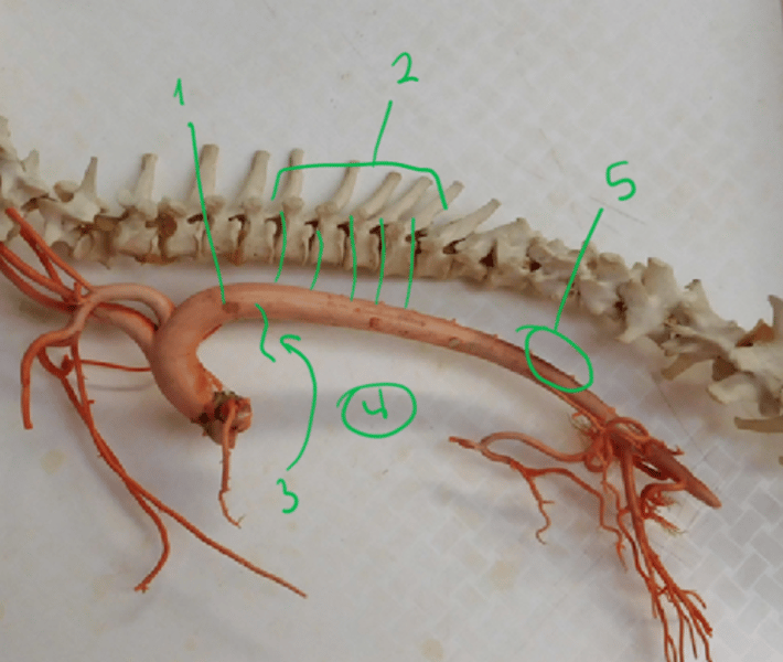
identify this structure (red) and its branches (orange).
1. aorta thoracica - this continues from the arcus aortae, then passes through the hiatus aorticus of the diaphragm. It gives off branches within the thoracic cavity:
2. a. intercostales dorsales: paired branches, dorsally of aorta. Runs into intercostal spaces, supplies muscles, vertebrae, spinal cord.
3. a. bronchoesophagea - supplies the esophageus and lungs.
4. inconstant branches - mediastinum, pericardium+esophageus.
5. a. costoabdominalis dorsalis - runs along caudal border of last rib.
which branch is only in horse from the aorta thoracica, supplying diaphragm?
w1: a. phrenica cranialis - only in horse, supplies the diaphragm.
what part of the descending aorta is caudal to the diaphragm? what are its branches?
1. aorta abdominalis - courses beneath vertebrae next to vena cava caudalis and stops at the aa. iliaca internae + the a. sacralis mediana, at the level of the last lumbar vertebrae.
- Its branches:
a. mesenterica cranialis - 2nd. unpaired, supplies intestine.
a. renalis - supplies kidneys, a. renalis dextri is a bit more cranially.
aa. adrenales mediae - located in middle part of adrenal glands, caudal to a. mesenterica cranialis. Supplies the adrenal glands. In su and car.
a. celiaca -1st.
a. mesenterica caudalis
gonadal arteries - aa. testiculares or aa. ovaricae.
aa. lumbales
aa. iliaca communes - a. iliaca interna and a. iliaca externa
a. phrenica caudalis - arising from phrenicoabdominal a.
paired. supplies diaphragm. Absent in Eq.
a. abdominalis cranialis - Arising also from phrenicoabdominal. artery in dog. Supplying adrenal glands and muscles of lumbar region. Absent in Eq.
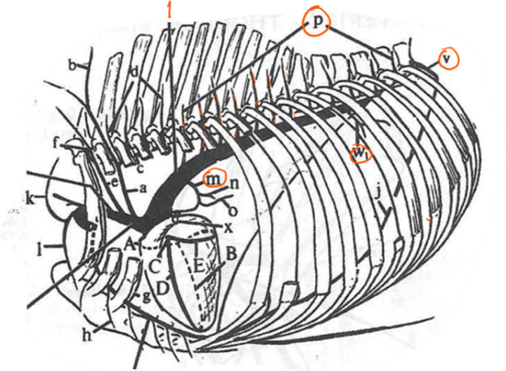
what are the typical branches of a. celiaca seen in ru?
f: a. lienalis is a branch of a. celiaca, going to spleen, pancreas + stomach.
in ru: the a. lienalis splits into: (k) a. ruminalis dextra, (i) a. ruminalis sinistra and the (j) a. reticularis.
a: a. gastrica sinistra - left surface of stomach.
in ru: gives off a. reticularis accessoria and (g) a. gastroepiploica sinistra.
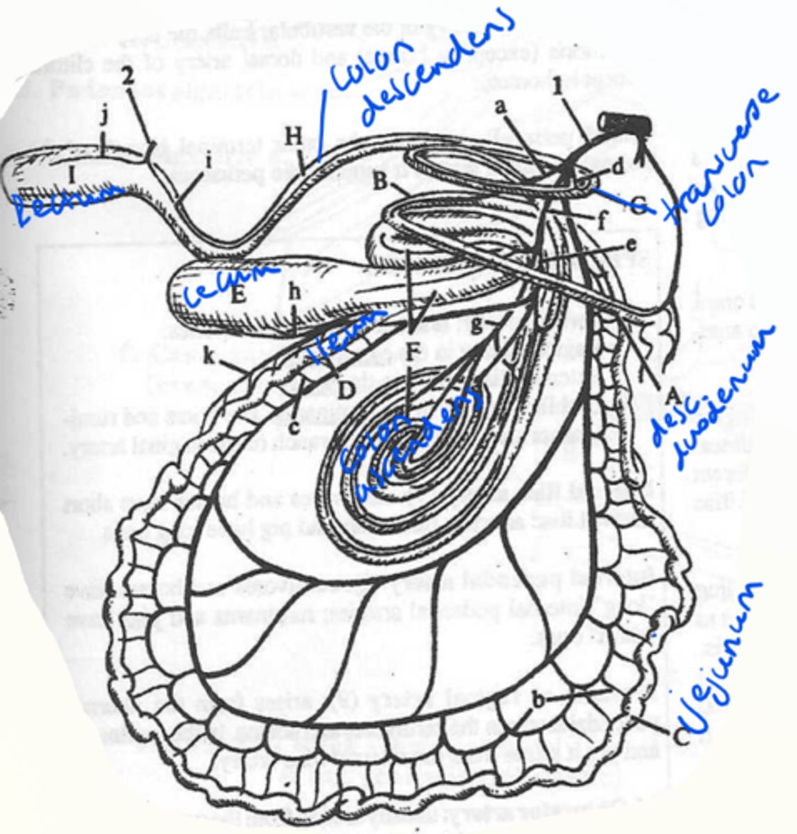
what are the branches of a. mesenterica cranialis? (cow - pic)
2nd unpaired, supplies the main part of intestine. largest visceral branch of abdominal aorta, arising caudal to a. celiaca.
a: pancreaticoduodenalis caudalis - supplies pancreas + duodenum
in ru: rami pancreatici (pancreatic branches also)
b: aa. jejunales
f: a. ileocolica - ileum, cecum + ascending colon
e: a. colica dextra - caudal part of asc. colon
d: a. colica media - transverse colon + cranial part of descending colon.
h: a. cecalis - supplies cecum + ileum. - shows branch in pic.
what does the aa. lumbales supply?
paired segmental branches, supplying lumbar part of spinal cord, abd. wall + lumbar region.
what is the direct continuation of aorta abdominalis in the end?
a. sacralis mediana - continuation, caudally on ventral surface of sacrum. It supplies the vertebral bodies and spinal cord.
what is the terminal division of aorta abdominalis?
quadrification of aorta:
2. 2x a. iliaca interna
3. 2x. a. ilaca externa
4. a. sacralis mediana
what is the main artery supplying head and neck?
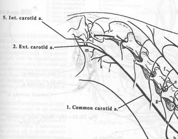
a. carotis communis dextra et sinistra. Located ventrolaterally to the trachea. It has many branches.
which artery is number 3?
a. palatina ascendens - palate, in small RU. A branch of a. carotis communis.
which arteries are these coming from a. carotis communis?
1. a. thyroidea caudalis
2. a. thyroidea cranialis
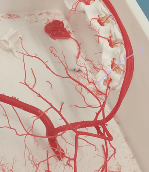
locate the a. carotis interna.
(2) a. carotis interna is the thinner branch coming from the (1) a. carotis communis in the place of its terminal division. It reaches the cranial cavity through the foramen lacerum.
Ru: rudimentary.
helps on the formation of the circulus arteriosus cerebri (arterial circle of brain) on ventral surface of brain, thus supplies the brain. In ru+su this blood supply is ensured by branches of a. maxillaris.
locate the a. carotis externa.
(2) a. carotis externa - largest branch from a. carotis communis. It bends dorsally in a curve and then continues along the border of r. mandibulae. It curves rostrally and continues as the (11) a. maxillaris on medial surface of mandible. It gives of several branches.
what are the branches of a. temporalis superficialis?
a. temporalis superficialis - branch of a. carotis communis.
branches:
1. transversa faciei - ventr. to arcus zygomaticus. Replaces a. facialis in small ru.
2. a. auricularis rostralis - ramifies around the auricle.
identify the branches.
1. a. occipitalis
2. a. lingualis
3. a. auricularis caudalis
4. a. transversa faciei - before it, should have a. facialis but not in small ru as this is together with a. transversa faciei in ru.
5. a. auricularis rostralis
6. a. temporalis superficialis
what are the other branches of a.maxillaris except for the a. alveolaris inferior?
1. muscular branches
2. aa. temporales profundae - temporal region
3. a. buccalis - buccinator muscle + buccal glands.
4. a. opthalmica externa - eyeball + accessory organs of eye.
5. a. infraorbitalis - thorugh foramen maxillare into canalis infraorbitalis, where it supplies upper teeth with its branches.
6. a. malaris - ventrally of orbit, reaching medial angle of eye.
7. a. palatina descendens - directed rostroventrally + supplies hard/soft palate.
locate v. maxillaris, what are its branches?
v. maxillaris - same position as artery. Perforates the parotid gland and the branches correspond to the branches of a. carotis externa and a. maxillaris.
v. auricularis caudalis, v. temporalis superficialis, v. auricularis rostralis, v. transversa facialis etc.
what vein runs with the a. carotis communis in a deeper position of the neck?
V. jugularis interna - present in car, su, bo and some eq.
- empties into the caudal part of v. jugularis externa.
- Collects venous blood from the thyroid gland, pharynx, tongue and partly from the brain.
identify the branches.
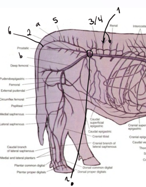
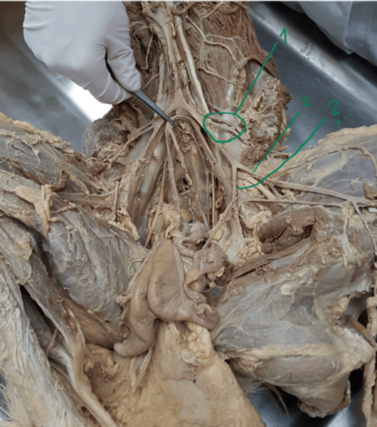
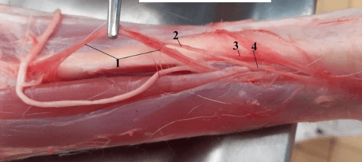
1. a. femoralis - distal continuation of a. iliaca externa.
2. a. circumflexa femoris lateralis - supplies m. quadriceps femoris.
3. a. saphena - running with nerve + vein, located on medial surface of crus going to plantar surfaces of digits.
4. genus descendens - directed to medial surface of knee.
5. a. nutricia ossis femoris - distally to the previous branch and enters femur.
6. a. femoris caudalis - caudally, supplies popliteal region muscle, going into back.
venous blood from the pelvic cavity and pelvic limb is collected by means of? what are its tributaries/branches?
vv. iliacae communes. (right and left common iliac veins) - empty into v. cava caudalis. Each is formed by the fusion of v. iliaca interna (a) et externa (b)
tributaries are:
1. vv. lumbales - blood from level of last lumbar vertebrae.
2. v. circumflexa ilium profunda - with artery.
3. v. testicularis sinistra - in bull+ram
4. v. ovarica sinistra - in cow/sheep
5. v. sacralis mediana - from sacrum
6. v. caudalis mediana - from tail, absent in eq.
what are the tributaries/branches of v. iliaca interna?
1. v. iliolumbalis - with a.
2. v. obturatoria
3. v. prostatica - absent in Eq
4. v. vaginalis - absent in Eq.
5. v. glutea cranialis - collects blood from gluteal region.
6. v. glutea caudalis - collects from caudal part of gluteal r.
7. v. pudenda interna - collects blood from perineal region, penis + clitoris.
what are the branches of v. iliaca externa?
v. iliaca externa accompanies artery on its caudal surface. Has the same tributaries as the artery. - v.circumflexa ilium profunda for ex.
- its terminal fusion is formed by v. profunda femoris & v. femoralis.
- collecting area + tributaries of v. profunda femoris corresponds to a. it collects blood from the plantar and dorsal surfaces of digits.
what are the tributaries/branches of v. femoralis?
v. femoralis - accompanies artery.
1. v. circumflexa ilium superficialis (only in dog)
2. v. saphena medialis - collects venous blood from plantar surfaces of digits.
3. v. genus descendens - acc. artery.
4. vv. femorales caudales.
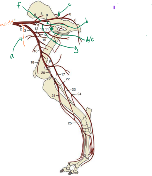
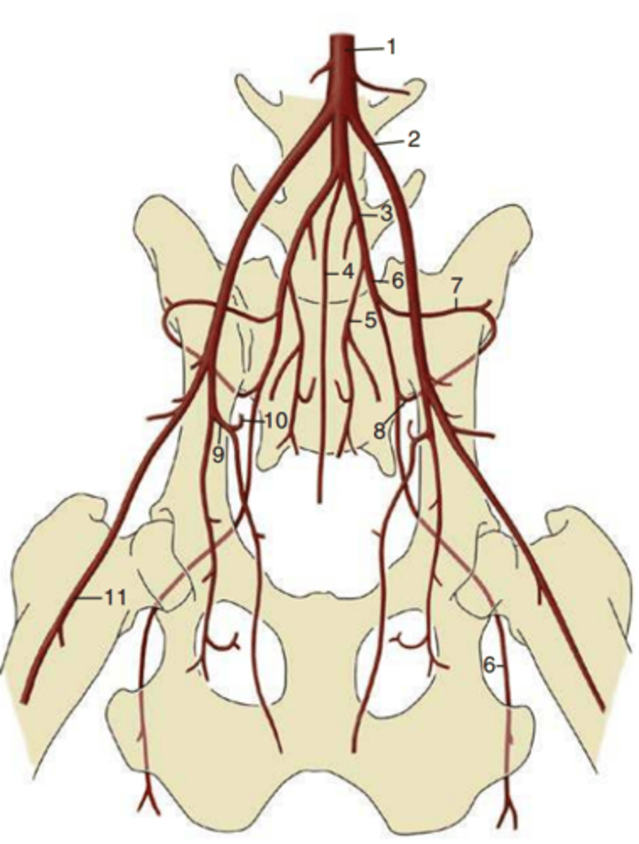
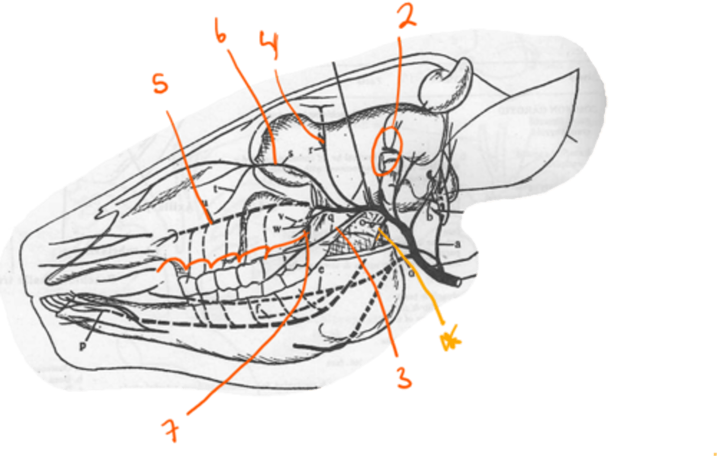
what artery runs caudally on medial surface of wing of ilium and body of ilium (1)? And what are its branches?
1. a. iliaca interna - branches:
a: a. umbilicalis - blood supply of cranial p. of urinary b, deferent duct, ureter + uterus.
b: iliolumbalis - laterally on wing of ilium. supplies inner lumbar n. + thigh m.
c: a. glutea cranialis - supplies deep gluteal muscle after leaving pelvic caivty.
d: a. prostatica - supplies accessory genital gl. + urethra, ureter, urinary b. + def. duct.
e: a. vaginalis - supplies uterus.
f: a. glutea caudalis - thigh m.
g: a. pudenda interna - s. penis, clitoris
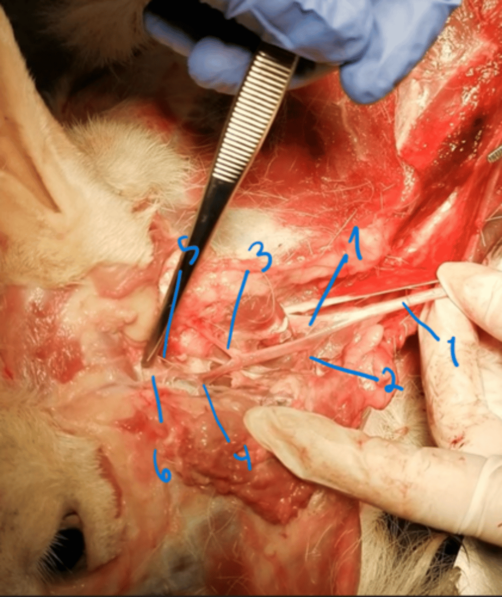
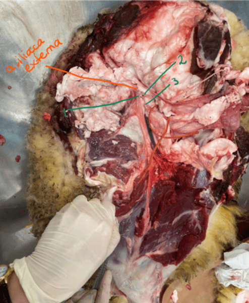
locate the arteria iliaca externa et interna.
1. a. iliaca externa - the big ones at the sides (most external)
2. a. iliaca interna - the smaller ones in the middle (most internal)
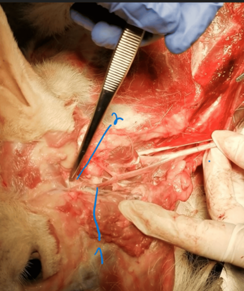
explain the branches that can be seen in this picture from a. iliaca interna.
1. a. umbilicalis - first branch coming from it, going to the uterus in female, blood supply of cranial part of urinary b, deferent duct and ureter too.
2. a. iliolumbalis - are the small "strings" going into the back bone, behind the main branch. Running laterally on wing of ilium, supplies inner lumbar n. + thigh m.
3. a. glutea cranialis - after continuation-1st branch, supply deep gluteal m. after leaving pelvic c.
then:a. prostatica/vaginalis, glutea caudalis + pudenda interna go deeper.
what is the branches of a. iliaca externa and what does it divide itself into?
this artery runs over abdominal roof to pubis. branches:
1. a. circumflexa ilium profunda - directed cranially to tuber coxae, supplies muscles of abdomen.
2. a. cremasterica/uterina - only in eq, supplies scrotum/uterus.
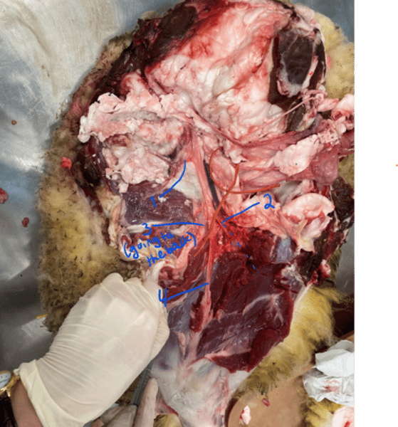
- a. iliaca externa divides itself at the level of pubis into a. profunda femoris (the internal one) and a. femoralis (most external, goes out to limb)
explain how the a. profunda femoris is located. What are its branches?
a. profunda femoris runs under pubis and supplies medial + laterocaudal group of thigh muscles, and organs of regio pubica. branches:
1. truncus pudendoepigastricus - supplies abdominal m., external male organs + mammary gl.
2. a. obturatoria - directed to foramen obturatum, should go into the back.
3. a. circumflexa femoris medialis - direct continuation, supplies medial + laterocaudal group of thigh muscles.
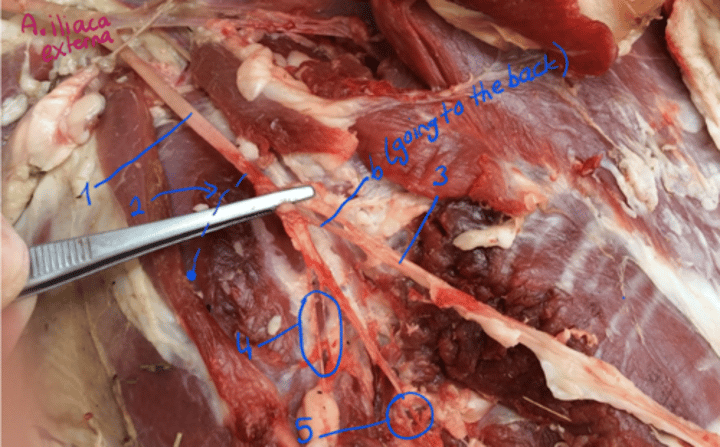
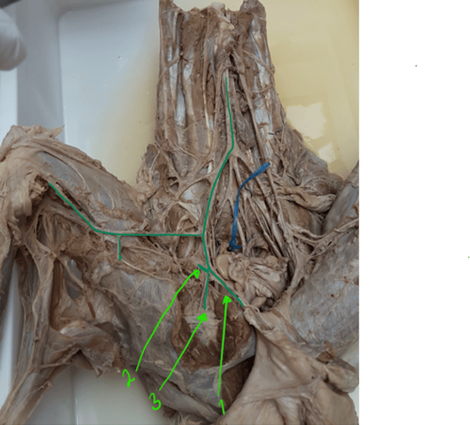
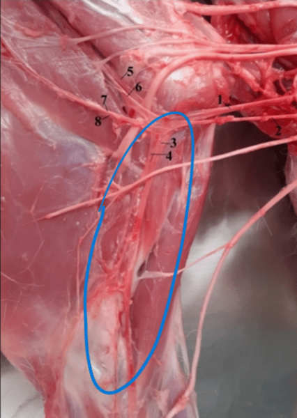
what are these arteries? (seen from lateral view)
a. iliaca externa - dividing into a. femoralis profunda going to the right (2 and 3) - where 2 is truncus pudendoepigastricus and then the second branch is a. femoralis - going down the leg (1)
identify these branches of a. femoralis (lateral view) class picture.
1. a. ilium profunda - not in book but mentioned in class.
2. a. saphena - runs together with nerve and vein of same vein, in caudodistal direction. Located on medial surface of crus and extends to the plantar surface of digits.
3. a. circumflexa femoralis lateralis - origin at same level as the a. profunda femoralis, supplies the m. quadriceps femoris
identify the branches of a. femoralis. (ventral view)
1. a. femoralis
2. circumflexa femoris lateralis - should be here, at same lecel as the a. profunda femoralis. supplies m. quadriceps femoris.
3. femoris caudalis - directed caudally.
4. a. genus descendens - to medial surface of knee.
5. a. saphena - running on medial surface of crus, extending to plantar surfaces of digits.
identify these branches of a. femoralis.
1. a. genus descendens
2. a. saphena
3. a. femoris caudalis
- the continuation into the knee should be the a. poplitea.
identify artery which is the direct continuation of a. femoralis. What does it divide into?
a. poplitea - after origin of a. femoralis caud. Runs between the heads of m. gastrocnemius and supplies the surrounding muscles and joint capsule of the stifle joint. It divides distally to the proximal epiphysis of tibia into the:
1: a. tibialis cranialis - passes cranial surface of tibia as direct continuation.
- from lvl. tarsal joint, it becomes a. dorsalis pedis - distally on dorsal surface of metatarsal r., supply digits.
2: a. tibialis caudalis - runs on caudal surface of tibia.
which vein is formed by the fusion of v. tibialis cranialis et caudalis?
v. poplitea which empties into the v. femoralis in the area of stifle joint.
v. tibialis cranialis receives the v. dorsalis pedis at the level of tarsal joint.
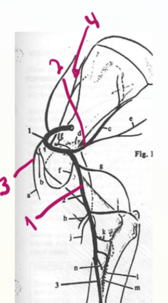
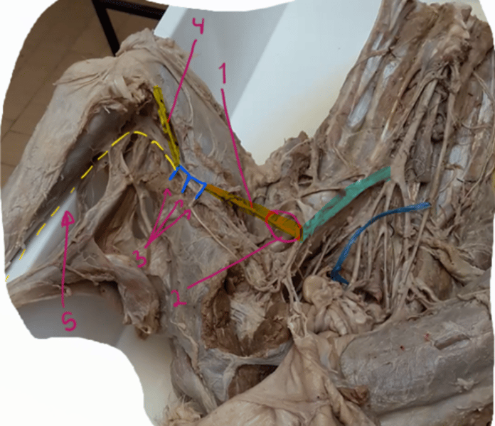
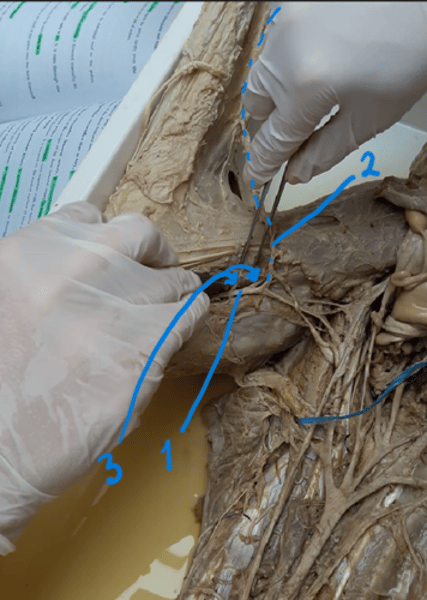
what is the main artery that supplies the thoracic limb? and what does it divide into + its branches.
A. axillaris, which is the direct continuation of a. subclavia. It runs distally over the medial surface of the arm. at flexor aspect of shoulder joint, it divides into:
1. a. subscapularis - dorsally
2. a. brachialis - distally
branches:
3. a. thoracica externa - supplies pectoral m.
4. a. suprascapularis - running along cranial margin of scapula, passes between m. subscapularis and m. supraspinatus, supplying them.
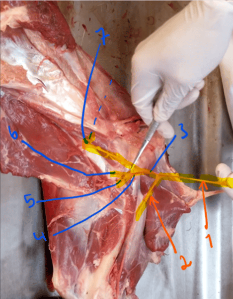
identify these branches.
1. a. axillaris
2. a. brachialis
3. a. subscapularis - directed dorsally along caudal margin of scapula. Branches are:
4. a. circumflexa humeri caudalis - turns on lateral surface of shoulder joint, supplies m. triceps brachii.
5. a. thoracodorsalis - supplies m. latissimus dorsi
6. a. collateralis radialis - distal direction and supplies the craniolateral muscles of forearm + distal surfaces of digits.
7. a. circumflexa scapulae - turns lat. supplies lateral muscles of shoulder.
which artery is the distal continuation of the a. axillaris from the shoulder joint, going straight to the elbow joint, crossing the humerus medially?
A. brachialis. Branches:
1. a. circumflexa humeri cranialis - level of collum humeri. Supplies shoulder joint + mm.
2. a. profunda brachii - caudally directed, supp. m. triceps brachii.
3. a. collateralis ulnaris - arises prox. to the medial surface of distal epiphysis of humerus. runs along to carpal joint.
4. a. brachialis superficialis - in car.
5. a. bicipitalis - m. biceps brachii.
6. a. transversa cubiti - elbow joint.
7. a. interossea communis - enters space bw. radius + ulna.
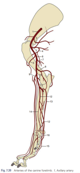
which artery of a. brachialis is present only in carnivores?
9. a. brachialis superficialis - present in car and is from the level of the elbow joint. it passes to the cranial border of the forearm.
8. a. collateralis ulnaris, 10. a. transversa cubiti, 11. a. interossea communis.
what does the a. interossea communis divide into?
- branch of a. brachialis. The a. interossea communis arises distally to the elbow joint and enters the space between radius and ulna. It curves distally and divides into the:
1. a. interossea cranialis
2. a. interossea caudalis
- supplies the wrist and palmar surfaces of digits.
what is the distal continuation of the a. brachialis after the arising of a. interossea communis?
1. a. interossea communis is shown.
2. a. mediana - the distal continuation, largest of forearm and continues over the flexor aspect of carpal joint to metacarpal region, helps on blood supply of palmar surfaces of digits.
3. a. profunda antebrachii - flexors on laterocaudal surface.
4. a. radialis - in middle part, runs to radius, supplies carpal, metacarpal region, palmar surfaces of digits + dorsal surfaces. It divides into r. dorsalis et. palmaris.
- a. radialis proximalis - only in Eq.
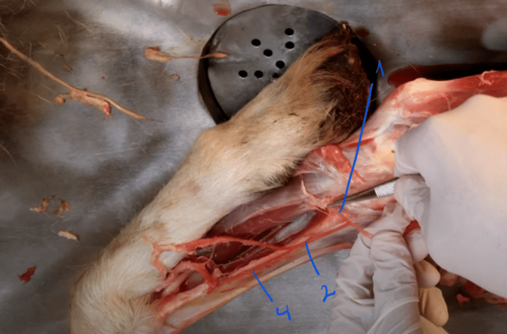
what branches do we see here in the region of radius?
1. a. mediana
2. a. radialis
3. r. dorsalis of a. radialis
4. r. palmaris of a. radialis
which arteries are clinically important in the aspect of pulse palpation?
a. mediana - eq
a. brachialis - bo
what are the 2 venous drainage systems of thoracic limb?
1. superficial system - formed by v. cephalica emptying into the v. jugularis
2. deep system - represented by the v. axillaris.
what is the main vein collecting venous blood from the thoracic limb into the v. subclavia?
V. axillaris - lying medially and ventrally to the artery.
Its tributaries are:
1. v. thoracica externa - with a, collecting blood from lateral thoracic wall.
2. v. subscapularis - with a.
3. v. thoracodorsalis - with a.
4. v. circumflexa humeri cranialis - absent in Ru.
what are the tributaries of the vein typically having this position? located caudally to the arteria brachialis.
V. brachialis - empties into the v. axillaris.
tributaries:
1. v. profunda brachii - runs with a. in muscles caudal to humerus.
2. v. bicipitalis - absent in Ru + Eq, arises in m. biceps brachii, empties in v. brachialis.
3. v. mediana cubiti - forms connection bw. v. brachialis et. cephalica.
4. v. collateralis ulnaris - runs with a.
5. v. interossea communis - same branches as a.
what is the tributaries to the v. mediana?
V. mediana empties into the v. brachialis. tributaries:
1. v. profunda antebrachii - collects blood from the flexors of carpal joint + digital joints.
2. v. radialis - collects blood from distal part of thoracic limb.
what vein represents the superficial venous drainage system of thoracic limb?
V. cephalica - connected with v. brachialis by means of v. mediana cubiti. It empties into the v. jugularis externa.
- blood sampling is done on v. cephalica in dogs and cats.
summary of all main arteries of thoracic limb.
1. a. axillaris
2. a. brachialis + a. subscapularis
3. a. circumflexa humeri cranialis - 1st division on brachialis.
4. a. circumflexa scapulae
5. a. circumflexa caudalis - gives off a. thoracodorsalis (may be as an individual branch) and a. collateralis radialis.
6. a. profunda brachii
7. a. collateralis ulnaris
8. a. bicipitalis
9. a. transversa cubiti
10. a. interossea communis
11. a. profunda antebrachii
12. a. radialis with its r. dorsalis et. ventralis.
What is the brain of vertebrates generally divided into?
1. Prosencephalon (presents most rostrally located part of prain, divided into telencephalon + diencephalon)
2. Mesencephalon (undivided)
3. Rhombencephalon (divided into metencephalon + myelencephalon)
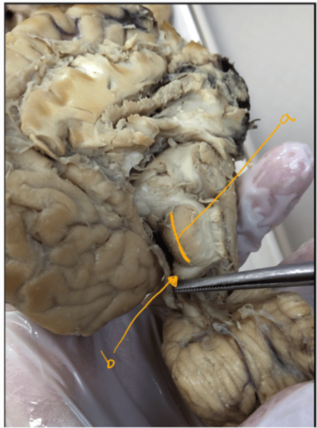
identify these structures of thalamencephalon.
- thalamencephalon of diencephalon (prosencephalon)
1. Metathalamus- lies laterally to the thalamus. Consist of: a) corpus geniculatum laterale, b) corpus geniculatum mediale - lies caudoventrally to the lateral one.
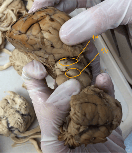
identify these structures of mesencephalon.
Shows a better image of the 2 pairs of eminences of tectum mesencephali (roof of midbrain)
a: colliculi rostrales (rostral colliculi) - higher rostral colliculi are in connection with the corpus geniculatum laterale.
b) colluculi caudales (caudal colliculi) - lower caudal colliculi are in connection with corpus geniculatum mediale.
How is the brain supplied with blood?
It is supplied by branches which originate from a. carotis interna, a. occipitalis, a. maxillaris and a. vertebralis.
what is the main function of the nervous system? What is it divided into?
- give the connection between the organism and the environment.
- regulates the functions of all organs in the living organism.
divided into:
1. central nervous system (Nervosum centrale) - brain + spinal cord
2. peripheral nervous system (nervosum periphericum) - cranial nerves, spinal nerves and the autonomic system.