Looks like no one added any tags here yet for you.
What is a nervous system required to do?
Produce effective responses to a stimulus from the environment.
Environmental factors
Can be either external or internal
External stimuli
Light
Temperature
Chemical
Touch
Vibration
Internal stimuli
Chemical (pH, ions, molecules)
Blood pressure
Temperature
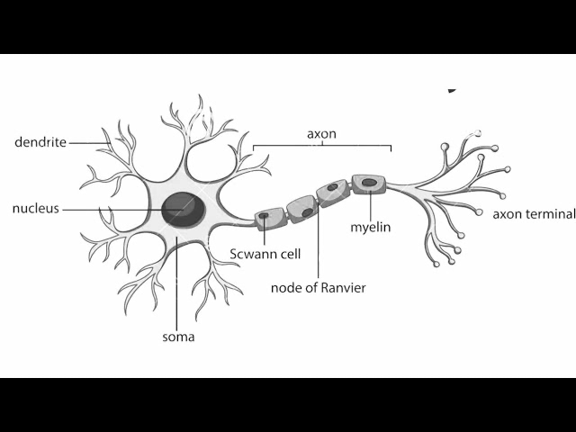
Neuron/neurone
basic cell types of nervous system
How can the parts of the neuron vary?
Cell body (soma) – size and shape
Dendrites – number, branching and length
Axon – length, diameter, branching, myelinated or unmyelinated
Synaptic terminals – number and structure
Synaptic transmission – chemical or electric
Variability in the morphology of neurones
different shapes and sizes of neurons in the nervous system
The structure of a neurone directly reflects its function
Interneurones
Located between neurones and form a connection between other neurones
Found in both invertebrate and vertebrate nervous systems
May be local or send their axons for long distances within a nervous system (projection interneurones)
Increase the number of synapses - and therefore the complexity of neuronal circuits
Diagram of neuron
Hydra
Hydra – an example of a freshwater invertebrate
A simple nerve net with no central nervous system.
Permits movement of the body and tentacles in water.
Action potentials (AP) can be conducted in all directions (AP conduction is bidirectional).
Sea anemone and corals - invertebrate nervous system
Slow but coordinated movement of polyps
Tentacles to catch prey
Body movements for defence
Tentacles/oral disc 4000x more sensitive than the ‘column’
Jellyfish - invertebrate nervous system
More complex nerve nets
Spontaneous rhythmic activity (slow state and startle)
Contractions of the margin of the ‘bell’ produce a propulsive force forwards
Starfish - invertebrate nervous system
A modified nerve net with control of limb movements coordinated by the neural ring
The radial nerves can control the movements of each limb individually
Movement and feeding = complex movements
What is bilateral symmetry?
Bilateral symmetry is when an organism has a mirror image on both sides, allowing for directional movement.
How does bilateral symmetry affect movement?
It allows for forward movement, with a head-first direction for better coordination.
What is cephalization?
Cephalization is the concentration of sensory organs and nervous tissue at the front of the body, forming a head.
Cosenqueces of cephalisation
1.Increase in number of nerve cells.
2.Concentration of nerve cells into ganglia; ganglia into brains, nerves into nerve cords.
3. Development of functional speciality:
AFFERENT neurons – towards the CNS.
EFFERENT neurons – away from CNS.
4. Localisation of specific functions in different parts of the
nervous system.
5. Development of interneurones and more complex
synaptic contacts.
6. Development of head bearing sense organs.
7. Development of a ventral nerve cord.
What is a segment?
A segment is a unit of anatomical structure that can be
repeated along the length of an animal
How does cephalization help survival?
It allows for faster processing of sensory information and better responses to the environment.
What is the central nervous system (CNS)?
The CNS consists of the brain and nerve cord, allowing for more complex coordination and behavior.
How did bilateral symmetry and cephalization affect behavior?
They allowed for complex behaviors like hunting and social interaction due to improved sensory and motor control.
Consequences of segmentation
1.Development of segmental ganglia with sufficiently complex
neural circuitry to control locomotion in individual segments
2. Coordination of movement (and/or limb movement) between
adjacent segments
e.g. in annelid worms or earth worms
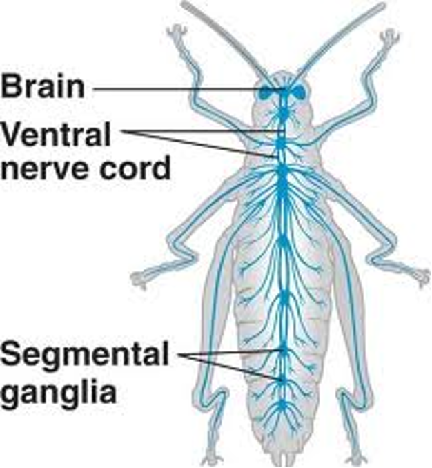
Arthopods - invertebrates
Arthropods have connectives = ganglia joined by connecting nerves
Arthropods also have an autonomic nervous system (ANS) which innervates the viscera of the body.
What type of nervous system do arthropods have?
Arthropods have a centralized nervous system (CNS) consisting of a brain and a ventral nerve cord.
How does coordinated movement occur in arthropods?
•Receiving sensory information from a defined part of a body segment whose activity it regulates directly.
•Activating dorsal/ventral or left/right limb muscles appropriately in response to stimuli.
•Using central pattern generators (CPGs) – repeated rhythmic motor output independent of sensory stimulation.
•Interconnections between segmental ganglia (connectives) can propagate activity along the length of the ventral nerve cord – and along the length of the animal – coordinated by the ‘brains’.
How is the molluscan nervous system organized?
The molluscan nervous system is organized into ganglia:
Buccal – feeding
Cerebral – coordination
Pleural – respiration
Pedal – movement
Parietal – peripheral functions
Visceral – organ regulation
What are ganglia in the nervous system?
Ganglia are clusters of nerve cell bodies
What can octopi do?
Exhibit ‘human-like’ intelligence (when observed in captivity):
•Gets food, clears the front of its den and arranges rocks in order to cover the entrance before going to sleep (foresight, planning, use of tools).
•Opens childproof caps on pill bottles (persistence, thinking).
•Blowing jets of water from the funnel to send a pill bottle to the other end of the tank where the water flow sends it back - repeatedly (playing).
•Recognise their human ‘caretakers’ by moving towards them and squirting water at them (memory, affection).
•Solving difficult problems using objects of differing colours and shapes (thinking).
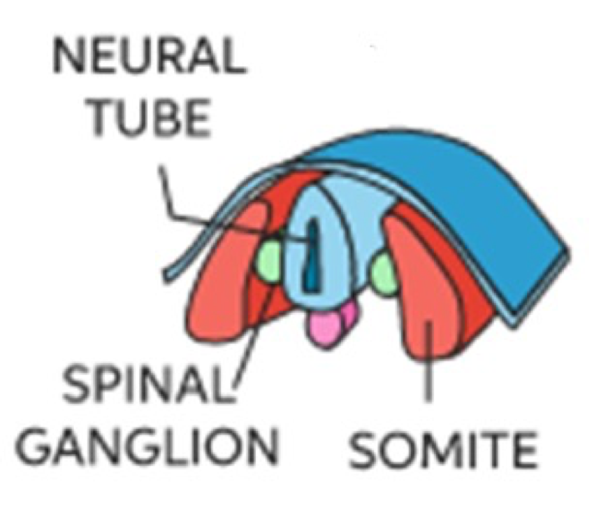
What does the neural tube develop into?
The entire central nervous system (CNS) (brain + spinal cord).
Diagram of neural tube
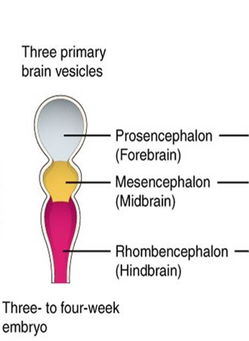
What are the three primary brain vesicles?
Prosencephalon (forebrain), Mesencephalon (midbrain), Rhombencephalon (hindbrain).
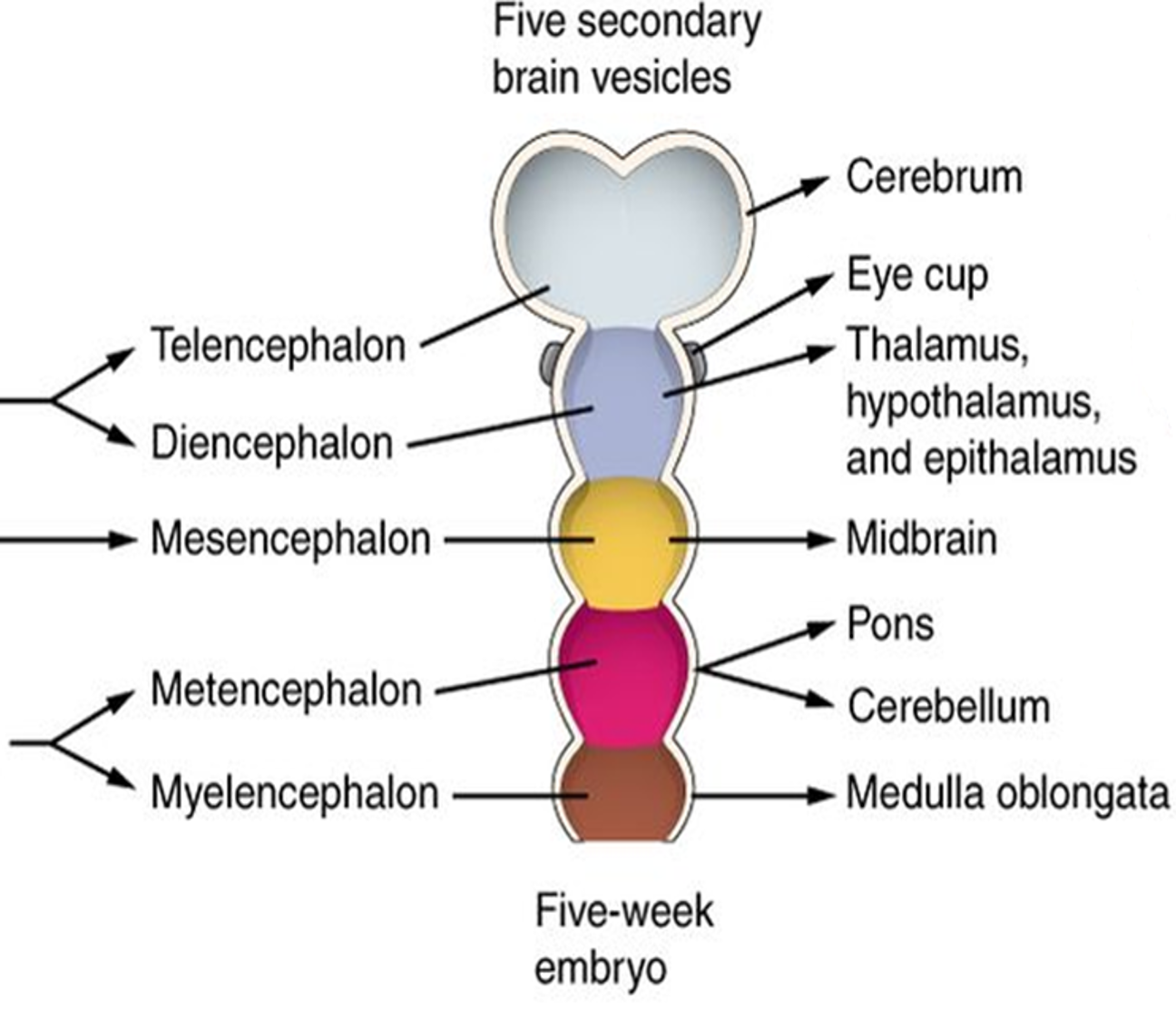
What are the five secondary brain vesicles?
Telencephalon, Diencephalon, Mesencephalon, Metencephalon, Myelencephalon
Which secondary vesicles develop from the prosencephalon (forebrain)?
Telencephalon and Diencephalon
Which secondary vesicle develops from the mesencephalon (midbrain)?
Mesencephalon (remains unchanged)
Which secondary vesicles develop from the rhombencephalon (hindbrain)?
Metencephalon and Myelencephalon
What does the telencephalon become?
Cerebrum
What does the diencephalon become?
Thalamus, hypothalamus, epithalamus, and eye cup
What does the mesencephalon become?
Midbrain
What does the metencephalon become?
Pons and cerebellum
What does the myelencephalon become?
Medulla oblongata
Which brain region is enlarged in lower vertebrates?
Olfactory bulbs due to reliance on smell. It helps detect food, predators, and mates.
What happens to the size of the cerebrum as vertebrates evolve?
It increases, allowing for more complex behaviors and cognitive functions.
What is the main function of the cerebrum in lower vertebrates?
Instinctive behaviors and motor control.
How many layers does the cerebral cortex have in lower vertebrates?
Three layers
How does the cerebral cortex in lower vertebrates compare to that in mammals?
Simpler (three layers in lower vertebrates vs. six layers in mammals).
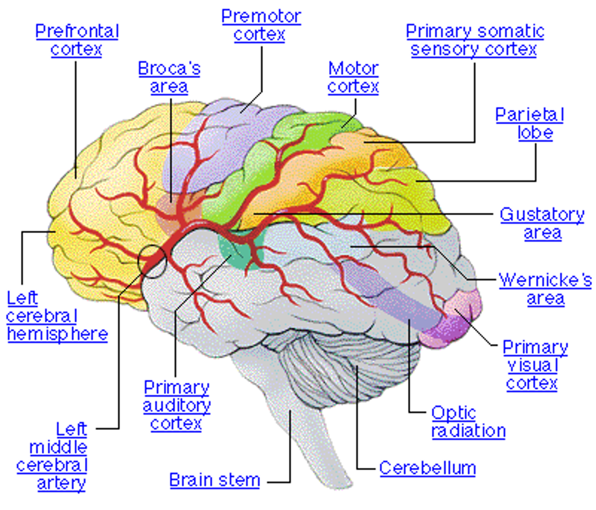
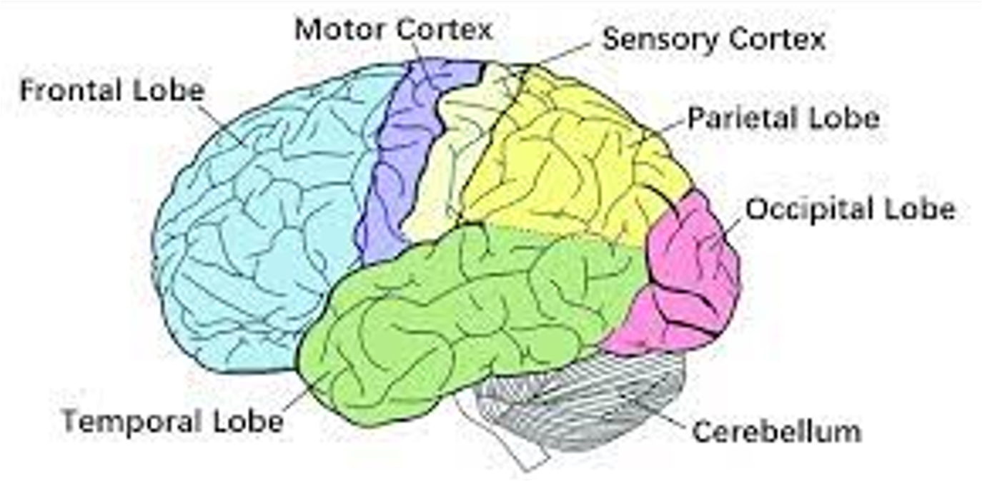
Key features of mammalian brains
•Folding of the cortex (gyri & sulci - cerebral cortex only found in higher vertebrates)
•Development of six layered neocortex
•Enlargement of the cerebellum
•Reduction of the olfactory system (especially in primates)
key developments of the human brain
•Development of frontal cortex
•Enlargement of cortical areas involved with unique human features:
manual dexterity
speech
facial expression
What happened to Phineas Gage, and how did it affect him?
A railway accident drove an iron rod through his frontal lobe. He survived, but his personality changed, making him unreliable, impulsive, and prone to inappropriate behavior.
What is cortical evolution in mammals?
Cortical evolution in mammals refers to the development and expansion of the cerebral cortex, particularly the neocortex, which is responsible for higher cognitive functions like reasoning, sensory processing, and motor control. This evolution has led to increased brain complexity, such as cortical folding, and is key in supporting sophisticated behaviors and cognitive abilities.
What are the key aspects of cortical evolution in mammals?
Cortical expansion: Increased size and complexity of the cortex, particularly in primates and cetaceans.
Neocortex development: Involved in higher-order functions like reasoning, language, and perception.
Cortical folding: More folds (sulci and gyri) increase surface area for neurons.
Layering: Six layers of the cortex support sophisticated processing.
Genetic and molecular factors: Genes influence brain size and organization.
Plasticity: Adaptability of the cortex based on experience and environment.
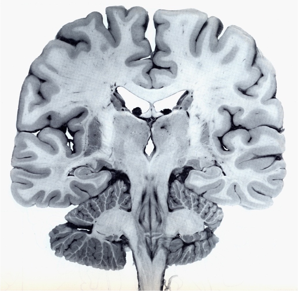
What is white matter and grey matter?
White matter: Composed of myelinated axons, which are responsible for transmitting signals between different parts of the nervous system. The myelin gives it a white appearance.
Grey matter: Contains collections of cell bodies, dendrites, and unmyelinated axons. It is where processing and integration of information take place in the brain and spinal cord.
What is the molecular structure of grey matter?
Neurons: Cell bodies contain the nucleus, rough endoplasmic reticulum, and Golgi apparatus for protein synthesis.
Synapses: Neurotransmitters like glutamate, GABA, and dopamine are released to transmit signals. Receptors (e.g., NMDA, AMPA) mediate synaptic communication.
Glial Cells: Astrocytes maintain the blood-brain barrier, microglia provide immune functions, and ependymal cellsproduce cerebrospinal fluid (CSF).
What is the molecular structure of white matter?
bundles of myelinated axons form tracts connecting nuclei
What are the layers (laminae) of the cortex and their characteristics?
The cortex is organized into layers (laminae), with six layers in most mammals, but some primitive three-layered areas remain (e.g., olfactory cortex).
Individual layers are characterized by:
The types of neurons they contain (e.g., pyramidal neurons, stellate neurons).
Their connections:
Afferent: Incoming connections (e.g., from the thalamus).
Efferent: Outgoing connections (e.g., to other cortical areas or spinal cord).
Intracortical: Communication between layers within the same cortex.
Three examples of the evolution of function of the vertebrate CNS
1.Swimming in fish
2. Walking on land by amphibians and reptiles
3. Birdsong
How do lampreys swim?
Lampreys swim using undulatory movements of their body, moving forwards and backwards.
What are central pattern generators (CPGs) and how do they function in lampreys?
CPGs are networks of neurons that produce rhythmic behaviors like swimming.
In lampreys, CPGs are located on both sides of the spinal cord in every segment.
Each side of the spinal cord generates the basic rhythmic drive for locomotion.
How do the left and right sides of the spinal cord coordinate in lampreys?
Connections between the left and right sides of the spinal cord ensure coordination of movement during swimming.
What controls the central pattern generators (CPGs) in lampreys?
The brainstem locomotor command centres control the CPGs.
These command centres are regulated by the basal ganglia (nuclei) in the cerebral hemisphere, which help manage locomotion.
What is the function of Mauthner neurones (M-neurones) in fish?
M-neurones mediate the fast escape or startle response in fish.
They detect vibrations from sensory input and trigger rapid movement for escape.
What are the characteristics of Mauthner neurones in fish and amphibians?
Most fish and amphibians have a large M-neurone on each side of the brainstem.
These neurones have large cell somas (100μm diameter) and their axon crosses the midline, extending throughout the spinal cord.
How do Mauthner neurones trigger movement in fish?
The axon of the M-neurone contacts interneurones and motor neurones at all spinal levels.
This causes unilateral muscle contraction, enabling rapid movement for fast escape responses.
How did the transition to walking on land evolve in animals?
The transition to walking on land started with amphibians, followed by salamanders, and then reptiles.
On land, animals had to overcome the pull of gravity instead of the friction of water, which required new adaptations, such as raising the body off the ground before taking steps.
What are the anatomical concepts involved in walking on land?
Antagonistic muscle groups like flexors and extensors work together to facilitate movement.
Hip and shoulder joints allow for efficient movement and support.
Multi-joint movements and significant involvement of the spinal cord are crucial for coordination and balance.
How did the cerebellum evolve during the transition to land?
The cerebellum evolved to support the new challenges of terrestrial life, including balance, coordination, and motor control.
What are the three main parts of the cerebellum and their functions in land animals?
Vestibulocerebellum: Responsible for balance.
Spinocerebellum: Helps with the body being raised off the ground.
Neocerebellum: Connected to the cerebral cortex for motor coordination.
How does the step cycle work in cats when walking on land?
In cats, flexors and extensors inhibit each other reciprocally, producing alternating stepping movements.
The step cycle is divided into four phases:
Flexion (F)
First Extension (E1)
Second Extension (E2)
Third Extension (E3)
What do the alternating stepping movements in cats demonstrate?
The alternating stepping movements in cats demonstrate the presence of central pattern generators (CPGs), which are neural circuits that control rhythmic movements like walking.
How do spinal reflexes contribute to walking?
Spinal circuits can work independently of the brain, allowing basic movement patterns like walking to occur through spinal reflexes.
How are antagonistic muscle groups involved in walking?
Flexors and extensors are inhibited or stimulated reciprocally, allowing for the coordinated movement of muscles needed for walking.
How is the movement of multiple joints coordinated during walking?
Coordination of multiple joints is achieved through sensory feedback that controls the rate of stepping. Cutting dorsal spinal roots interrupts this feedback pathway, showing its crucial role.
What role does supraspinal control play in walking?
Ascending sensory pathways transmit information from the spinal cord to the sensory cortex.
The motor cortex and other motor areas are stimulated, and descending motor pathways exert control over motor neurones in the spinal cord to regulate movement.
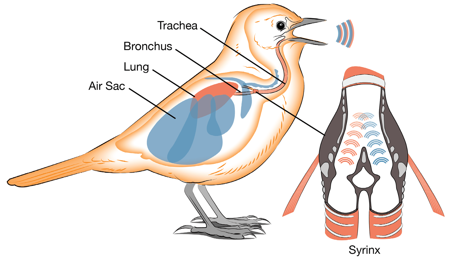
Why do birds sing, and what is involved in their songs?
Birdsong is composed of syllables that are characteristic of specific species.
It is a learned behavior, not instinctual.
The neuroanatomy of birdsong involves a complex network of nuclei and tracts in the bird's brain, which control the syrinx (the vocal organ), enabling the production of song.
Photoreceptors
•Photoreceptors are cells that contain molecules (opsins) which absorb photons in their external membrane.
What are the main types of photoreceptors in vertebrates and invertebrates?
Vertebrates have rods and cones; invertebrates have ommatidia in compound eyes.
What is the role of rods and cones in vertebrates?
Rods are responsible for dim-light vision (night vision), while cones are responsible for color vision and function in bright light.
How do photoreceptors in vertebrates and invertebrates respond to light?
In vertebrates, photoreceptors hyperpolarize (inhibit neurotransmitter release) in response to light. In invertebrates, photoreceptors depolarize (stimulate neurotransmitter release).
What photopigment do vertebrate rods and invertebrate photoreceptors use?
Vertebrate rods use rhodopsin, and invertebrate photoreceptors use various opsins for light detection.
What is the ciliary line in terms of photoreceptor cells?
The ciliary line refers to the photoreceptive part of the cell, which is derived from a modified cilium. This structure helps in detecting light and transmitting signals in the visual system.
What is the rhabdomere line in photoreceptor cells?
The rhabdomere line refers to the photoreceptive membranes that are derived from the cell body of photoreceptor cells. These membranes contain photopigments and play a key role in light detection.
What are the two classes of photoreceptors?
Ciliary photoreceptors
Rhabdomeric photoreceptors
What is the ciliary-derived visual system in vertebrates?
In vertebrates, the ciliary-derived visual system consists of ciliary photoreceptors (rods and cones) that detect light. These photoreceptors have a modified cilium structure and are responsible for converting light into electrical signals, which are processed by the brain for vision.
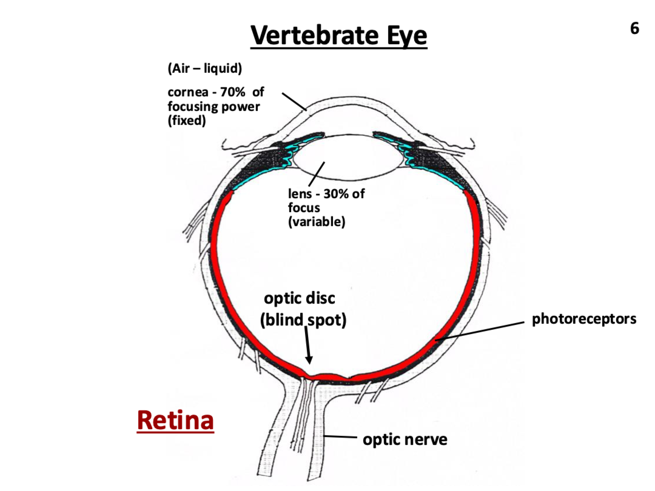
Vertebrate eye diagram
Cornea: Focuses light.
Iris: Controls pupil size to regulate light entry.
Lens: Focuses light and adjusts for distance.
Retina: Contains photoreceptors (rods and cones) to detect light.
Vitreous Humor: Maintains eye shape and transmits light to the retina.
Optic Nerve: Transmits visual signals to the brain.
Sclera: Provides structure and protection.
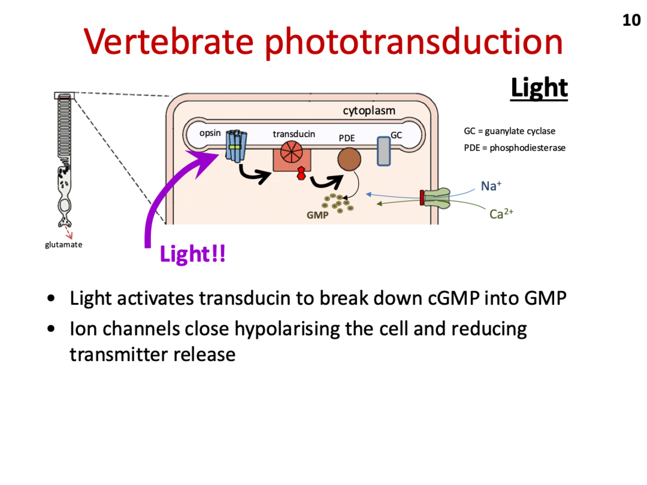
What is phototransduction?
Phototransduction is the process by which light is converted into electrical signals in the retina. It involves the activation of photoreceptor cells (rods and cones) that contain opsins.
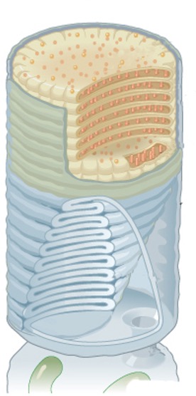
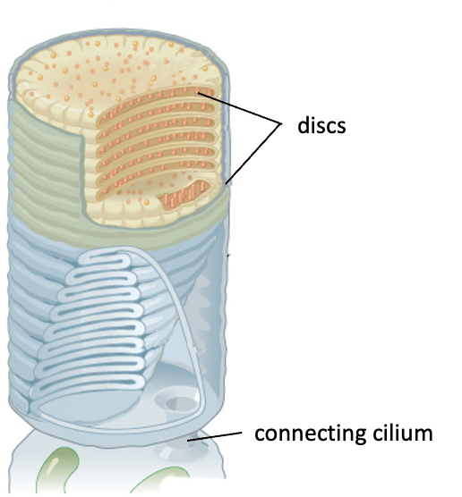
How do opsins and discs contribute to phototransduction?
Opsins are membrane-bound proteins in photoreceptor cells. The presence of discs in the photoreceptor cells greatly increases the number of opsins, enhancing light trapping efficiency and improving the cell's ability to detect light.
Why are rods more active in low light levels?
Rods have more discs in their outer segments, which contain opsin molecules. This allows them to capture more light, making them highly sensitive to low light levels.
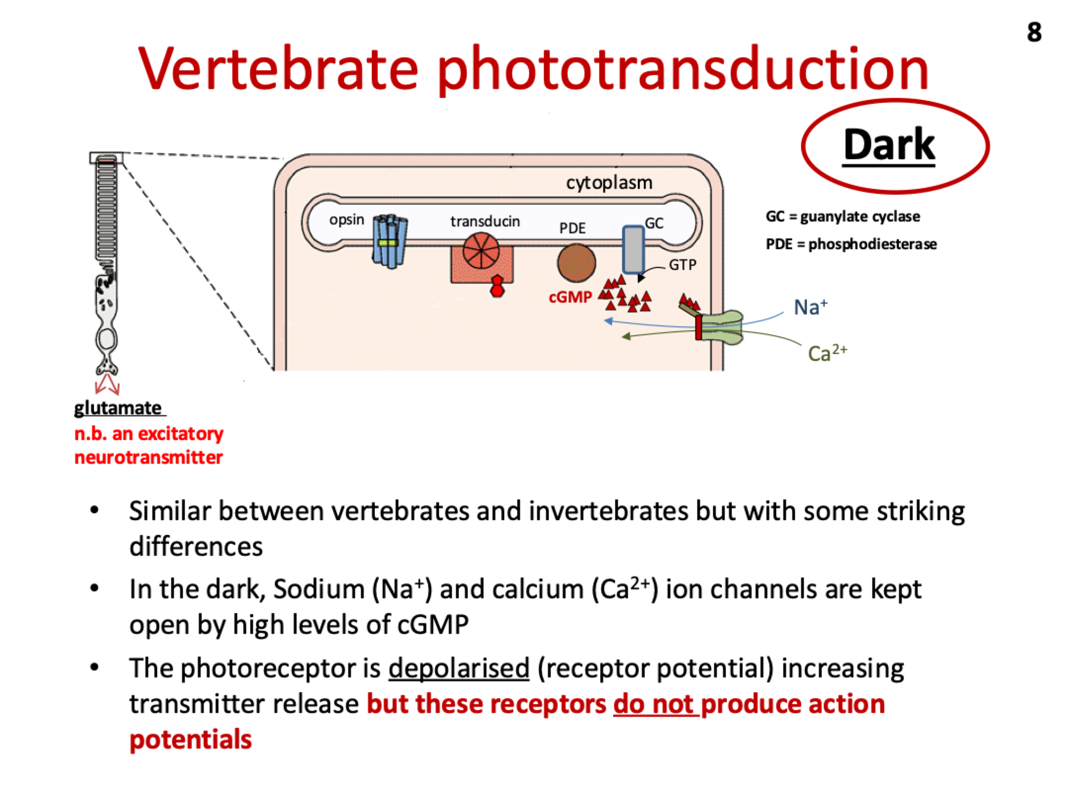
How does phototransduction work in vertebrates and what happens in the dark?
In the dark, high cGMP levels keep Na+ and Ca²⁺ channels open, causing depolarization and a steady release of neurotransmitters.
Light reduces cGMP, causing the channels to close and the photoreceptor to hyperpolarize, decreasing neurotransmitter release.
Vertebrate photoreceptors do not produce action potentials, only graded responses.
What happens in vertebrate phototransduction when light is absorbed?
Light activates transducin, which triggers phosphodiesterase (PDE) to break down cGMP into GMP.
The decrease in cGMP causes Na+ and Ca²⁺ ion channels to close, leading to hyperpolarization of the photoreceptor and a reduction in neurotransmitter release.
What are the four classes of photoreceptors in humans?
Rods (low light, no color) and Cones (red, green, blue; color vision in bright light).
What is the function of rods in the human eye?
Rods are active in low light (scotopic vision) but do not detect color.
What is the function of cones in the human eye?
Cones detect red, green, and blue light, allowing for color vision in bright light.
How does red light help with night vision?
Red light helps with dark adaptation by preserving rod function, improving vision in low light.
Do all vertebrates have trichromatic vision?
No, not all vertebrates have trichromatic vision. Some have dichromatic or other forms of color vision.
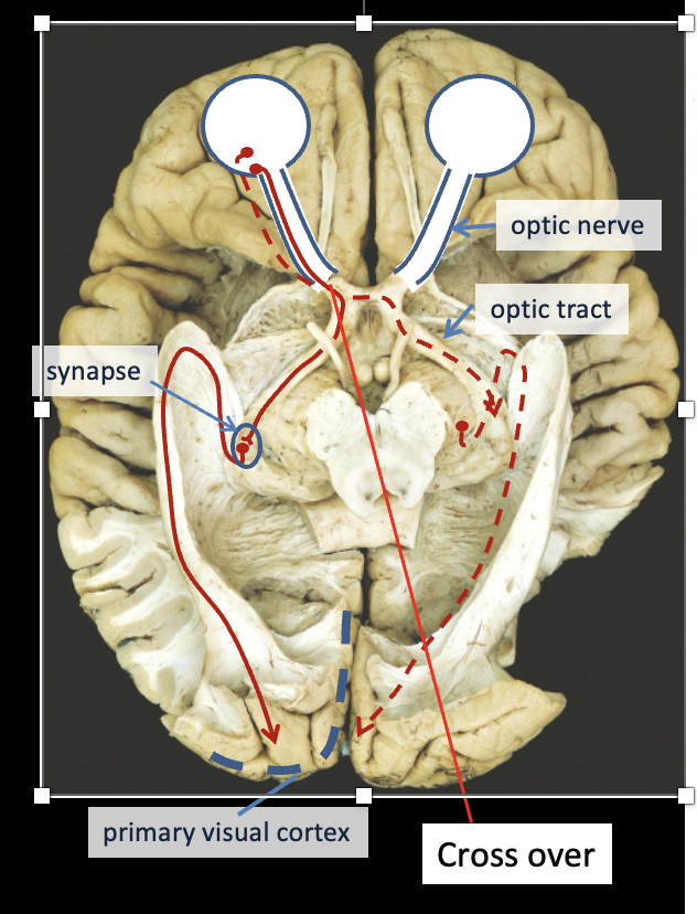
What are the key steps in the human visual pathway?
Light enters the eye and hits the retina.
Rods and cones in the retina convert light to electrical signals.
Signals travel along the optic nerve to the brain.
At the optic chiasm, the nasal retinal fibers cross, but the temporal fibers stay on the same side.
The information travels along the optic tract to the lateral geniculate nucleus (LGN) in the thalamus.
The LGN sends signals through the optic radiations to the primary visual cortex in the occipital lobe.
How is visual processing organized in the brain?
Visual processing is modular:
V1 processes basic visual features.
V4 handles color perception.
V5 processes motion and dynamic form.
How does visual processing differ in lower vertebrates (reptiles, amphibians, fish) compared to mammals?
In lower vertebrates, visual processing occurs mainly in the optic tectum (midbrain), with no visual processing in the forebrain. In mammals, the midbrain is involved in visual reflexes, while complex visual processing happens in the visual cortex of the forebrain.
Avian vision and behaviour
What is the rhabdomere-derived visual system in invertebrates?
In invertebrates, the rhabdomere-derived visual system consists of rhabdomeric photoreceptors. These photoreceptors have rhabdomeres (light-sensitive membranes) that are derived from the cell body. These systems are responsible for detecting light and enabling vision, especially in compound eyes.
How does phototransduction occur in invertebrates?
In invertebrates, light activates phospholipase C, which breaks down PIP2 into IP3 and DAG. This triggers an unknown mechanism that opens ion channels in the membrane, causing the photoreceptor to depolarize. Unlike in vertebrates, there are no action potentials generated.
INVERTEBRATE PHOTORECEPTORS
What are compound eyes?
Compound eyes are visual organs found in insects and crustaceans, composed of multiple photoreceptor units(ommatidia), each with its own lens.
What advantage does the shape of compound eyes provide?
Due to their bulbous shape, compound eyes provide nearly all-around vision, allowing insects and crustaceans to detect movement from multiple angles.