BIO 221 exam 1
 Call Kai
 Learn
Learn Practice Test
 Spaced Repetition
 Match
Match Flashcards
 Knowt Play
1/99

Earn XP
Description and Tags
molecular biology
Name | Mastery | Learn | Test | Matching | Spaced | Call with Kai | Chat |
|---|
No analytics yet
Send a link to your students to track their progress
100 Terms
types of weak chemical interactions (weak bonds)
electrostatic interaction
h bond
hydrophobic interactions (hydrophobic groups pushed together)
van der waals forces
components of an amino acid
amino group
carboxyl group
a-carbon
side chain

types of amino acids based on R group
nonpolar
uncharged polar
acidic
basic
disulfide bridge
SH oxidizes
stabilizes 3o or 4o structure
only in transmembrane or secreted proteins
uncharged polar amino acids
side chains include hydroxyl (OH) or amide (CONH2) groups
hydrophilic

acidic amino acids
negatively charged
side chain includes carboxyl group
hydrophilic
can form ionic or h bonds
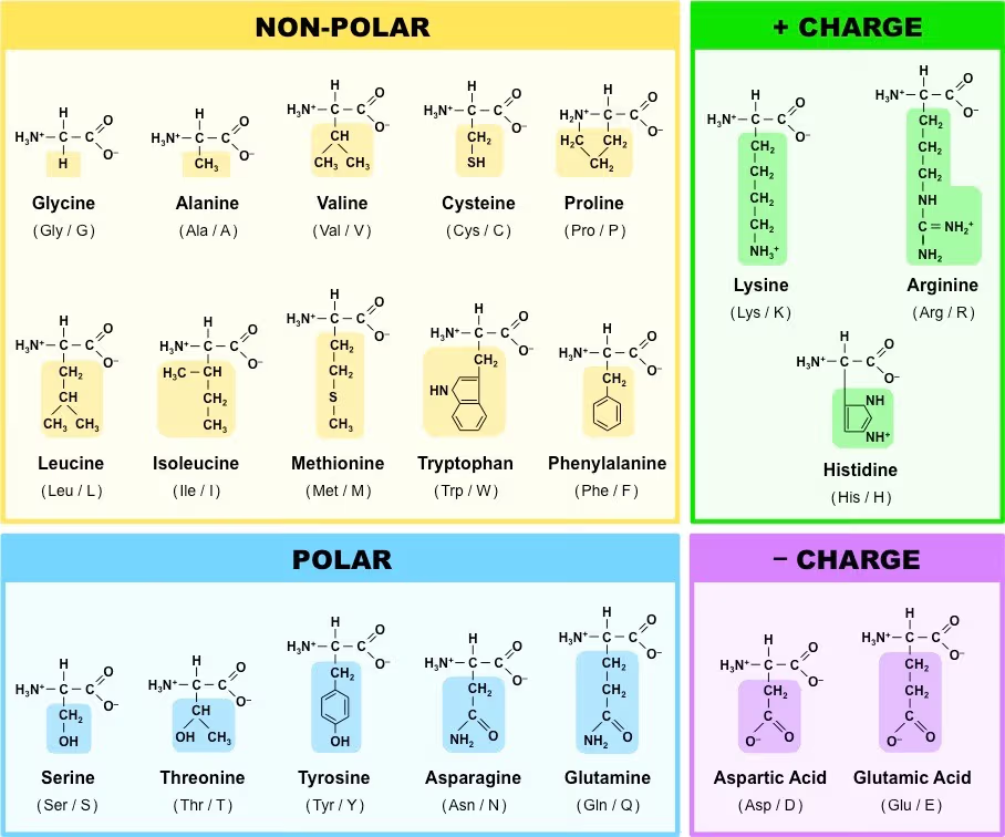
basic amino acids
positively charged
side chain includes amino group
hydrophilic
can form ionic or h bonds

special amino acids
glycine
proline
cysteine
glycine
r group is just singular H atom
lots of conformational freedom (flexible)
proline
r group bends around to form a ring by covalently bonding to alpha carbon
rigid. less conformational freedom
helix breaker
cysteine
r group can form disulfide bridges (SH)
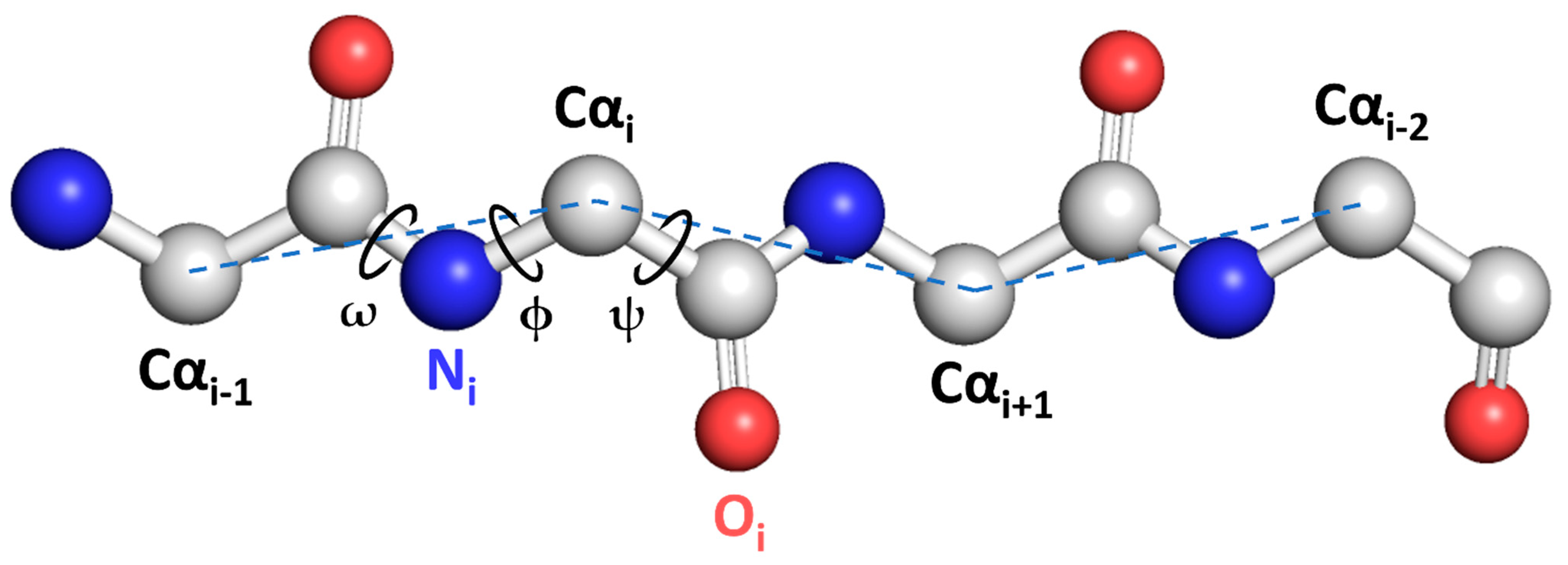
peptide bond
covalent linkage between amino acids
carboxyl to amino
rigid - partial double bond character
always written N → C
how do polypeptides adopt diff conformations if peptide bond cant rotate?
two other bonds can rotate:
phi (ϕ) - between N and alpha C (left)
psi (ψ) - between alpha C and carboxyl (right)
conformation
change spatial arrangements by rotation
configuration
to change you need to break covalent bonds
protein
polymer(s) of amino acids that is functional
linkers
transitional regions between 2° structures
turns and loops
very flexible
turns vs loops
turns allow for “reverse” direction (dramatic change in direction) - short
loop = “alternation” in dimensionality - longer section
hairpin turn (protein)
2 strands connected by a 2 residue turn
α helix
usually right handed
stabilized by h bonds
about 3.6 residues per turn
r groups pointed out
β pleated sheet
backbone nearly fully extended - maximize h bonds between strands
anti parallel or parallel
side chains point up or down
curve
coiled coil
supercoiling of α helices around each other (motif)
nonpolar side chains pointed inward
amphipathic
no intercolation
common representations of proteins
backbone, ribbon, cartoon, wire, space-filling

letter code for amino acids in peptide
letter = one letter code of AA
number = position (# from N terminus)
if there is a second letter it shows what the AA was mutated to
motif
recognizable folding pattern of 2 or more 2° structures
about 1000 diff motifs
ex: helix-turn-helix
domain
independently stable region of a protein associated with a particular function. can have multiple motifs or none
modular construction of proteins
idea that new proteins w diff functions can come from diff domain combinations
ligands
molecules that bind to proteins
include small molecules, other proteins, other macromolecules
allosteric regulation
ligand binds, changing the conformation of the protein, affecting its function
changes affinity for second ligand
regulate protein activity
reversible binding (noncovalent)
usually small molecule
positive and negative regulation
covalent modification
post translational
modification of AAs to change properties of protein
phosphorylation
acetylation
methylation
glycosylation
lipidation
addition of other proteins
disordered regions of proteins
unstructured
some are flexible hinges between domains
some allow promiscuous interaction
IDPs (intrinsically disordered proteins)
completely structurally disordered
functional promiscuity
scavengers: serve as reservoirs for ions or small molecules in solution
scaffolds that allow multiple other proteins to bind
number of bp in one turn of a dna double helix
10.5 bp
purines
guanine and adenine
pyrimidines
cytosine and thymine
uracil structure
pyrimidine
thymine without the methyl
two c=o

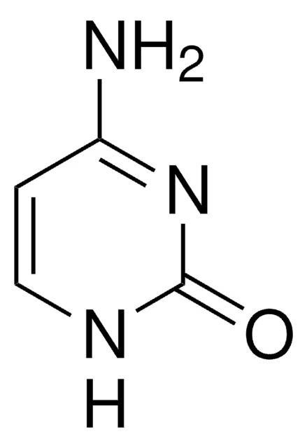
cytosine
pyrimidine
one C=O and one amine
thymine
pyrimidine
two c=o and one methyl

adenine
purine
one amine

guanine
purine
one amine and one c=o

composition of a nucleotide
sugar backbone (pentose)
phosphate
nitrogenous base

base flipping
with ATP, bases can flip out of the helix
necessary during repair and recombination
DNA transactions examples (protein-dna)
DNA replication
transcription of DNA
DNA packing by histones
DNA repair
code that proteins read in DNA double helix
A = H bond acceptors
D = H bond donors
H = nonpolar H (van der waals)
M = methyl groups (hydrophobic forces)
major and minor groove
major groove contains more information than the minor groove (can recognize A:T vs T:A)
minor groove not as accessible
B DNA
most common
right handed
high humidity
A DNA
low humidity or nonaqueous solutions
more compact
right handed
thought to help protect against damage
Z DNA
left handed
zigzag - bases arent straight
thought to be involved in gene regulation
formed to provide relief from supercoiling (torsional strain) while transcription occurs
formation can also signal DNA damage or instability
DNA double helix is never perfect
propeller twist - base pairs not in same plane
precise rotation per bp is not consistent
what denatures DNA?
heat - thermal energy disrupts h bonds
alkaline solution - OH destabilizes h bonds
formamide and urea - same
renaturation and reannealing
DNA from same source
DNA hybridization
annealing of ssDNA from two different sources
could be DNA and RNA
don’t need 100% complementarity to hybridize
hyperchromicity
increase in light absorbance (260nm) when DNA goes from ds to ss
the curve is steep at the melting point (Tm)
what influences Tm?
G:C content (more G:C = higher Tm because they are 3 h bonds)
higher ionic strength = higher Tm
alternation
when DNA is linear and then circular or the other way. ex: lambda phage is a dna virus of e coli that has linear dna that circularizes once it is injected into the bacterial cell
negative supercoil
underwound (fewer bases per turn)
constrained dna is usually in this form
easy to separate strands - good for replication and transcription
nucleosomes package dna to introduce negative supercoils
positive supercoils
less common than negative
overwound (more bases per turn)
resists unwinding of the helix - may be an adaptation to high heat
topoisomerase i
nicks DNA: only cuts one strand
adds and subtracts twists one at a time
then ligates ofc
topoisomerase ii
cuts both strands of DNA
requires ATP to rotate the DNA
adds or subtracts two twists at a time
then ligates
protein family
proteins with similar primary and tertiary structure and function
protein superfamily
two or more families with a little primary similarity but use same motifs and often have similar functions
DNA topoisomers
same chemical formula, different conformation because the strands are wrapped differently
can be separated by gel electrophoresis
EcoRI effect on supercoiled plasmid DNA
linearizes the plasmid DNA
DNAse I effect on supercoiled plasmid DNA
relaxes all supercoils but doesn’t religate (quick)
Topoisomerase effect on supercoiled plasmid DNA
relaxes supercoils and religates, but works slowly so it depends on the treatment time how much of the plasmid gets relaxed
RNA structure/properties
usually single stranded
Uracil not thymine
ribose not deoxyribose in the sugar-phosphate backbone
can naturally form complex structures by folding
stability of RNA
less stable than DNA because:
autocatalytic degradation (thru hydrolysis of backbone)
cytosine can undergo spontaneous deamination to become uracil (and its difficult for the cell to know that has happened)
uracil can mispair
uracil is more sensitive to UV than thymine
RNA function: genetic intermediate
mRNA
between gene and protein
RNA function: structural role
rRNA
structural component of ribosome (also enzymatic)
RNA function: adaptor role
tRNA
adaptor between codons and amino acids
RNA function: regulatory role
miRNA
gene regulation (where and when a gene product will be made)
RNA function: enzymatic role
ribozyme
usually involved in cleavage of other nucleotides
catalyzes addition of AAs in ribosome
RNA secondary structures
hairpin (5-10 NT loop at end)
bulge (one side)
internal loop (basically double bulge)
stem loop (>10 NT end loop)
junction (helices diverging from a single point)
pseudoknots (pairing between bases that are not contiguous - stabilizing)
tetraloop (base stacking - stabilizing)
benefits of non-Watson-Crick pairing in RNA
more self complementarity
stabilization
adds complexity to the structure
examples of ribozymes
RNA splicing - removing introns from certain mRNAs requires ribozymes
RNAse P - first ribozyme discovered. processes large tRNA precursor into tRNA
most ribozymes cleave other RNAs
some cleave themselves (hammerhead ribozyme)
evidence for RNA world hypothesis
RNA can serve as both a repository of information and as a catalyst
RNA recognition motif (RRM)
4 stranded antiparallel beta sheet with 2 alpha helices
many RNA binding proteins have this motif
surface of the sheet mediates the interaction
specifically 3 conserved residues
chromatin
the material that collectively composes the chromosome. compacts, protects, organizes DNA
histones
proteins that are major players in packaging DNA
small
positively charged
similar across eukaryotes
core and linker histones
nonhistone proteins
not as abundant as histones
regulate DNA packing, transcription, repair, replication, and recombination
nucleosomes
nucleosome core particle = NPC/core
8 histones in core and 1 linker histone
about 147 bp wrapped around core, 20+ in between cores
DNA wrapped around core in a left handed way - stabilize negative supercoils
histone names
H2A, H2B, H3, H4 = core
H1 = linker
structure of core histone
3 alpha helices separated by loops and an N-terminal tail that sticks out of the nucleosome
importance of N-terminal tails of histones
stabilize wrapping of DNA around nucleosome
highly regulated - can undergo methylation, phosphorylation, and acetylation that alters chromatin accessibility and recruits modifiers
structural basis for binding and bending DNA
14 contact sites b/w nucleosome and dna on the minor groove
142 h bonds b/w them
mostly to backbone, some to bases
h bonding facilitates bending because of the charge stabilization
heterochromatin
dense
limited expression
dark stain
associated with specific regions on chromosome (centromere and telomere)
eucromatin
open
potentially active
poor stain
less organized
higher expression
linker histone (H1)
binds to linker DNA and middle of core DNA
further tightens association- more compact
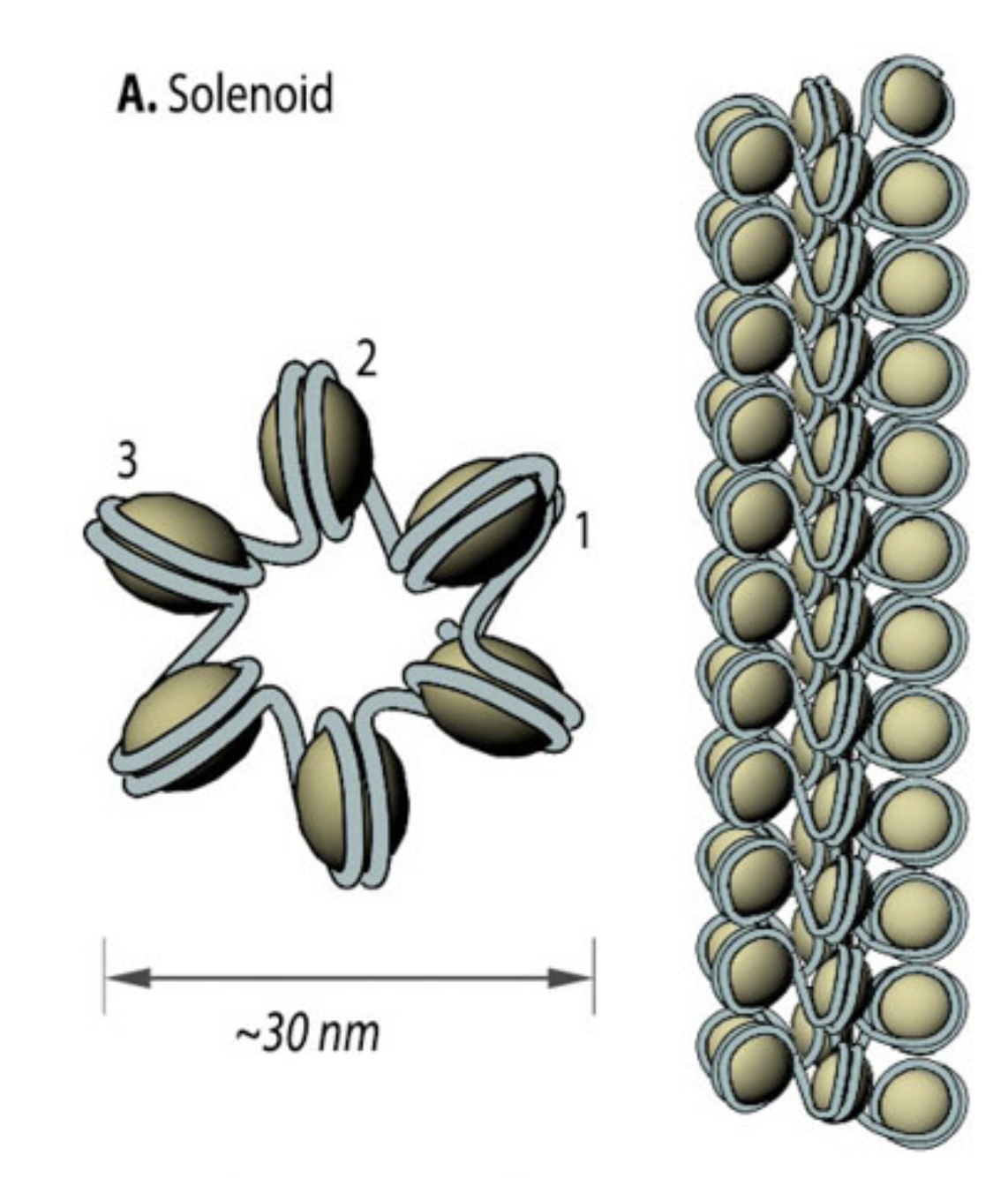
turns 10nm fibers into 30 nm
solenoid/superhelix model
structure of 30 nm fiber
flat surfaces of histones are adjacent
linker DNA buried
hole in the middle
zigzag model
structure of 30nm fiber
linker dna passes thru central axis (no hole)
exit and entry points more accessible

looped domains
further compaction of chromatin
30 nm fiber → 700 nm fiber
large loops and coils and folds
the width of a whole chromosome is 1400 nm
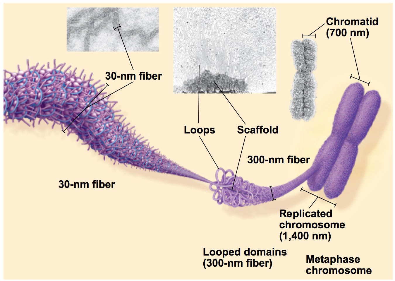
regulation of chromatin structure (chromatin remodelling)
overall to either condense or open up genomic dna to control gene expression
tails of histones get modified by enzymes
acetylation enzymes (histone modification)
histone acetyltransferase (HAT) +
histone deacetylase (HDAC) -
phosphorylation enzymes (histone modification)
histone kinase +
histone phosphatase -
methylation enzymes (histone modification)
histone methyltransferase +
histone demethylase -
takeaways from histone tail modifications
type of mod and position are important
acetylation = transcriptional activation
methylation = transcriptional silencing
what proteins recognize the histone tail modifications?
nucleosome-modifying enzymes (complexes)
nucleosome-remodeling complex
nucleosome modifying enzymes
recognize modifications and can add modifications (any)
some modifications can change chromatin structure, others recruit other proteins to do so
nucleosome remodeling complex
facilitate nucleosome movement (using ATP)
mediate the following:
sliding - movement along dna
transfer or ejection
dimer exchange - histone dimers are replaced with variants (numerous variants exist, ex: CENP-A)
CENP-A
replaces H3 in nucleosome of centromere with one with an extended tail that can attach to kinetochore proteins