cell cycle
1/42
There's no tags or description
Looks like no tags are added yet.
Name | Mastery | Learn | Test | Matching | Spaced | Call with Kai | Chat |
|---|
No analytics yet
Send a link to your students to track their progress
43 Terms
introduction
modern cell theory states that all new cells are derived from pre-existing cells through cell division
cell division involves both nuclear division and cytokinesis
two types of nuclear division
mitosis: nuclear division that produces
two genetically identical daughter nuclei → same DNA
each containing the same number of chromosomes as the parent nucleus
meiosis: nuclear division that produces
four daughter nuclei which are not genetically identical
each containing half the number of chromosomes as the parent nucleus
meiosis is also known as reduction division, since the number of chromosomes in the cell is halved (haploid)
two types of cells are obtained from cell division:
somatic cells: all body cells except the reproductive cells → mitosis gives rise to somatic cells
gametes: reproductive cells, such as sperms and eggs → meiosis gives rise to gametes
chromosome structure
chromatin which is highly condensed to become a distinct visible structure
observed in a dividing cell → a cell undergoing mitosis or meiosis
sister chromatids
each replicated chromosome appears as a double arm structure which consists of two sister chromatids joined together
each chromatid is made up of one DNA molecule
sister chromatids are genetically identical (same nucleotide sequence) as the DNA is replicated during S phase of interphase
centromere
the region at which the two sister chromatids of a single replicated chromosome are joined
spindle fibre/mitotic fibre
structure that consists of fibres made up of microtubules (proteins synthesised by ribosomes) and associated proteins (tubulin proteins)
able to lengthen or shorten → by adding or removing the tubulin proteins
centrosome
microtubule organizing centre (MTOC) in animal cells
made up of a pair of centrioles
role of centrioles during mitosis (function)
the centrioles organise the spindle fibres
to separate sister chromatids during anaphase of mitosis
each pole of the cell has a pair of centrioles
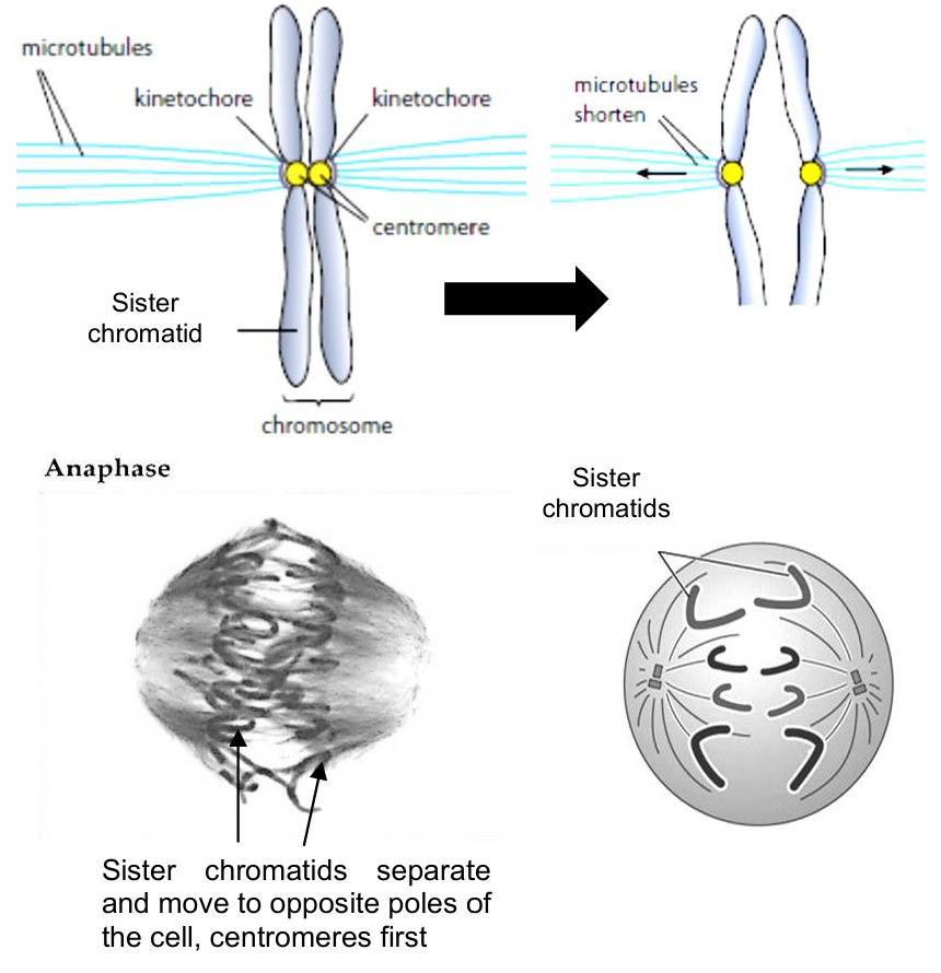
kinetochore
disc-shaped protein structure which binds to the chromatids at the centromere
location where spindle fibres (microtubules) attach during nuclear division
the spindle fibres extend from the kinetochores to the poles to the centrosome
cell cycle
for cell division to occur, cells undergo a sequence of events, known as the cell cycle
cell cycle refers to the period from which the cell is formed by cell division (i.e. start of G1 phase) to the point that the cell itself divides (i.e., end of cytokinesis)
cell cycle comprises 3 phases: interphase (majority of the time), mitosis/meiosis (nuclear division), cytokinesis
the duration of the cell cycle is highly variable
e.g.: root tip cells of onion divide once every 20 hours, while epithelial cells in the human intestine divide once every 10 hours
most types of cells never divide again after they become specialised → exits the cell cycle ⇒ protects integrity of DNA in cell
e.g. guard cells in plants and many cells of the mammalian nervous system
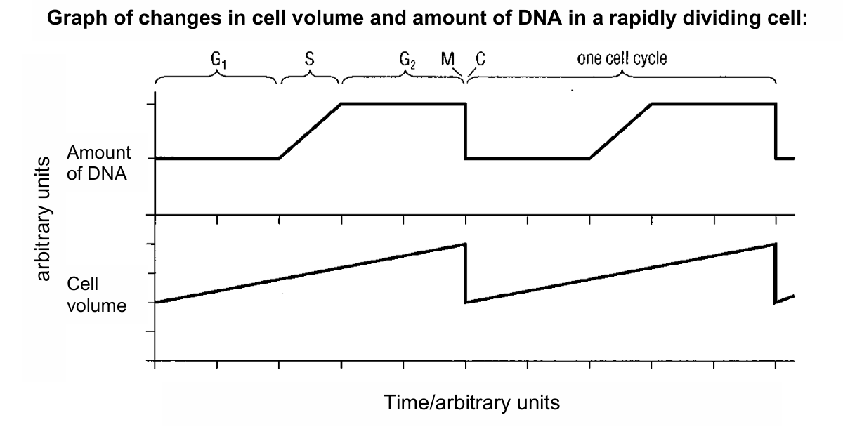
interphase
in this phase, the cell prepares for cell division
volume of cell increases as DNA is replicated and organelles are synthesised during interphase
all non-dividing specialised cells (e.g., white blood cell) exit the cell cycle and enter the G0 (resting) phase after the M phase (mitosis) → where they do not proceed further in the cell cycle
interphase comprises 3 subphases
G1 (first “gap”) phase
occurs after cytokinesis
duration is highly variable (lasting from a few hours to months or years)
cell grows and synthesizes proteins, enzymes, and RNA
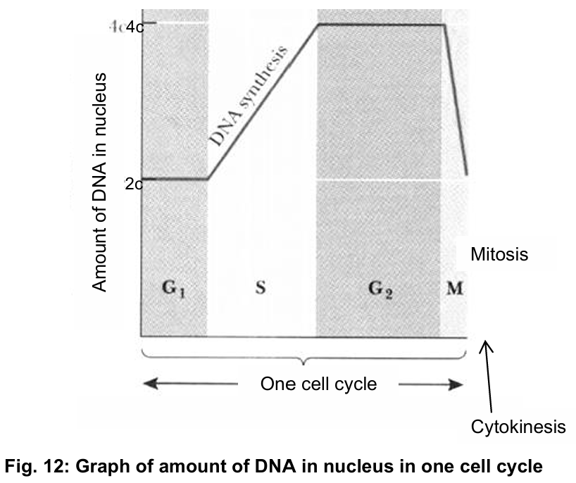
S (synthesis) phase
replication of DNA occurs → doubles the amount of DNA in the cell
one DNA molecule is replicated to form 2 DNA molecules, each DNA consisting of one daughter strand and one parental DNA strand
G2 (second “gap”) phase
cell continues to grow and synthesize proteins; it includes ribosomal proteins and proteins which make up the spindle fibres
formation of new organelles in preparation for cell division
centrosome duplicates (each daughter cell has one) in preparation for cell division
mitosis
comprises of 4 stages: prophase, metaphase, anaphase and telophase (PMAT)
overview
at G1 phase of interphase:
1 DNA molecule is found in 1 chromosome → before DNA replication
at S phase of interphase:
DNA molecule replicates to form two genetically identical DNA molecules
DNA molecules are joined at the centromere to form 1 chromosome
at prophase of mitosis:
the 2 DNA molecules will condense to form a chromosome with two sister chromatids
each sister chromatid is one DNA molecule
at telophase of mitosis and after cytokinesis:
sister chromatids are separated into two daughter cells
each daughter cell contains 1 chromosome, which is made up of 1 DNA molecule
prophase → mitosis
chromosomes become visible due to condensation of chromatin
each chromosome consists of two sister chromatids, joined at the centromere [due to DNA replication]
the nucleolus disappears → not an organelle/physical structure
in animal cells, the centrosomes (duplicated) migrate to opposite poles of the cell
spindle fibres extend from each pole towards the equator of the cell
nuclear envelope breaks down, due to the nuclear envelope fragmenting into vesicles
![<ol><li><p>chromosomes become visible due to condensation of chromatin</p></li><li><p>each chromosome consists of two sister chromatids, joined at the centromere [due to DNA replication]</p></li><li><p>the nucleolus disappears → not an organelle/physical structure</p></li><li><p>in animal cells, the centrosomes (duplicated) migrate to opposite poles of the cell</p></li><li><p>spindle fibres extend from each pole towards the equator of the cell</p></li><li><p>nuclear envelope breaks down, due to the nuclear envelope fragmenting into vesicles</p></li></ol><p></p>](https://assets.knowt.com/user-attachments/bd441cb9-131c-4b42-856c-6342aa5841fe.png)
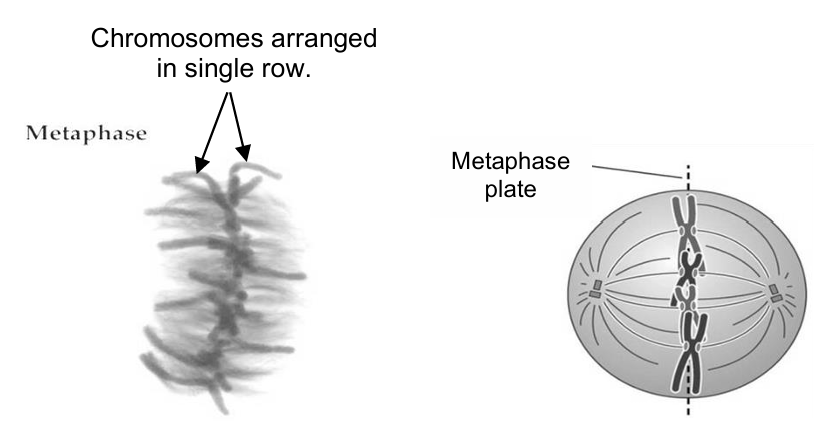
metaphase → mitosis
spindle fibres attach to the kinetochore at the centromere of the chromosome
chromosomes arrange in a single row, at the metaphase plate / equator of the cell
anaphase → mitosis
the centromere of each chromosome divides, causing the sister chromatids of each chromosome to separate
the sister chromatids move to opposite poles of the cell, centromeres first
this is due to the shortening of the spindle fibres
the cell elongates as non-kinetochore spindle fibres (do not interact with kinetochore proteins on chromosomes) lengthen
telophase → mitosis
the sister chromatids reach the respective poles of the cell and become the chromosomes of the daughter cells
the chromosomes uncoil and become chromatin → less visible
nucleolus in each nucleus reappears
nuclear envelope reforms around the chromosomes at each pole, due to the fusion of nuclear membrane vesicles
the spindle fibres break down
significance of mitosis
mitosis maintains genetic stability of an organism or a cell from one generation to the next
two daughter cells formed are genetically identical to the parent cell
daughter cells have the same number and type of chromosomes as the parent cell
mitosis occurs during the growth and development of a multicellular organism.
e.g. development of a fertilized egg (zygote) into an adult human being
mitosis occurs during the replacement of cells of worn-out tissues of the body
e.g. skin cells are constantly dying and are replaced with new identical cells
mitosis is the basis of asexual reproduction
e.g. vegetative propagation in plants
production of offspring that are identical to parents allows a population to rapidly colonise/spread in a habitat
mitosis occurs during an immune response
e.g. proliferation / cloning of activated B lymphocytes and T lymphocytes
how mitosis maintains genetic stability from one generation of cells to the next
replication of DNA occurs in the parent cell before mitosis begins
amount of DNA is doubled during S phase of interphase and halved after cytokinesis
chromosomes are arranged at the equator of the cell during metaphase
sister chromatids separate during anaphase and are evenly distributed between the two nuclei during telophase
what would happen if mitosis did not occur properly in a cell?
abnormal number of chromosomes in daughter cells → chromosomal mutation → may lead to cancer
rate of mitosis
length of cell cycle depends on:
type of cell
e.g. epithelial cells lining the intestine divide every 8 to 10 hours, however, nerve cells and red blood cells do not divide
environmental factors
e.g. food, temperature, and oxygen supply
meiosis
consists of 2 successive nuclear divisions
meiosis I → first meiotic division
meiosis II → second meiotic division
each meiotic division is divided into 4 stages: prophase, metaphase, anaphase and telophase
DNA replication during S phase of interphase (precedes meiosis)
homologous chromosomes are not normally condensed to form chromosomes during interphase
after the chromosomes replicate once, the diploid cell divides twice to yield four haploid daughter cells
meiosis is also known as reduction division, since the number of chromosomes in the cell is halved
ploidy
refers to the number of sets of chromosomes within the nucleus of a cell
diploid (2n)
a diploid cell has two sets of chromosomes, one set derived from each parent
the total number of chromosomes in a diploid cell is represented as 2n
human somatic cells have a diploid number of 46 chromosomes (2n = 46) ⇒ 23 pairs of chromosomes: 22 pairs of homologous chromosomes plus a pair of sex chromosomes in all human somatic cells
haploid (n)
haploid cell has only one set of chromosomes
only one member of each pair of chromosomes is present → either X or Y chromosomes
the total number of chromosomes in a haploid cell is represented as n
human gametes (ovum, sperm) are haploid, and they contain 23 chromosomes (n = 23)
homologous chromosomes
refers to a pair of chromosomes with the following structural features: same arm length and shape, same centromere position, same sequence of genes along the chromosome and same staining pattern (in a karyotype)
a pair of homologous chromosomes is similar but not genetically identical
they contain the same number and type of genes (e.g., genes that code for characteristics like eye colour), but they may be of different alleles (e.g. one allele
codes for blue eyes while the other allele codes for brown eyes)
inherit one chromosome of each homologous pair from each parent
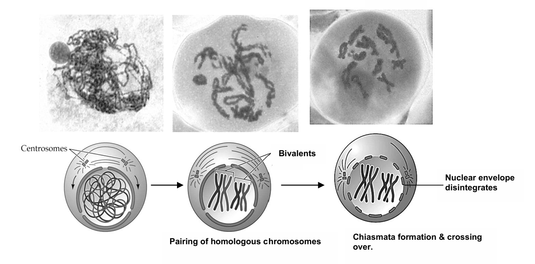
bivalents
describes homologous chromosomes that pair up during prophase I of meiosis, in a process known as synapsis
non-sister chromatids
chromatids of a pair of homologous chromosomes
have the same number and sequence of genes but may carry different alleles
non-sister chromatids should only be used when describing crossing over during prophase I of meiosis, not when describing other processes in mitosis or meiosis
synapsis
occurs during prophase I of meiosis
homologous chromosomes pair up (form bivalents) and are physically connected to each other
crossing over
exchange of corresponding sections between chromatids of a pair of homologous chromosomes when they are in synapsis
chiasma/chiasmata (plural)
x-shaped structure formed between chromatids (non-sister chromatids) of a pair of homologous chromosomes
the site(s) where corresponding sections of homologous chromosomes break and rejoin (form recombinant chromatids)
enables exchange of genetic material to occur between homologous chromosomes, in a process known as crossing over ⇒ chromatids are no longer genetically identical
prophase I → meiosis
chromosomes become visible due to condensation of chromatin
homologous chromosomes pair up in a process known as synapsis, and each pair of homologous chromosomes constitutes a bivalent
chiasmata may form between chromatids of a pair of homologous chromosomes, and crossing over occurs
centrosomes migrate to opposite poles of the cell
spindle fibres extend from each pole towards the equator of the cell
the nucleolus disappears and nuclear envelope breaks down, due to the nuclear membrane fragmenting into vesicles
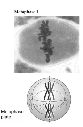
metaphase I → meiosis
spindle fibres attach to the kinetochore at the centromere of the chromosome
the homologous chromosomes arrange in two rows at the metaphase plate / equator of the cell
the arrangement of each pair of homologous chromosomes is completely independent of the arrangement of other pairs
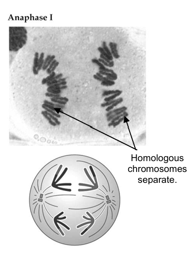
anaphase I → meiosis
homologous chromosomes separate and move to opposite poles of the cell, centromeres first
this is due to the shortening of the spindle fibres
the cell elongates as non-kinetochore spindle fibres lengthen
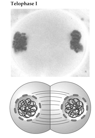
telophase I → meiosis
chromosomes reach opposite poles of the cell
nucleolus in each nucleus reappears
nuclear envelope reforms around each group of chromosomes at each pole, due to the fusion of nuclear membrane vesicles
the spindle fibres break down
end of meiosis I
at the end of meiosis I and after cytokinesis, two daughter cells are formed → each daughter cell possesses haploid number of chromosomes
the nuclei of the daughter cells (at the end of Meiosis I) may enter interphase (chromosomes uncoil) but DNA replication does not occur → only happens once in both mitosis and meiosis
in some cells of certain species, there is neither telophase I nor interphase, and the cell passes from anaphase I into prophase II directly ⇒ their chromosomes do not uncoil
cytokinesis (division of the cytoplasm) usually occurs simultaneously with telophase I, forming two haploid daughter cells
prophase II → meiosis
in cells where Telophase I and Interphase occur, the nucleolus disappears, and nuclear envelope breaks down, due to the nuclear membrane fragmenting into vesicles
if centrosomes are present, they migrate to opposite poles of the cell
the spindle fibres develop at right angles/perpendicular to the spindle axis of meiosis I
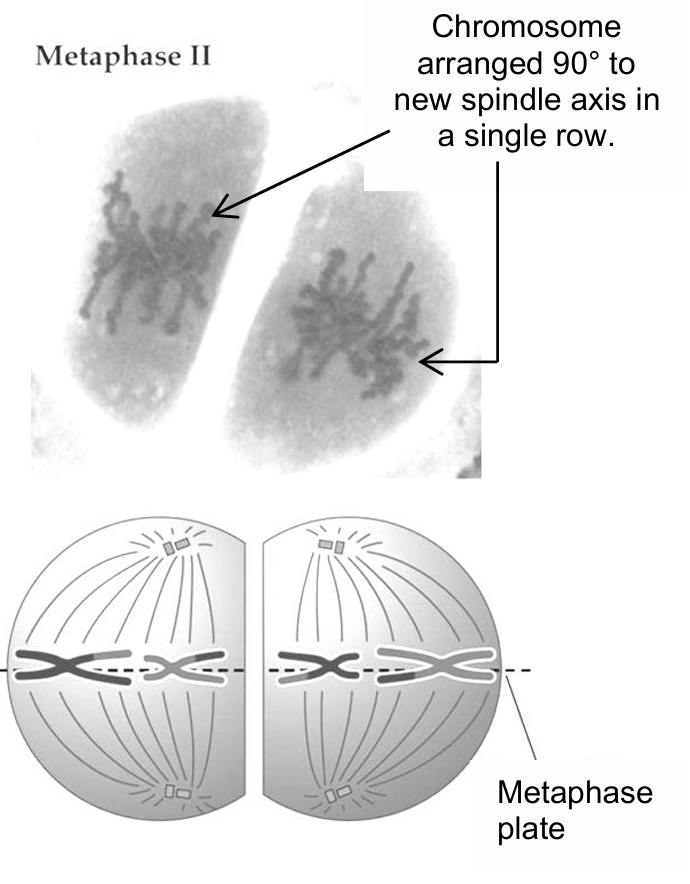
metaphase II → meiosis
spindle fibres attach to the kinetochore at the centromere of the chromosome
chromosomes arrange themselves 90° to the new spindle axis in a single row, at the metaphase plate/equator of the cell
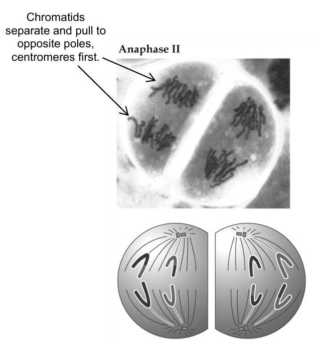
anaphase II → meiosis
the centromere of each chromosome divides, causing the chromatids of each chromosome to separate
the chromatids move to opposite poles of the cell, centromeres first
this is due to the shortening of the spindle fibres
telophase II → meiosis
the chromatids reach the opposite poles of the cell and become the chromosomes of the daughter cells
the chromosomes uncoil and become chromatin
nucleolus in each nucleus reappears
nuclear envelope reforms around the chromosomes at each pole, due to the fusion of nuclear membrane vesicles
the spindle fibres break down
at the end of meiosis II and cytokinesis, the parent cell has divided to four daughter cells → each daughter cell possesses a haploid number of chromosomes
significance of meiosis
meiosis gives rise to genetic variation between gametes through crossing over of homologous chromosomes and the independent assortment of bivalents
meiosis prevents doubling of chromosome numbers upon fusion of gametes
in sexually reproducing species, meiosis produces four haploid gametes, which are genetically non-identical
each gamete has half the number of chromosomes of the parent cell
during fertilization, the nuclei of one male and one female haploid gamete fuse to restore the diploid number (2n) of chromosomes in the zygote
if meiosis did not occur, fusion of gametes would result in a doubling in the number of chromosomes for each successive sexually reproduced generation
how meiosis brings about genetic variation:
crossing over between homologous chromosomes during prophase I → there is an exchange of genetic material between homologous chromosomes
independent assortment of chromosomes
independent arrangement of homologous chromosomes at the equator of the cell during metaphase I and their subsequent separation during anaphase I
random arrangement of the non-identical chromatids at the equator during metaphase II, and the subsequent separation of the non-identical chromatids during
anaphase II [when crossing over occurs in prophase I, the sister chromatids are no longer genetically identical]
both crossing over and independent assortment of chromosomes will lead to new combination of alleles
all 4 resultant cells are different from one another → with more pairs of homologous chromosomes, the number of possible combinations of alleles becomes enormous
a human, with 22 homologous pairs of chromosomes + 1 pair of sex chromosomes, has a potential of 223 = 8,388,608 different combinations of alleles
![<ul><li><p>crossing over between homologous chromosomes during prophase I → there is an exchange of genetic material between homologous chromosomes</p></li><li><p>independent assortment of chromosomes</p><ul><li><p>independent arrangement of homologous chromosomes at the equator of the cell during metaphase I and their subsequent separation during anaphase I</p></li><li><p>random arrangement of the non-identical chromatids at the equator during metaphase II, and the subsequent separation of the non-identical chromatids during<br>anaphase II [when crossing over occurs in prophase I, the sister chromatids are no longer genetically identical]</p></li></ul></li><li><p>both crossing over and independent assortment of chromosomes will lead to new combination of alleles</p></li><li><p>all 4 resultant cells are different from one another → with more pairs of homologous chromosomes, the number of possible combinations of alleles becomes enormous</p><ul><li><p>a human, with 22 homologous pairs of chromosomes + 1 pair of sex chromosomes, has a potential of 2<sup>23 </sup>= 8,388,608 different combinations of alleles</p></li></ul></li></ul><p></p>](https://assets.knowt.com/user-attachments/792161e1-ed70-4949-a356-e936befd90ed.png)
genetic variation
genetic variation: differences in the DNA sequences between individuals of a species
essential for evolution by providing a varied population of individuals
allows natural selection of individuals best adapted to survive under certain environmental condition, ensuring that the species would be able to survive even when environmental conditions change
meiosis, random fertilisation of gametes and mutation can lead to genetic variation
random fertilisation of gametes also contributes to genetic variation of an individual ⇒ fusion of a male and a female gamete is due to chance
differences between mitosis and meiosis
mitosis | meiosis |
|---|---|
DNA replicates once, and nucleus divides once | DNA replicates once, but there are two successive nuclear divisions |
homologous chromosomes do not pair up during prophase | homologous chromosomes associate to form bivalents in prophase I |
chiasmata are not formed | chiasmata likely to form |
crossing over does not occur | crossing over likely to occur |
chromosomes form a single row at the equator of the cell during metaphase | homologous chromosomes form two rows at the equator of the cell during metaphase I |
homologous chromosomes are not separated during anaphase | homologous chromosomes are separated during anaphase I |
two daughter cells are formed | four haploid daughter cells are formed |
daughter cells have the same number of chromosomes as the parent cell | daughter cells have only half the number of chromosomes found in the parent cell |
daughter cells are genetically identical to parent cell in absence of mutation | daughter cells are genetically different from parent cell |
cytokinesis
cytokinesis: cytoplasmic division of a cell between the two nuclei, bringing about the separation into two daughter cells
cytokinesis and mitosis/meiosis are separate processes
cytokinesis accompanies mitosis, usually beginning of telophase
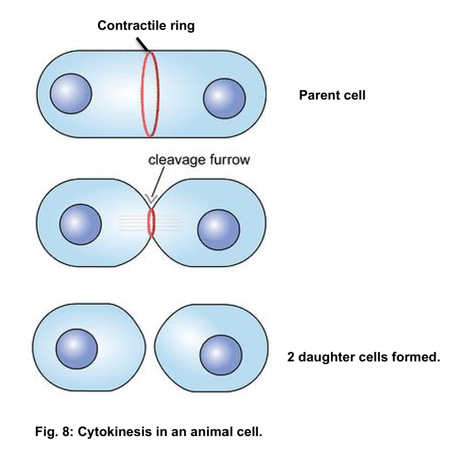
cytokinesis in animal cells
a contractile ring (protein), made of actin filaments, surrounds the dividing cell
as the contractile ring contracts, it pulls the cell surface membrane inwards to form a cleavage furrow
cleavage furrow deepens and eventually separates to form 2 daughter cells
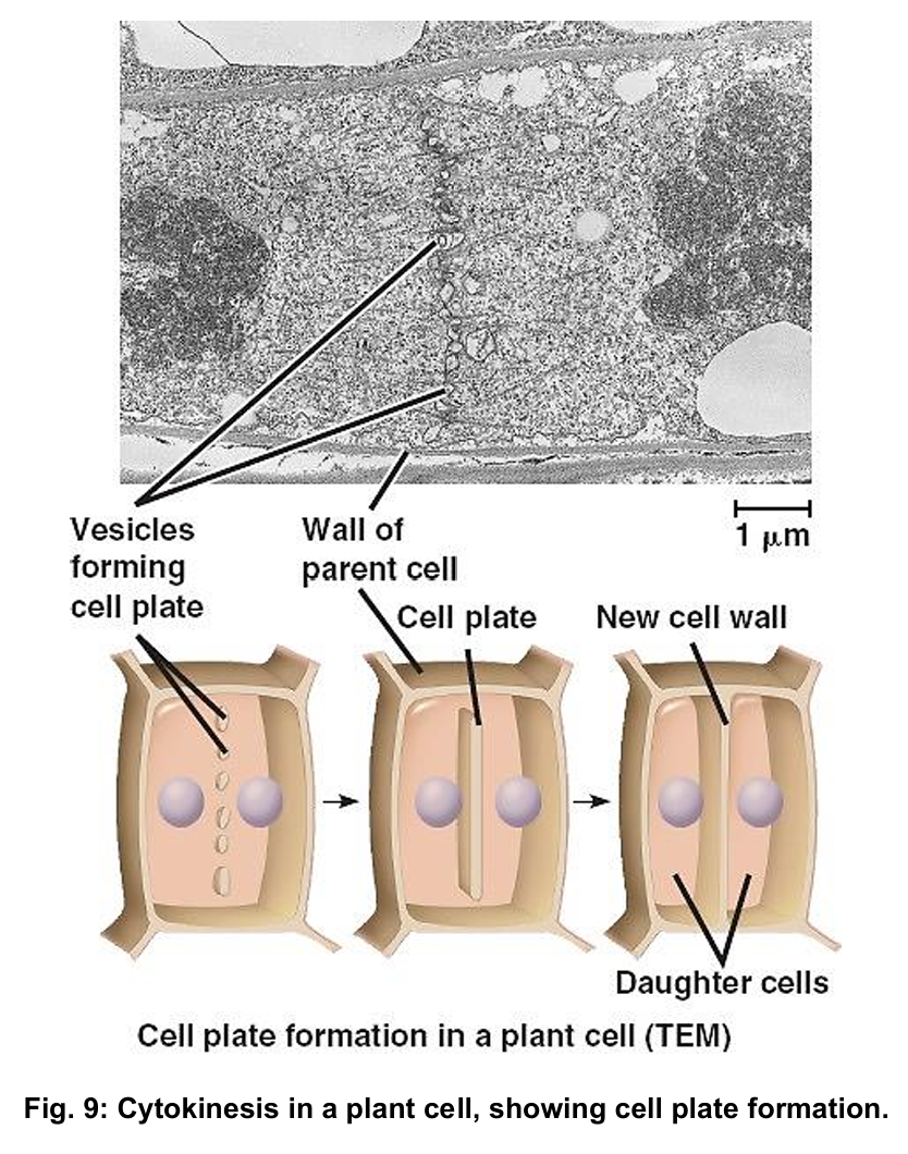
cytokinesis in plant cells
a series of Golgi vesicles appear in the middle of the parent cell
the contents of the Golgi vesicles are used to form the cell walls that separates the daughter cells
the membranes of the Golgi vesicles (phospholipid bilayer) form the new cell surface membrane
the Golgi vesicles fuse to form the cell plate, which grows outwards
the cell plate eventually fuses with the parent cell wall and cell surface membrane, separating the two daughter cells