Implantation and placentation
 Call Kai
 Learn
Learn Practice Test
 Spaced Repetition
 Match
Match Flashcards
 Knowt Play
1/62

There's no tags or description
Looks like no tags are added yet.
Name | Mastery | Learn | Test | Matching | Spaced | Call with Kai | Chat |
|---|
No analytics yet
Send a link to your students to track their progress
63 Terms
define placenta formation
foetal membranes becoming closely attached to the uterine wall to facilitate physiological exchange of gases, nutrients and waste products
what is the amnion?
the innermost fluid filled membrane
what is the function of the amnion?
allows symmetrical growth + foetus protection
temperature control and foetal movement
what is the function of the the yolk sac in:
reptiles and birds
marsupials
higher mammals
surrounds the yolk
becomes the definitive placenta
nutritional role in early pregnancy
what does the allantois come from
hind gut
what does the allantois become?
connection with the umbilical cord
what does the chorion become?
foetal contribution to placenta
what are all placentas at some stage of development?
chorionic
what are the 3 stages of implantation
apposition
adhesion
attachment or invasion
what does apposition mean?
blastocyst or foetal membrane become closely apposed during uterine lining
what is adhesion?
a biochemical interaction b/w trophoblast and epithelium
what determiens whether a placenta is attached or implanted?
whether it invades the trophoblasts or not
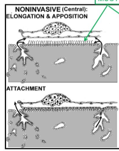
What type of implantation is this?
non-invasive

What type of implantation is this?
invasive
what inhibits trophoblast attachment to the endothelium?
MUC1
what are the 5 groups of classification of placental structure
foetal extraembryonic membranes
source of nutrition
maternal tissue shed with foetus at parturition
histological structure
macroscopic: nutrient exchange area
what is involved in foetal extraembryonic membrane placenta classification?
whether it’s
chorionic
yolk sac
allantoic in origin
what are the 2 sources of nutriton of a placenta
hemotrophic
histotrophic
what is a hemotrophic placenta?
gets its nutrition from the endometrium
what is a hemotrophic source of placental nutrition?
nutrition from circulating maternal blood
what do we classify a placenta as in terms to the maternal tissue shed?
deciduate vs non-deciduate
what are the 3 possible foetal tissue layers b/w mother and foetal circulation
endothelium
connective tissue
trophoblast
what are the 3 possible maternal layers in histological classification of the placenta?
epithelium
connective tissue
endothelium
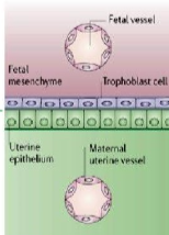
Describe this histological classification of the placenta
epitheliochorial
3 foetal layers: endothelium, connective and trophoblast
3 maternal layers: epithelial, connective and endothelial
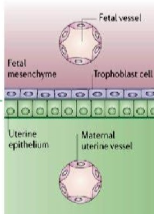
Identify this placental histological classification
epitheliochorial

State the classification of this placenta
endotheliochorial

describe this histological structure of the placenta
Endotheliochorial:
3 foetal layers: endothelial, connective, endothelial
1 maternal: endothelial

Describe what this placental hitsological classification is
Haemochorial
3 embryonic layers: endothelial, connective and trophoblast
0 maternal layers
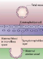
identify thsi placental classification
haemochorial
what are the 4 types of macroscopic chorionic villi distributions
diffuse
cotyledonary
zonary
discoid
what species has diffuse chorionic villi?
sow
mare
what species has cotyledonary chorionic villi
cow and ewe
what species has zonary chorionic villi
bitch
queen
what species has discoid chorionic villi
primates
what are chorionic villi
areas of nutrient exchange = funcitonal units of the placenta
describe sow placentation
epitheliochorial
diffuse
non-deciduate
describe the ewe and cow placenta
synepitheliochorial
cotyledonary
non-deciduate
why do we put ‘syn’ before cow and ewe placenta?
some trophoblasts develop into binucleate giant cells
some of these migrate and fuse to maternal epithelial cells = syncytium
what are BNC cells
binucleate giant cells
where do BNC cells come from?
trophoblasts developing from nonnucleate into binucleate and increasing in size
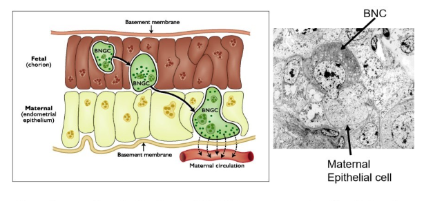

What cell type is identified here?
top = BNC
bottom = maternal epithelial cell

What is being shown taking place?
the fusion of the BNC to the maternal epithelial cell forming the syncytium
what are the 2 types of placentome
convex
concave
which species have convex placentomes
cow
giraffe
what species have concave placentomes
sheep
goats
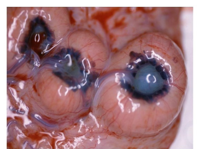
What can be identified in this?
centre = cotyledone
outer = caruncle
describe the placentation in the horse
epitheliochorial placentation
diffue
non-deciduate
what is interesting to note about cotyledones in the equine placenta
they are present but they’re microscopic

what is interesting to note about the placentation of the horse around day 24
has both a choriovitelline and chorioallantoic placenta
a small gap forms between the two = chorionic girdle
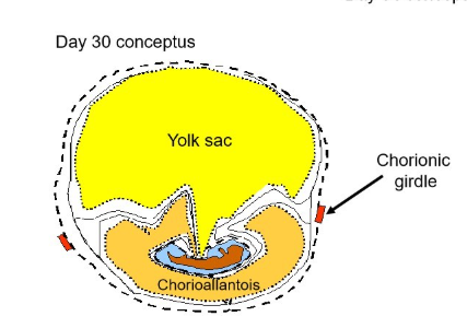
What do we call the area where the chorioallantois and choriovitelline meet?
chorionic girdle
how does the equine placenta change from day 35 (in terms of the 2 placentas)
the yolk sac regresses and the allantoi takes over until about day 50 when it’s just a remnant attached to the endometrium
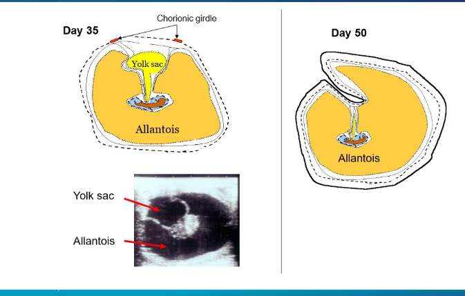
what do the points of attachment of the chorionic girdle develop into in the horse?
endometrial cups
by what day do we see equine endometrial cups, do they remain there?
day 50
no
what hormone are endometrial cups linked to?
eCG = equine chorionic gonadotrophin
what eventually happens to the endometrial cups?
surrounded adn eventually destroyed by immune cells
describe dog placentation
endothelialchorial placentaton
zonary
deciduate
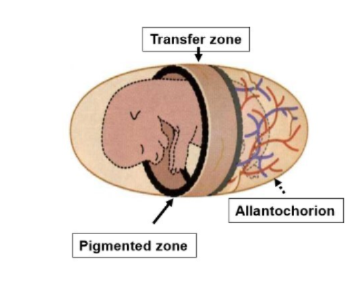
what are the parts of the zonary placenta
pigmented zone (dark band)
transfer zone (between the pigmented zones)
describe human/primate placentation
haemochorial
invasive
deciudal
what species have haemochorial placentation
human
rodents
lagomorphs
most primates
what is decidualisation
process of endometrial stromal cells proliferating and enlargening to become decidual cells
what do decidual cells become?
the decidua is the mother placenta
what are 2 roles of decidual tissue
communication b/w mother and foetus
limiting trophoblast invasion

What type of placental histology is this?
haemochorial
note the close proximity of the maternal blood to the foetal blood (only 3 layers of cells between them).